---
title: "Abdominal Wall Mass"
docid: "d51e2268-67b6-4a60-9222-f5a86f61ddec"
authors:
- key: "c1df94ab-4a9f-44c4-add7-1f174fb9ac45"
value: "Siva P. Raman, MD"
breadcrumbs:
-
name: "Gastrointestinal"
slug: "gastrointestinal"
treeNodeId: "b52263f7-5978-4a22-a17d-7260e0033943"
-
name: "Differential Diagnosis"
slug: "differential-diagnosis"
treeNodeId: "a0fd80ff-6231-49d3-94b8-ea083449979d"
-
name: "Abdominal Wall"
slug: "abdominal-wall"
treeNodeId: "08db01f7-2961-47f7-954d-2a5fca7e707d"
-
name: "Anatomically Based Differentials"
slug: "anatomically-based-differentials"
treeNodeId: "1525b44f-9d47-4ff4-8330-693211bd5eb5"
-
name: "Abdominal Wall Mass"
slug: "abdominal-wall-mass"
treeNodeId: null
category: "Gastrointestinal"
documentVersionId: "258ccc6a-0370-4b08-adc4-dfc3ef9a1ce6"
imageCount: 18
lastUpdated: "07/15/22"
pageDescription: "Abdominal Wall Mass"
pageKeywords: "Gastrointestinal, Differential Diagnosis, Abdominal Wall, Anatomically Based Differentials, Abdominal Wall Mass"
pageTitle: "Abdominal Wall Mass | STATdx"
enhancedTitle: "Abdominal Wall Mass"
type: "DDX"
references: true
breadcrumbs:
- "Gastrointestinal"
- "Differential Diagnosis"
- "Abdominal Wall"
- "Anatomically Based Differentials"
- "Abdominal Wall Mass"
---
# ESSENTIAL INFORMATION
- ## Key Differential Diagnosis Issues
- Given limitations of clinical examination, imaging plays important role in differentiating true soft tissue masses from hernias, vascular abnormalities, and normal variants
- Most soft tissue masses have nonspecific appearance and may require biopsy or excision for diagnosis
- ## Helpful Clues for Common Diagnoses
- **Abdominal Wall Hernias**
- **Inguinal hernia**
- Most common external hernia, which extends into groin anterior to horizontal plane of pubic tubercle
- Divided into direct (arises anteromedial to inferior epigastric vessels) and indirect (arises superolateral to inferior epigastric vessels) subtypes
- **Ventral hernia**
- Broad term describing acquired or congenital hernias through anterior and lateral abdominal wall
- Midline hernias include epigastric (above umbilicus) and hypogastric (below umbilicus) hernias
- Incisional hernias occur at prior surgical incision sites
- **Umbilical hernia**
- Hernias arising at midline in upper 1/2 of umbilical ring, which can be congenital or acquired
- Very common incidental finding on imaging, although usually small and asymptomatic
- **Spigelian hernia**
- Hernia through defect lateral to rectus sheath (inferior and lateral to umbilicus) often covered by external oblique muscle and aponeurosis
- **Femoral hernia**
- Groin hernia extending medial to femoral vessels with frequent compression of femoral vein
- Most common in older female patients with very high risk of strangulation and incarceration
- **Lumbar hernia**
- Hernia through defect in lumbar muscle or thoracolumbar fascia
- Can be congenital or acquired with many acquired due to incisions in flank region for renal surgery
- **Abdominal Wall Abscess**
- Loculated fluid collection (± internal gas) with peripheral enhancement and surrounding edema/fat stranding
- Differentiate drainable abscess from diffuse, nondrainable edema/fluid (cellulitis/phlegmon)
- Presence of gas-containing abdominal wall abscess in close contiguity with bowel tethered to abdominal wall raises possibility of enterocutaneous fistula
- **Sebaceous Cyst**
- Common incidental finding, appearing as small, round/oval, well-encapsulated cyst near skin surface
- Should be low density and nonenhancing without surrounding subcutaneous edema/fat stranding
- **Lipoma**
- Common incidental mass in subcutaneous tissues and between muscle planes, demonstrating uniform fat density with no internal soft tissue component
- Differentiate from liposarcoma, which demonstrates internal complexity and soft tissue component
- Confident diagnosis may be difficult on US, but mass should have similar echogenicity to subcutaneous fat
- **Keloid**
- Benign fibrotic scar tissue or tissue overgrowth at site of soft tissue injury (i.e., surgical incision or trauma)
- Usually asymptomatic but can be painful or pruritic
- No clear imaging features to allow differentiation of large keloid from other soft tissue masses
- **Hematoma**
- Heterogeneous, high-density blood products, which gradually evolve and become lower in density over time
- More diffuse subcutaneous blood products may reflect subcutaneous ecchymosis
- **Paraumbilical Varices**
- Common portosystemic collaterals in patients with severe cirrhosis and portal hypertension
- Serpiginous enhancing structures that connect to recanalized paraumbilical vein near falciform ligament
- May be visible/palpable at skin (i.e., caput medusae)
- **Injection Site**
- Common incidental finding usually secondary to injection of heparin, insulin, or other medications
- Small nodular foci with ectopic gas, blood, or fluid
- May chronically evolve into injection granulomas, appearing as rounded or linear foci of soft tissue or calcification (most common in buttocks)
- **Calcified Scar**
- Heterotopic ossification (myositis ossificans traumatica) can occur at abdominal incision sites and is most common in linear alba after midline abdominal incision
- Ossified scar in incision can resemble rib (with both cortex and medulla)
- **Muscle Asymmetry (Mimic)**
- May be mistaken for mass and are common secondary to prior surgery, paralysis, myopathy, etc.
- **Melanoma**
- 5th most common new cancer in US, but imaging typically not utilized for diagnosis of primary tumor
- Most commonly multiple small subcutaneous nodules, although rarely presents as solitary abdominal wall mass
- Homogeneous enhancement ± hyperintense on T1 MR
- ## Helpful Clues for Less Common Diagnoses
- **Endometriosis**
- Endometriosis implants may be seen within incision sites after prior C-section or hysterectomy
- Typically appears as solid, spiculated subcutaneous mass with variable enhancement (usually hypointense on T1 and hyperintense on T2 MR)
- May be associated with clinical history of cyclical pain (corresponding with menstruation) at incision site
- **Calcinosis Syndromes**
- Dystrophic: Calcifications may be due to tissue injury response, such as implanted medical device, connective tissue diseases (scleroderma, dermatomyositis, CREST), severe pancreatitis, or fat necrosis
- Metastatic: Most often in patients with calcium-phosphate imbalance (renal failure, milk-alkali syndrome)
- Tumoral calcification: Large globular deposits of calcification near joints
- **Soft Tissue Metastases**
- Most common malignancies to metastasize to soft tissues are melanoma and renal cell carcinoma
- Soft tissue nodule or mass(es) in subcutaneous fat or muscle with enhancement similar to primary tumor
- Easily overlooked on CT if careful survey of soft tissues not undertaken, but often more apparent on PET
- Tumor may also be implanted at site of surgery (probably more common with laparoscopic surgery) or biopsy
- Surgical seeding can also occur with benign lesions, including uterine fibroids and ectopic splenic tissue
- **Lymphoma and Leukemia**
- Cutaneous T-cell lymphoma (a.k.a. mycosis fungoides or Sézary syndrome)
- Skin 2nd most common site of extranodal lymphoma (after GI tract)
- Skin involvement may be difficult to appreciate on imaging unless unusually nodular or mass-like
- Subcutaneous panniculitis-like T-cell lymphoma
- Manifests as site of soft tissue induration/infiltration or as discrete nodules
- Leukemia cutis (i.e., chloroma or granulocytic sarcoma)
- Primary B-cell cutaneous lymphomas more likely to present as solitary isolated skin lesion
- Posttransplant lymphoproliferative disorders (PTLD) can rarely manifest in subcutaneous soft tissues
- **Desmoid**
- Benign locally aggressive neoplasm, which can be intraabdominal or extraabdominal (e.g., abdominal wall)
- Abdominal wall lesions most frequently arise from rectus or oblique muscles, especially at incision sites
- Major risk factors include prior surgery, trauma, Gardner syndrome, and familial adenomatous polyposis
- Variable appearance but typically solid, well-defined, hypoenhancing, heterogeneously high signal on T2 and low signal on T1 MR
- **Sarcoma**
- Malignant mesenchymal soft tissue tumors, which encompass wide range of different histologic subtypes
- May be difficult to differentiate from other soft tissue masses based on imaging alone, although most sarcomas tend to be larger and more heterogeneous with frequent necrosis (± distant metastatic disease)
- Different subtypes of sarcomas cannot be differentiated on imaging with any accuracy
- **Rhabdomyolysis**
- Muscle necrosis in response to wide variety of causes, including crush injury, seizures, statin medications, etc.
- Involved muscles on CT generally appear either normal or abnormally hypodense (due to edema)
- MR more sensitive, with muscles demonstrating T2 hyperintensity and enlargement, as well as hyperenhancement (can appear ring-like or mass-like)
- Commonly leads to severe renal damage due to release of myoglobin into bloodstream
- **Pancreatic Panniculitis**
- Subcutaneous fat necrosis seen with pancreatitis and pancreatic adenocarcinoma (due to ↑ serum lipase)
- Manifest as small nodular foci of predominantly fat density on CT and hyperechoic on US
- **Kaposi Sarcoma**
- Most common AID-related vascular neoplasm in Western world, presenting as either diffuse infiltration of skin or discrete subcutaneous nodules
## References
# Selected References
1. [Ballard DH et al: Imaging of abdominal wall masses, masslike lesions, and diffuse processes. Radiographics. 40(3):684-706, 2020](http://www.ncbi.nlm.nih.gov/pubmed/?term=32330085%5Bpmid%5D)
1. [Draghi F et al: Abdominal wall sonography: a pictorial review. J Ultrasound. 23(3):265-78, 2020](http://www.ncbi.nlm.nih.gov/pubmed/?term=32125676%5Bpmid%5D)
1. [Kania LM et al: Interpreting body MRI cases: classic findings in pelvic MRI. Abdom Radiol (NY). 45(9):2916-30, 2020](http://www.ncbi.nlm.nih.gov/pubmed/?term=32607649%5Bpmid%5D)
1. [Mao A et al: Post-cesarean section abdominal wall endometrioma. Cureus. 12(8):e10088, 2020](http://www.ncbi.nlm.nih.gov/pubmed/?term=33005511%5Bpmid%5D)
1. [Youssef AT: The ultrasound of subcutaneous extrapelvic endometriosis. J Ultrason. 20(82):e176-80, 2020](http://www.ncbi.nlm.nih.gov/pubmed/?term=33365153%5Bpmid%5D)
1. [Hensen JH et al: Abdominal wall endometriosis: clinical presentation and imaging features with emphasis on sonography. AJR Am J Roentgenol. 186(3):616-20, 2006](http://www.ncbi.nlm.nih.gov/pubmed/?term=16498086%5Bpmid%5D)
1. [Zafar HM et al: Anterior abdominal wall hernias: findings in barium studies. Radiographics. 26(3):691-9, 2006](http://www.ncbi.nlm.nih.gov/pubmed/?term=16702448%5Bpmid%5D)
1. [Aguirre DA et al: Abdominal wall hernias: imaging features, complications, and diagnostic pitfalls at multi-detector row CT. Radiographics. 25(6):1501-20, 2005](http://www.ncbi.nlm.nih.gov/pubmed/?term=16284131%5Bpmid%5D)
1. [Shadbolt CL et al: Imaging of groin masses: inguinal anatomy and pathologic conditions revisited. Radiographics. 21 Spec No:S261-71, 2001](http://www.ncbi.nlm.nih.gov/pubmed/?term=11598262%5Bpmid%5D)
## Images
### Selected Images
 containing loops of nonobstructed small bowel.](images/app.statdx.com_image_thumbnail_dcf3e531-1181-43fa-8a2a-588ef14eb1de_annotated_true_size_900_quality_90_7a3efacf7c9ea3cb8a17271f24a81f1ac6d54288.jpg)
**Abdominal Wall Hernias**
*Axial CECT shows a right inguinal hernia containing loops of nonobstructed small bowel.*

**Abdominal Wall Hernias**
*Axial CECT shows a right inguinal hernia containing loops of nonobstructed small bowel.*

**Sebaceous Cyst**
*Axial CECT shows an encapsulated, near water density mass in the left buttock. Sebaceous cysts are a common incidental finding and, when demonstrating a classic appearance, do not require further follow-up or evaluation.*

**Lipoma**
*Coronal CECT shows a large, fat-containing mass within the right lateral abdominal wall, compatible with a simple lipoma. Note the absence of any complexity or soft tissue component within the mass.*

**Hematoma**
*Axial CECT shows an acute, high-density subcutaneous hematoma in a patient with recent trauma.*

**Hematoma**
*Axial CECT in a patient with cirrhosis and portal hypertension shows subcutaneous varices overlying the anterior abdominal wall, representing a caput medusae.*

**Paraumbilical Varices**
*Axial T1 C+ MR shows an enhancing mass in the left anterior pelvic wall, found to represent a scar endometrioma in this patient status post prior laparoscopic pelvic surgery.*

**Endometriosis**
*Sagittal CECT shows a soft tissue mass intimately associated with the umbilicus, ultimately found at biopsy to represent endometriosis.*

**Soft Tissue Metastases**
*Axial CECT shows a hypodense mass in the midline anterior abdominal wall, proven to represent a metastasis from the patient's known primary colon cancer.*

**Soft Tissue Metastases**
*Axial CECT shows a hypodense mass in the abdominal wall musculature, representing a metastasis from the patient's known colon cancer.*
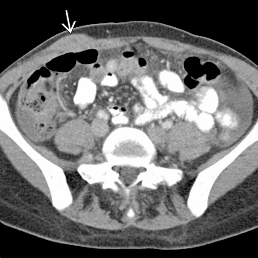
**Lymphoma and Leukemia**
*Axial CECT shows a biopsy-proven chloroma in the right anterior abdominal wall in a patient with known leukemia.*

**Desmoid**
*Axial CECT shows multiple large, hypodense masses in the pelvic subcutaneous soft tissues in a patient with known familial polyposis, representing desmoid tumors.*

**Desmoid**
*Axial CECT shows a hypodense mass in the right anterior abdominal wall, ultimately found to represent a desmoid tumor.*

**Sarcoma**
*Axial CECT shows a large, rapidly growing mass in the left anterior abdominal wall, representing a primary soft tissue sarcoma (malignant fibrous histiocytoma).*

**Sarcoma**
*Axial T1 C+ FS MR shows a highly invasive, large tumor in the buttock , which enhances significantly. This lesion proved on biopsy to be a high-grade epithelioid sarcoma.*
### Additional Images

**Abdominal Wall Hernias**
*Axial CECT shows a right inguinal hernia containing loops of nonobstructed small bowel.*

**Abdominal Wall Hernias**
*Axial CECT shows a right inguinal hernia containing loops of nonobstructed small bowel.*

**Sebaceous Cyst**
*Axial CECT shows an encapsulated, near water density mass in the left buttock. Sebaceous cysts are a common incidental finding and, when demonstrating a classic appearance, do not require further follow-up or evaluation.*

**Lipoma**
*Coronal CECT shows a large, fat-containing mass within the right lateral abdominal wall, compatible with a simple lipoma. Note the absence of any complexity or soft tissue component within the mass.*

**Hematoma**
*Axial CECT shows an acute, high-density subcutaneous hematoma in a patient with recent trauma.*

**Hematoma**
*Axial CECT in a patient with cirrhosis and portal hypertension shows subcutaneous varices overlying the anterior abdominal wall, representing a caput medusae.*

**Paraumbilical Varices**
*Axial T1 C+ MR shows an enhancing mass in the left anterior pelvic wall, found to represent a scar endometrioma in this patient status post prior laparoscopic pelvic surgery.*

**Endometriosis**
*Sagittal CECT shows a soft tissue mass intimately associated with the umbilicus, ultimately found at biopsy to represent endometriosis.*

**Soft Tissue Metastases**
*Axial CECT shows a hypodense mass in the midline anterior abdominal wall, proven to represent a metastasis from the patient's known primary colon cancer.*

**Soft Tissue Metastases**
*Axial CECT shows a hypodense mass in the abdominal wall musculature, representing a metastasis from the patient's known colon cancer.*

**Lymphoma and Leukemia**
*Axial CECT shows a biopsy-proven chloroma in the right anterior abdominal wall in a patient with known leukemia.*

**Desmoid**
*Axial CECT shows multiple large, hypodense masses in the pelvic subcutaneous soft tissues in a patient with known familial polyposis, representing desmoid tumors.*

**Desmoid**
*Axial CECT shows a hypodense mass in the right anterior abdominal wall, ultimately found to represent a desmoid tumor.*

**Sarcoma**
*Axial CECT shows a large, rapidly growing mass in the left anterior abdominal wall, representing a primary soft tissue sarcoma (malignant fibrous histiocytoma).*

**Sarcoma**
*Axial T1 C+ FS MR shows a highly invasive, large tumor in the buttock , which enhances significantly. This lesion proved on biopsy to be a high-grade epithelioid sarcoma.*
### Additional Images
 with extensive varices
  in the parastomal region. These develop in patients with portal hypertension (e.g., following colectomy for primary sclerosing cholangitis with cirrhosis).](images/app.statdx.com_image_thumbnail_b593a259-7880-4bc0-893a-3cede1985b33_annotated_true_size_900_quality_90_d23259d664dcbe385fadc731b06bfaabbe8cb8ed.jpg)
**Paraumbilical Varices**
*Axial CECT shows a colostomy with extensive varices in the parastomal region. These develop in patients with portal hypertension (e.g., following colectomy for primary sclerosing cholangitis with cirrhosis).*

**Paraumbilical Varices**
*Axial CECT shows prominent parastomal varices .*

**Paraumbilical Varices**
*Axial CECT shows a cirrhotic liver and a large parumbilical varix .*

**Paraumbilical Varices**
*Axial CECT shows continuation of the parumbilical varix with collaterals in the rectus muscles and subcutaneous fat (caput medusae).*
in the parastomal region. These develop in patients with portal hypertension (e.g., following colectomy for primary sclerosing cholangitis with cirrhosis).](images/app.statdx.com_image_thumbnail_b593a259-7880-4bc0-893a-3cede1985b33_annotated_true_size_900_quality_90_d23259d664dcbe385fadc731b06bfaabbe8cb8ed.jpg)
**Paraumbilical Varices**
*Axial CECT shows a colostomy with extensive varices in the parastomal region. These develop in patients with portal hypertension (e.g., following colectomy for primary sclerosing cholangitis with cirrhosis).*

**Paraumbilical Varices**
*Axial CECT shows prominent parastomal varices .*

**Paraumbilical Varices**
*Axial CECT shows a cirrhotic liver and a large parumbilical varix .*

**Paraumbilical Varices**
*Axial CECT shows continuation of the parumbilical varix with collaterals in the rectus muscles and subcutaneous fat (caput medusae).*