---
title: "Aberrant Right Subclavian Artery"
docid: "a20702fe-5409-44a5-a804-cb071023bade"
authors:
- key: "770e1d77-2287-436e-910b-48232afc7842"
value: "Prabhakar Rajiah, MBBS, MD, FACR, FRCR, FACC, FAHA, FSCCT"
- key: "e915766e-8102-46e4-a33e-c83f8ae12f29"
value: "Harold Goerne, MD"
breadcrumbs:
-
name: "Cardiac"
slug: "cardiac"
treeNodeId: "fa90100b-619c-430e-8074-b5b9789bab39"
-
name: "Diagnosis"
slug: "diagnosis"
treeNodeId: "5c92cf4f-e9d5-4059-9c13-22255c51c121"
-
name: "Aorta"
slug: "aorta"
treeNodeId: "4d206a6b-1a82-467c-9199-0df25ab749d7"
-
name: "Aberrant Right Subclavian Artery"
slug: "aberrant-right-subclavian-artery"
treeNodeId: null
category: "Cardiac"
documentVersionId: "69195035-5819-4996-b27d-b107c62d7c60"
imageCount: 12
lastUpdated: "01/28/25"
pageDescription: "Aberrant Right Subclavian Artery"
pageKeywords: "Cardiac, Diagnosis, Aorta, Aberrant Right Subclavian Artery"
pageTitle: "Aberrant Right Subclavian Artery | STATdx"
enhancedTitle: "Aberrant Right Subclavian Artery"
type: "DX"
references: true
breadcrumbs:
- "Cardiac"
- "Diagnosis"
- "Aorta"
- "Aberrant Right Subclavian Artery"
---
# KEY FACTS
- ## Terminology
- Aberrant right subclavian artery (ARSA) as last branch of left-sided aortic arch, isthmus, or proximal descending aorta
- Course: Retroesophageal 80%; intertracheoesophageal 15%; pretracheal 5%
- ## Imaging
- Barium esophagogram shows persistent esophageal narrowing at level of extrinsic compression with oblique course
- CTA or MRA help to understand origin and course of supraaortic trunks
- No vascular ring formed by left aortic arch and ARSA
- Loose vascular ring if ARSA originates from Kommerell diverticulum
- Complete vascular ring if ARSA with circumflex right descending thoracic aorta with right ductus
- ## Top Differential Diagnoses
- Major aortopulmonary collateral arteries (MAPCAs)
- Right arch with aberrant left subclavian artery
- Retroesophageal diverticulum
- ## Pathology
- Regression of right 4th arch between right subclavian and right common carotid arteries, including right ductus arteriosus
- ## Clinical Issues
- Most common congenital aortic arch abnormality (0.5-2.0%)
- 90-95% asymptomatic; incidental imaging finding
- Adults: Dysphagia (lusoria), dyspnea, back pain
- Infants: Cough, stridor, aspiration pneumonia
- Surgery for symptoms, aneurysm, or large diverticulum
# TERMINOLOGY
- ## Abbreviations
- Aberrant right subclavian artery (ARSA)
- ## Synonyms
- Lusoria artery (arteria lusoria)
- Incomplete vascular ring
- ## Definitions
- Aberrant origin of right subclavian artery as last branch of left-sided aortic arch or proximal descending aorta
- Normally RSA originates from brachiocephalic artery
# IMAGING
- ## General Features
- ### Best diagnostic clue
- ARSA as last branch of left-sided aortic arch or proximal descending aorta
- Retroesophageal/retrotracheal course of ARSA
- ### Location
- ARSA takeoff from distal aortic arch, isthmus, or proximal descending aorta
- Course: Retroesophageal 80%; intertracheoesophageal 15%; pretracheal 5%
- ### Size
- Usually normal vessel caliber
- Dilated proximal segment in Kommerell diverticulum
- Aneurysm may be present
- ### Morphology
- No vascular ring formed by left aortic arch and ARSA
- Trachea and esophagus are in middle between aortic arch and ARSA
- Loose vascular ring if ARSA originates from Kommerell diverticulum
- Right ductus/ligamentum arteriosum completes ring
- Ductus arising from junction between diverticulum and normal-sized subclavian artery
- Seen in 15-30% of ARSA
- Complete vascular ring if ARSA with circumflex right descending thoracic aorta and right ductus
- Arch itself crosses midline, posterior to esophagus
- Right ductus extends from descending aorta to right pulmonary artery, completing vascular ring
- ## Radiographic Findings
- Chest x-ray may show enlargement of superior mediastinum
- Barium esophagogram shows persistent esophageal narrowing at level of extrinsic compression with oblique course
- ## CT Findings
- ### CTA
- Excellent modality to demonstrate aortic arch configuration and branching pattern
- MPR, MIP, and VRT reconstructions help to understand origin and course of supraaortic trunks
- Evaluates esophageal and tracheal compression
- NECT: May be seen incidentally
- 4D CT: May show dynamic esophageal compression during cardiac cycle due to distension of ARSA during systole
- ## MR Findings
- ### MRA
- Arch vessel branching pattern is (from right to left): Right common carotid artery; left common carotid artery; left subclavian artery; right subclavian artery (RSA)
- Course: Obliquely from caudal left to cranial right
- Aneurysm of ARSA: 1.5x d of distal subclavian artery
- Left ductus may be seen; no vascular ring
- Since there is vasculature on only 3 sides of trachea and esophagus
- Posterior esophageal compression in 10%
- Usually in 4th or 5th decade
- Stretching, sclerosis/calcification, aneurysm of ARSA
- Complete vascular ring may be seen if
- Kommerell diverticulum: Dilation of origin of ARSA
- Diameter of orifice of diverticulum and distance from furthest aortic wall to end of diverticulum measured
- Circumflex right descending thoracic aorta with arch behind esophagus
- ### SSFP cine
- May show dynamic esophageal compression during cardiac cycle due to vessel distension during systole
- ## Imaging Recommendations
- ### Best imaging tool
- CTA or MRA
- ### Protocol advice
- ECG gating is not necessary if only evaluation of vascular ring is desired
- High-pitch helical mode of dual source CT scanner (Flash) mode, provide anatomic information
# DIFFERENTIAL DIAGNOSIS
- ## Major Aortopulmonary Collateral Arteries
- Collateral vessels arising from descending aorta that end in lungs in patients with pulmonary artery atresia
- [Right Arch With Aberrant Left Subclavian Artery](/document/right-aortic-arch/5f186c96-4cc3-453e-840d-12ebfad13115)
- Mirror image of left arch with ARSA
- Caused by persistent right 4th arch and regression of left 4th arch in between left common carotid and left subclavian arteries
- Aberrant left subclavian artery originates as last branch from right aortic arch and courses behind esophagus to reach left
- 90% have left ductus vascular ring
- 2nd most common cause of complete vascular ring
- ## Retroesophageal Diverticulum
- Outpouching at origin of aberrant left subclavian artery in right aortic arch; seen in 60% of these cases
# PATHOLOGY
- ## General Features
- ### Etiology
- Embryologic insult during formation of aortic arches
- Right 4th arch regresses between RSA and right common carotid artery, including ductus arteriosus
- Distal right dorsal aorta forms proximal RSA
- Distal portion from 7th right intersegmental artery
- Kommerell diverticulum: Remnant of dorsal aortic arch
- Persistence of right 6th arch component that forms right ductus arteriosus
- Left circumflex aorta
- Regression of right 4th arch between right common carotid and right subclavian arteries
- Persistence of right 6th arch component that forms ductus
- Right-sided descending aorta
- Distal left dorsal aorta forms definitive distal aortic arch
- ### Associated abnormalities
- ARSA is present in 35% of patients with Down syndrome
- Kommerell diverticulum: 14.9%
- Aneurysm of distal RSCA: 12.8%
- Others: Coarctation; patent ductus arteriosus; ventricular septal defect; truncus bicaroticus; type B interrupted arch
- ## Staging, Grading, & Classification
- Retroesophageal course: 80%
- Interesophageotracheal course: 15%
- Pretracheal course: 5%
# CLINICAL ISSUES
- ## Presentation
- ### Most common signs/symptoms
- 90-95% asymptomatic; usually incidental finding in imaging studies
- ### Other signs/symptoms
- Adults: Dysphagia (lusoria), dyspnea, back pain, arm claudication, abnormal chest x-ray
- Infants: Cough, stridor, occasionally aspiration pneumonia
- Rare: Ruptured diverticulum; dissection
- ## Demographics
- Age: Any; mean at detection 41.8 ± 26.6 years
- Sex: F:M = 3:1
- Epidemiology
- Most common congenital aortic arch abnormality
- Prevalence 0.5-2.0% of population
- ## Natural History & Prognosis
- Caution required during esophageal surgeries
- Caution required in thyroid and parathyroid surgeries
- Right inferior laryngeal nerve passes directly from vagus nerve at level of larynx to neck
- ## Treatment
- Surgery for symptoms, aneurysm, or large diverticulum
- Diverticulum orifice > 3-cm or > 5-cm depth between wall adjacent to trachea and opposite aortic wall
6f7d98b5-f858-4f63-8153-640ab0b652fd
## References
# Selected References
1. [Robb CL et al: Subclavian artery: anatomic review and imaging evaluation of abnormalities. Radiographics. 42(7):2149-65, 2022](http://www.ncbi.nlm.nih.gov/pubmed/?term=36053845%5Bpmid%5D)
1. [Mazzaccaro D et al: Analysis of origin of the supra-aortic trunks from the aortic arch. J Vasc Surg. 67(2):399-408, 2018](http://www.ncbi.nlm.nih.gov/pubmed/?term=28830708%5Bpmid%5D)
1. [Hanneman K et al: Congenital variants and anomalies of the aortic arch. Radiographics. 37(1):32-51, 2017](http://www.ncbi.nlm.nih.gov/pubmed/?term=27860551%5Bpmid%5D)
1. [Polednak AP: Prevalence of the aberrant right subclavian artery reported in a published systematic review of cadaveric studies: the impact of an outlier. Clin Anat. 30(8):1024-8, 2017](http://www.ncbi.nlm.nih.gov/pubmed/?term=28514512%5Bpmid%5D)
1. [Allen D et al: Arteria lusoria: an anomalous finding during right transradial coronary intervention. Case Rep Cardiol. 2016:8079856, 2016](http://www.ncbi.nlm.nih.gov/pubmed/?term=27478652%5Bpmid%5D)
1. [Tanaka A et al: Kommerell's diverticulum in the current era: a comprehensive review. Gen Thorac Cardiovasc Surg. 63(5):245-59, 2015](http://www.ncbi.nlm.nih.gov/pubmed/?term=25636900%5Bpmid%5D)
1. [Etesami M et al: Computed tomography in the evaluation of vascular rings and slings. Insights Imaging. 5(4):507-21, 2014](http://www.ncbi.nlm.nih.gov/pubmed/?term=25008430%5Bpmid%5D)
1. [Mahmodlou R et al: Aberrant right subclavian artery: a life-threatening anomaly that should be considered during esophagectomy. J Surg Tech Case Rep. 6(2):61-3, 2014](http://www.ncbi.nlm.nih.gov/pubmed/?term=25598945%5Bpmid%5D)
1. [Roofthooft MT et al: Down syndrome and aberrant right subclavian artery. Eur J Pediatr. 167(9):1033-6, 2008](http://www.ncbi.nlm.nih.gov/pubmed/?term=18172685%5Bpmid%5D)
1. [Donnelly LF et al: Aberrant subclavian arteries: cross-sectional imaging findings in infants and children referred for evaluation of extrinsic airway compression. AJR 178:1269-74, 2002](http://www.ncbi.nlm.nih.gov/pubmed/?term=11959744%5Bpmid%5D)
## Images
### Selected Images
 , left internal carotid artery
  , left external carotid artery
  , left subclavian artery
  , and aberrant right subclavian artery (ARSA)
  . Mild aortic coarctation
  is also noted.](images/app.statdx.com_image_thumbnail_87b003df-dee1-4d51-890f-1f947af7b590_annotated_true_size_900_quality_90_181333cebaf67cc8ff05e7b3df5c1183cab2cecf.jpg)
*CTA cinematic rendering reconstruction shows 5 branches from the aortic arch. From the proximal to distal, the branches are: Right common carotid artery , left internal carotid artery , left external carotid artery , left subclavian artery , and aberrant right subclavian artery (ARSA) . Mild aortic coarctation is also noted.*
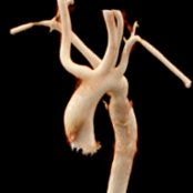
*CTA cinematic rendering reconstruction shows 5 branches from the aortic arch. From the proximal to distal, the branches are: Right common carotid artery , left internal carotid artery , left external carotid artery , left subclavian artery , and aberrant right subclavian artery (ARSA) . Mild aortic coarctation is also noted.*
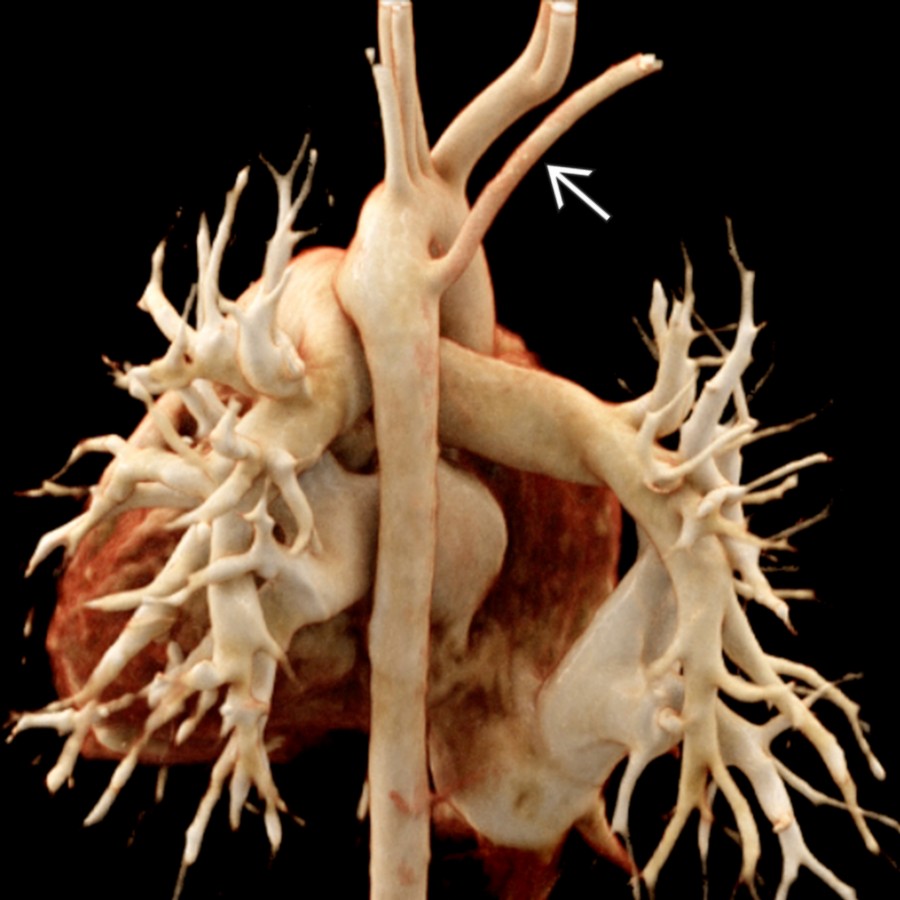
*CTA cinematic rendering reconstruction shows ARSA that originates as the last branch from the aortic arch and courses behind the esophagus to reach the right.*

*Coronal oblique MIP CTA shows a prominent Kommerell diverticulum , which is a dilatation of a proximal ARSA that originates in the proximal descending aorta .*

*Axial oblique MIP reconstruction from a CTA shows an ARSA with retroesophageal course causing esophageal compression between aortic arch, trachea , and ARSA.*
### Additional Images

*Axial oblique MIP reconstruction from a CTA shows an ARSA that causes esophageal compression .*

*3D reconstruction of the airways and lungs from a CT in the same patient shows severe esophageal luminal narrowing from an ARSA.*

*CTA cinematic rendering reconstruction shows 5 branches from the aortic arch. From the proximal to distal, the branches are: Right common carotid artery , left internal carotid artery , left external carotid artery , left subclavian artery , and aberrant right subclavian artery (ARSA) . Mild aortic coarctation is also noted.*

*CTA cinematic rendering reconstruction shows 5 branches from the aortic arch. From the proximal to distal, the branches are: Right common carotid artery , left internal carotid artery , left external carotid artery , left subclavian artery , and aberrant right subclavian artery (ARSA) . Mild aortic coarctation is also noted.*

*CTA cinematic rendering reconstruction shows ARSA that originates as the last branch from the aortic arch and courses behind the esophagus to reach the right.*

*Coronal oblique MIP CTA shows a prominent Kommerell diverticulum , which is a dilatation of a proximal ARSA that originates in the proximal descending aorta .*

*Axial oblique MIP reconstruction from a CTA shows an ARSA with retroesophageal course causing esophageal compression between aortic arch, trachea , and ARSA.*
### Additional Images

*Axial oblique MIP reconstruction from a CTA shows an ARSA that causes esophageal compression .*

*3D reconstruction of the airways and lungs from a CT in the same patient shows severe esophageal luminal narrowing from an ARSA.*
 and right vertebral artery
  , left internal carotid artery , left external carotid artery
  , left vertebral artery , left subclavian artery , and ARSA .](images/app.statdx.com_image_thumbnail_ee1ce812-91ce-4836-a7d8-26c425081e90_annotated_true_size_900_quality_90_5423ed25cea6e5dcb724ee235086d4bb1aa9edb7.jpg)
*MRA 3D posterior oblique view shows 6 branches from the aortic arch. From proximal to distal the branches are: A common trunk dividing into right common carotid artery and right vertebral artery , left internal carotid artery , left external carotid artery , left vertebral artery , left subclavian artery , and ARSA .*

*Sagittal T2-weighted MR of the cervical spine shows a round structure between the spine and esophagus , consistent with an aberrant right subclavian artery causing esophageal compression.*

*Coronal MIP reconstruction from a MRA shows an ARSA and a normal left subclavian artery , both originating at the same level in the distal aortic arch .*

*CTA cinematic rendering reconstruction shows a right-sided aortic arch with mirror-image branching and aberrant left subclavian artery , which originates as the last branch from the arch.*

*Coronal MIP CTA reconstruction in a 56-year-old man shows an ARSA originating from the proximal descending thoracic aorta and extending towards the right.*

*Axial MIP CTA reconstruction in a 56-year-old man shows an ARSA originating from the proximal descending thoracic aorta and extending towards the right, with compression of the esophagus .*
, left vertebral artery , left subclavian artery , and ARSA .](images/app.statdx.com_image_thumbnail_ee1ce812-91ce-4836-a7d8-26c425081e90_annotated_true_size_900_quality_90_5423ed25cea6e5dcb724ee235086d4bb1aa9edb7.jpg)
*MRA 3D posterior oblique view shows 6 branches from the aortic arch. From proximal to distal the branches are: A common trunk dividing into right common carotid artery and right vertebral artery , left internal carotid artery , left external carotid artery , left vertebral artery , left subclavian artery , and ARSA .*

*Sagittal T2-weighted MR of the cervical spine shows a round structure between the spine and esophagus , consistent with an aberrant right subclavian artery causing esophageal compression.*

*Coronal MIP reconstruction from a MRA shows an ARSA and a normal left subclavian artery , both originating at the same level in the distal aortic arch .*

*CTA cinematic rendering reconstruction shows a right-sided aortic arch with mirror-image branching and aberrant left subclavian artery , which originates as the last branch from the arch.*

*Coronal MIP CTA reconstruction in a 56-year-old man shows an ARSA originating from the proximal descending thoracic aorta and extending towards the right.*

*Axial MIP CTA reconstruction in a 56-year-old man shows an ARSA originating from the proximal descending thoracic aorta and extending towards the right, with compression of the esophagus .*