---
title: "ADEM"
docid: "a3fafeb7-5861-4364-beb8-c0e30220564e"
authors:
- key: "99e1aff7-f42c-43a0-95ae-d89c8551aa01"
value: "Kevin R. Moore, MD"
- key: "a25c450b-3d34-4f64-bba3-cc0834813df6"
value: "Miral D. Jhaveri, MD, MBA"
- key: "b2e6dabb-ee1c-42a4-a332-9f0814c1c607"
value: "Surjith Vattoth, MD, FRCR"
breadcrumbs:
-
name: "Brain"
slug: "brain"
treeNodeId: "6d8829f1-14d7-45af-8675-255189aa526a"
-
name: "Diagnosis"
slug: "diagnosis"
treeNodeId: "51c00394-446e-4a38-94af-d3b1d14d34e8"
-
name: "Pathology-Based Diagnoses"
slug: "pathology-based-diagnoses"
treeNodeId: "d9d3a8ed-f21b-4831-8c77-591a3500ef77"
-
name: "Infectious, Inflammatory, and Demyelinating Disease"
slug: "infectious-inflammatory-and-demyel-"
treeNodeId: "7210f860-fe5f-4a2d-81cc-4fe06c769607"
-
name: "Inflammatory and Demyelinating Disease"
slug: "inflammatory-and-demyelinating-dis-"
treeNodeId: "62ab4dc3-dbf6-45a9-8532-f0e962aa62dc"
-
name: "ADEM"
slug: "adem"
treeNodeId: null
category: "Brain"
documentVersionId: "c2a39730-fd89-4f20-9447-d7fb297710c6"
imageCount: 22
lastUpdated: "08/07/20"
pageDescription: "ADEM"
pageKeywords: "Brain, Diagnosis, Pathology-Based Diagnoses, Infectious, Inflammatory, and Demyelinating Disease, Inflammatory and Demyelinating Disease, ADEM"
pageTitle: "ADEM | STATdx"
enhancedTitle: "ADEM"
type: "DX"
references: true
breadcrumbs:
- "Brain"
- "Diagnosis"
- "Pathology-Based Diagnoses"
- "Infectious, Inflammatory, and Demyelinating Disease"
- "Inflammatory and Demyelinating Disease"
- "ADEM"
---
# KEY FACTS
- ## Terminology
- Autoimmune-mediated white matter (WM) demyelination of brain &/or spinal cord, usually with remyelination
- ## Imaging
- Best diagnostic clue: Multifocal WM and deep gray lesions days to weeks following infection/vaccination
- May involve both brain and spinal cord; WM > gray matter, but usually both affected
- Both supratentorial and infratentorial lesions
- Multifocal punctate to large flocculent FLAIR hyperintensities
- Deep/juxtacortical WM > periventricular WM
- Do not usually involve callososeptal interface
- Most lesions show increased signal on DWI (T2 shine-through)
- Punctate, ring, incomplete ring, peripheral enhancement
- Absence of enhancement does not exclude diagnosis
- MRS: ↓ NAA within lesions; may see ↑ Cho, ↑ lactate
- ## Top Differential Diagnoses
- Multiple sclerosis
- Autoimmune-mediated vasculitis
- Acute hypertensive encephalopathy, PRES
- Fabry disease
- Behçet disease
- ## Pathology
- > 30 different infectious agents and immunizations reported
- Anti-MOG (myelin oligodendrocyte glycoprotein) IgG antibodies found more commonly in younger patients
- ## Clinical Issues
- Mean age 5-8 years, but can occur at any age
- Male predominance (M:F = 1:0.6-0.8), unlike MS
- Usually monophasic, self-limited
- Complete recovery within 1 month: 50-60%
- Mortality: 10-30%
- ## Diagnostic Checklist
- Imaging findings often lag behind symptom onset, resolution
# TERMINOLOGY
- ## Abbreviations
- Acute disseminated encephalomyelitis (ADEM)
- ## Definitions
- Autoimmune-mediated white matter (WM) demyelination of brain &/or spinal cord, usually with remyelination
# IMAGING
- ## General Features
- ### Best diagnostic clue
- Multifocal WM/basal ganglia lesions days to weeks following infection/vaccination
- 93% within 3 weeks of infection, 5% within 1 month of vaccination
- ### Location
- May involve both brain and spinal cord; WM > gray matter, but usually both affected
- Deep/juxtacortical WM > periventricular WM
- Both supratentorial and infratentorial lesions
- ### Size
- Tumefactive lesions may be large, but with less mass effect than expected from tumor size
- ### Morphology
- Punctate to flocculent
- Tumefactive, mass-like lesions possible
- ## CT Findings
- ### NECT
- Initial CT normal in 40%
- ### CECT
- Multifocal punctate or ring-enhancing lesions
- ## MR Findings
- ### T2WI
- Hyperintensities may be better visualized in brainstem and posterior fossa on T2
- ### FLAIR
- Multifocal punctate to large, flocculent FLAIR hyperintensities
- Bilateral but asymmetric
- Involve peripheral WM-gray matter junction subcortical WM
- Thalami and basal ganglia frequently involved, typically symmetric
- Can involve brainstem and posterior fossa
- Do not usually involve callososeptal interface
- ### DWI
- Variably hyperintense lesions on DWI (trace) images
- Apparent diffusion coefficient (ADC) may be increased or decreased
- Most lesions show increased signal (T2 shine-through)
- Diffusion restriction uncommon, suggests worse prognosis
- Diffusivity normal within normal-appearing WM (NAWM), unlike MS
- ### T1WI C+
- Punctate, ring, incomplete ring, peripheral enhancement
- Cranial nerve(s) may enhance
- Absence does not exclude diagnosis
- ### MRS
- NAA low within lesions; lactate may be elevated
- Choline often elevated in acute lesions
- NAA normalizes with resolution of symptoms/MR abnormalities
- Magnetization transfer ratio (MTR)
- ADEM MTR normal within NAWM, unlike MS
- ## Imaging Recommendations
- ### Best imaging tool
- Contrast-enhanced MR
- Initial imaging often normal but more sensitive than CT
- May appear identical to MS; repeat MR necessary to distinguish with certainty
- ### Protocol advice
- Limited rapid interval follow-up may be provided by FLAIR alone
- ## Nuclear Medicine Findings
- Tc-99m-HMPAO SPECT shows more extensive hypoperfusion than T2 lesions
# DIFFERENTIAL DIAGNOSIS
- [Multiple Sclerosis](/document/pediatric-multiple-sclerosis-brain/f2592b04-f800-4235-9eea-a43f2bf4adfe)
- Predilection for periventricular WM (callososeptal interface), involves subcortical U fibers, commonly in posterior fossa
- Lesions often more symmetric than ADEM
- Relapsing-remitting course common
- [Autoimmune-Mediated Vasculitis](/document/miscellaneous-vasculitis/5a4d4cbd-67e3-4722-8a44-8d411cbb98f0)
- Multifocal gray matter-WM lesions
- Bilateral, usually cortical/subcortical, basal ganglia/thalami
- Ring-enhancing lesions may mimic infection
- [Acute Hypertensive Encephalopathy, PRES](/document/acute-hypertensive-encephalopathy--/efc6f9c2-dad9-4eb8-bad2-421bfaf1ec57)
- Typically posterior circulation in cortex/subcortical WM
- May affect deep gray nuclei
- [Aging Brain With Hyperintense WM Lesions](/document/normal-aging-brain/2a315550-b2ea-4afe-a2ef-f93a2209f276)
- Atherosclerotic brain changes in 50% patients > 50 years old
- Found in normotensive patients; more common in hypertensives patients
- Present in 10-30% of cognitively normal elderly patients
- MR: Scattered, asymmetric WM lesions, without enhancement
- Often periatrial; posterior fossa uncommon
- Spares callososeptal interface, subcortical U fibers
- [Fabry Disease](/document/fabry-disease/83fd222a-9b37-4087-afab-34ba74525887)
- Synonym: Angiokeratoma corporis diffusum universalis
- X-linked recessive; incidence 1/40,000
- Deficiency α-galactosidase A; overaccumulation of glycosphingolipids within lysosomes
- MR: Scattered, asymmetric WM lesions without enhancement
- May involve brainstem and posterior fossa
- Spares callososeptal interface and subcortical U fibers
- Cranial MR sensitive to identify neurologic involvement in asymptomatic patients
- Present with renal failure/heart disease
- [Behçet Disease](/document/behet-disease/4e447bb6-0f14-40e1-929a-4c1465feec0a)
- MR: Scattered, asymmetric, subcortical WM lesions without cortical involvement
- Nodular enhancement in acute phase
- Predilection for midbrain
- ADC ↑, similar to ADEM
- Classic triad: Oral and genital ulcerations with uveitis
# PATHOLOGY
- ## General Features
- ### Etiology
- Autoimmune-mediated severe acute demyelination
- Following nonspecific upper respiratory tract infection, often viral
- > 30 different infectious agents and immunizations reported
- After specific viral illness: Epstein-Barr, influenza A, mumps, coronavirus
- Especially after exanthematous diseases of childhood (chickenpox, measles)
- After vaccination: Diphtheria, influenza, rabies, smallpox, tetanus, typhoid
- Spontaneous (no known cause)
- ### Genetics
- ADEM associated with DRB1*01 and DRB1*017(03) in Russian population
- ### Associated abnormalities
- Acute hemorrhagic leukoencephalopathy variant associated with ulcerative colitis and asthma
- Anti-MOG (myelin oligodendrocyte glycoprotein) IgG antibodies found more commonly in younger patients
- ## Gross Pathologic & Surgical Features
- None, unless hemorrhage (rare) or tumefactive edema
- ## Microscopic Features
- Acute myelin breakdown
- Perivenous inflammation; lymphocytic infiltrates
- Relative axonal preservation; atypical astrogliosis
- Virus generally not found, unlike viral encephalitides
- Similar to experimental allergic encephalomyelitis, supporting autoimmune-related etiology
# CLINICAL ISSUES
- ## Presentation
- ### Most common signs/symptoms
- Usually preceded by prodromal phase: Fever, malaise, myalgia
- Multifocal neurologic symptoms, 2 days to 4 weeks after viral illness/immunization
- Initial symptoms: Headache, fever, drowsiness
- Cranial nerve palsies, seizures, hemiparesis
- Decreased consciousness (from lethargy to coma)
- Behavioral changes
- ### Other signs/symptoms
- Seizures in 10-35%
- ### Clinical profile
- Cerebrospinal fluid (CSF) normal in 60%
- If abnormal (lymphocyte pleocytosis, elevated protein)
- Usually lacks CSF oligoclonal bands
- ## Demographics
- ### Age
- Children > adults
- Mean age 5-8 years, but can occur at any age
- ### Sex
- Male predominance (M:F = 1.0:0.6-0.8), unlike MS
- ### Epidemiology
- Rare, yet most common para-/postinfectious disorder
- Most common in winter and spring
- Exact epidemiology unknown, but increasingly reported
- ## Natural History & Prognosis
- Usually monophasic, self-limited
- Variable prognosis
- Complete recovery within 1 month (50-60%)
- Neurologic sequelae (most commonly seizures) (20-30%)
- Mortality (10-30%)
- Relapses are rare
- "Relapsing disseminated encephalomyelitis"
- May not be separate entity from relapsing-remitting MS
- Typically delay between symptom onset and imaging findings
- Varicella and rubella ADEM have preferential patterns
- Varicella ADEM characterized by cerebellar ataxia and mild pyramidal dysfunction
- Rubella ADEM characterized by acute explosive onset, seizures, coma, and moderate pyramidal signs
- Rare manifestations of ADEM
- Acute hemorrhagic leukoencephalopathy (2%)
- Young patients with abrupt symptom onset
- Fulminant, often ending in death
- Bilateral striatal necrosis (usually in infants, may be reversible)
- ## Treatment
- Immunosuppressive/immunomodulatory therapy
- MR may show prompt improvement after therapy
- Plasma exchange therapy
- 40% of patients failing steroid treatment may show marked improvement
# DIAGNOSTIC CHECKLIST
- ## Image Interpretation Pearls
- Imaging findings often lag behind symptom onset, resolution
3aec74c8-de53-4430-a00a-d8165ad210c7
## References
# Selected References
1. [Carvalho K et al: Acute disseminated encephalomyelitis (ADEM) associated with mosquito-borne diseases: Chikungunya virus X yellow fever immunization. Rev Soc Bras Med Trop. 53:e20190160, 2020](http://www.ncbi.nlm.nih.gov/pubmed/?term=31994659%5Bpmid%5D)
1. [Molero-Senosiain M et al: Neuro-ophthalmological manifestations as complication of an infection with Mycoplasma pneumoniae and subsequent development of disseminated acute encephalitis. Arch Soc Esp Oftalmol. 95(5(:254-8, 2020](http://www.ncbi.nlm.nih.gov/pubmed/?term=32147128%5Bpmid%5D)
1. [Stokes Brackett AC et al: Multiphasic acute disseminated encephalomyelitis and differential with early onset multiple sclerosis. Intractable Rare Dis Res. 9(1):61-3, 2020](http://www.ncbi.nlm.nih.gov/pubmed/?term=32201679%5Bpmid%5D)
1. [Baumann M et al: MRI of the first event in pediatric acquired demyelinating syndromes with antibodies to myelin oligodendrocyte glycoprotein. J Neurol. 265(4):845-55, 2018](http://www.ncbi.nlm.nih.gov/pubmed/?term=29423614%5Bpmid%5D)
1. [Aubert-Broche B et al: Monophasic demyelination reduces brain growth in children. Neurology. 88(18):1744-50, 2017](http://www.ncbi.nlm.nih.gov/pubmed/?term=28381515%5Bpmid%5D)
1. [Bester M et al: Neuroimaging of multiple sclerosis, acute disseminated encephalomyelitis, and other demyelinating diseases. Semin Roentgenol. 49(1):76-85, 2014](http://www.ncbi.nlm.nih.gov/pubmed/?term=24342677%5Bpmid%5D)
1. [Longoni G et al: White matter changes in paediatric multiple sclerosis and monophasic demyelinating disorders. Brain. 140(5):1300-15, 2017](http://www.ncbi.nlm.nih.gov/pubmed/?term=28334875%5Bpmid%5D)
1. [Koelman DLH et al: Acute disseminated encephalomyelitis: prognostic value of early follow-up brain MRI. J Neurol. 264(8):1754-62, 2017](http://www.ncbi.nlm.nih.gov/pubmed/?term=28695361%5Bpmid%5D)
1. [Marziali S et al: Acute disseminated encephalomyelitis following Campylobacter jejuni gastroenteritis: Case report and review of the literature. Neuroradiol J. 30(1):65-70, 2017](http://www.ncbi.nlm.nih.gov/pubmed/?term=27888275%5Bpmid%5D)
1. [Kanekar S et al: A pattern approach to focal white matter hyperintensities on magnetic resonance imaging. Radiol Clin North Am. 52(2):241-61, 2014](http://www.ncbi.nlm.nih.gov/pubmed/?term=24582339%5Bpmid%5D)
1. [Daida K et al: Cytomegalovirus-associated encephalomyelitis in an immunocompetent adult: a two-stage attack of direct viral and delayed immune-mediated invasions. case report. BMC Neurol. 16(1):223, 2016](http://www.ncbi.nlm.nih.gov/pubmed/?term=27855658%5Bpmid%5D)
1. [Pohl D et al: Acute disseminated encephalomyelitis: updates on an inflammatory CNS syndrome. Neurology. 87(9 Suppl 2):S38-45, 2016](http://www.ncbi.nlm.nih.gov/pubmed/?term=27572859%5Bpmid%5D)
1. [Yuan JL et al: Acute Disseminated Encephalomyelitis following Vaccination against Hepatitis B in a Child: A Case Report and Literature Review. Case Rep Neurol Med. 2016:2401809, 2016](http://www.ncbi.nlm.nih.gov/pubmed/?term=27478662%5Bpmid%5D)
1. [Baumann M et al: Clinical and neuroradiological differences of paediatric acute disseminating encephalomyelitis with and without antibodies to the myelin oligodendrocyte glycoprotein. J Neurol Neurosurg Psychiatry. 86(3):265-72, 2015](http://www.ncbi.nlm.nih.gov/pubmed/?term=25121570%5Bpmid%5D)
1. [Karussis D: The diagnosis of multiple sclerosis and the various related demyelinating syndromes: a critical review. J Autoimmun. 48-49:134-42, 2014](http://www.ncbi.nlm.nih.gov/pubmed/?term=24524923%5Bpmid%5D)
1. [Mariotto S et al: Clinical spectrum and IgG subclass analysis of anti-myelin oligodendrocyte glycoprotein antibody-associated syndromes: a multicenter study. J Neurol. 264(12):2420-30, 2017](http://www.ncbi.nlm.nih.gov/pubmed/?term=29063242%5Bpmid%5D)
1. [Nakamura Y et al: Anti-MOG antibody-positive ADEM following infectious mononucleosis due to a primary EBV infection: a case report. BMC Neurol. 17(1):76, 2017](http://www.ncbi.nlm.nih.gov/pubmed/?term=28420330%5Bpmid%5D)
1. [Tenembaum SN: Acute disseminated encephalomyelitis. Handb Clin Neurol. 112:1253-62, 2013](http://www.ncbi.nlm.nih.gov/pubmed/?term=23622336%5Bpmid%5D)
1. [Wingerchuk DM et al: Acute disseminated encephalomyelitis, transverse myelitis, and neuromyelitis optica. Continuum (Minneap Minn). 19(4 Multiple Sclerosis):944-67, 2013](http://www.ncbi.nlm.nih.gov/pubmed/?term=23917095%5Bpmid%5D)
1. [Callen DJ et al: Role of MRI in the differentiation of ADEM from MS in children. Neurology. 72(11):968-73, 2009](http://www.ncbi.nlm.nih.gov/pubmed/?term=19038851%5Bpmid%5D)
1. [Noorbakhsh F et al: Acute disseminated encephalomyelitis: clinical and pathogenesis features. Neurol Clin. 26(3):759-80, ix, 2008](http://www.ncbi.nlm.nih.gov/pubmed/?term=18657725%5Bpmid%5D)
1. [Rossi A: Imaging of acute disseminated encephalomyelitis. Neuroimaging Clin N Am. 18(1):149-61; ix, 2008](http://www.ncbi.nlm.nih.gov/pubmed/?term=18319160%5Bpmid%5D)
1. [Tenembaum S et al: Acute disseminated encephalomyelitis. Neurology. 68(16 Suppl 2):S23-36, 2007](http://www.ncbi.nlm.nih.gov/pubmed/?term=17438235%5Bpmid%5D)
1. [Menge T et al: Acute disseminated encephalomyelitis: an update. Arch Neurol. 62(11):1673-80, 2005](http://www.ncbi.nlm.nih.gov/pubmed/?term=16286539%5Bpmid%5D)
1. [Yeh EA et al: Detection of coronavirus in the central nervous system of a child with acute disseminated encephalomyelitis. Pediatrics. 113(1 Pt 1):e73-6, 2004](http://www.ncbi.nlm.nih.gov/pubmed/?term=14702500%5Bpmid%5D)
1. [Dale RC: Acute disseminated encephalomyelitis. Semin Pediatr Infect Dis. 14(2):90-5, 2003](http://www.ncbi.nlm.nih.gov/pubmed/?term=12881796%5Bpmid%5D)
1. [Garg RK: Acute disseminated encephalomyelitis. Postgrad Med J. 79(927):11-17, 2003](http://www.ncbi.nlm.nih.gov/pubmed/?term=12566545%5Bpmid%5D)
1. [Idrissova ZhR et al: Acute disseminated encephalomyelitis in children: clinical features and HLA-DR linkage. Eur J Neurol. 10(5):537-46, 2003](http://www.ncbi.nlm.nih.gov/pubmed/?term=12940836%5Bpmid%5D)
1. [Okamoto K et al: MR features of diseases involving bilateral middle cerebellar peduncles. AJNR Am J Neuroradiol. 24(10):1946-54, 2003](http://www.ncbi.nlm.nih.gov/pubmed/?term=14625215%5Bpmid%5D)
1. [Sener RN: Neuro-Behcet's disease: diffusion MR imaging and proton MR spectroscopy. AJNR Am J Neuroradiol. 24(8):1612-4, 2003](http://www.ncbi.nlm.nih.gov/pubmed/?term=13679280%5Bpmid%5D)
1. [Stonehouse M et al: Acute disseminated encephalomyelitis: recognition in the hands of general paediatricians. Arch Dis Child. 88(2):122-4, 2003](http://www.ncbi.nlm.nih.gov/pubmed/?term=12538312%5Bpmid%5D)
1. [Inglese M et al: Magnetization transfer and diffusion tensor MR imaging of acute disseminated encephalomyelitis. AJNR Am J Neuroradiol. 23(2):267-72, 2002](http://www.ncbi.nlm.nih.gov/pubmed/?term=11847052%5Bpmid%5D)
1. [Murthy JM: Acute disseminated encephalomyelitis. Neurol India. 50(3):238-43, 2002](http://www.ncbi.nlm.nih.gov/pubmed/?term=12391446%5Bpmid%5D)
1. [Tenembaum S et al: Acute disseminated encephalomyelitis: a long-term follow-up study of 84 pediatric patients. Neurology. 59(8):1224-31, 2002](http://www.ncbi.nlm.nih.gov/pubmed/?term=12391351%5Bpmid%5D)
1. [Bizzi A et al: Quantitative proton MR spectroscopic imaging in acute disseminated encephalomyelitis. AJNR Am J Neuroradiol. 22(6):1125-30, 2001](http://www.ncbi.nlm.nih.gov/pubmed/?term=11415908%5Bpmid%5D)
1. [Honkaniemi J et al: Delayed MR imaging changes in acute disseminated encephalomyelitis. AJNR Am J Neuroradiol. 22(6):1117-24, 2001](http://www.ncbi.nlm.nih.gov/pubmed/?term=11415907%5Bpmid%5D)
1. [Straussberg R et al: Improvement of atypical acute disseminated encephalomyelitis with steroids and intravenous immunoglobulins. Pediatr Neurol. 24(2):139-43, 2001](http://www.ncbi.nlm.nih.gov/pubmed/?term=11275464%5Bpmid%5D)
1. [Dale RC et al: Acute disseminated encephalomyelitis, multiphasic disseminated encephalomyelitis and multiple sclerosis in children. Brain. 123 Pt 12:2407-22, 2000](http://www.ncbi.nlm.nih.gov/pubmed/?term=11099444%5Bpmid%5D)
1. [Rust RS: Multiple sclerosis, acute disseminated encephalomyelitis, and related conditions. Semin Pediatr Neurol. 7(2):66-90, 2000](http://www.ncbi.nlm.nih.gov/pubmed/?term=10914409%5Bpmid%5D)
1. [Schaefer PW et al: Diffusion-weighted MR imaging of the brain. Radiology. 217(2):331-45, 2000](http://www.ncbi.nlm.nih.gov/pubmed/?term=11058626%5Bpmid%5D)
1. [Kocer N et al: CNS involvement in neuro-Behcet syndrome: an MR study. AJNR Am J Neuroradiol. 20(6):1015-24, 1999](http://www.ncbi.nlm.nih.gov/pubmed/?term=10445437%5Bpmid%5D)
## Images
### Selected Images

*Axial FLAIR MR shows peripheral, confluent areas of hyperintensity predominantly involving the subcortical white matter (WM) in this child with ADEM. The bilateral but asymmetric pattern is typical of ADEM.*

*Axial FLAIR MR shows peripheral, confluent areas of hyperintensity predominantly involving the subcortical white matter (WM) in this child with ADEM. The bilateral but asymmetric pattern is typical of ADEM.*

*Axial T1 C+ MR in the same patient shows marked, irregular enhancement of nearly all lesions. As ADEM is a monophasic illness, enhancement of the majority of lesions is typical; all lesions have a similar time course. Enhancement of multiple sclerosis (MS) lesions is more variable.*

*Axial T1 C+ MR shows an incomplete ring of peripheral enhancement, typical of a demyelinating process. Other contrast enhancement patterns include ovoid or punctate homogeneous enhancement.*

*Axial DWI MR shows increased signal in areas of FLAIR hyperintensity. The foci were hypointense on ADC images, indicating diffusion restriction. Both WM and gray matter involvement is present. Diffusion restriction is an uncommon imaging finding and is associated with a worse prognosis.*
 appearance. Enhancement of several lesions was present on postcontrast T1 images (not shown).](images/app.statdx.com_image_thumbnail_f74d958e-f845-47e7-884e-331bf3dcb299_size_168_quality_85_10b7c767_20251018T064936Z.jpg)
*Axial T2 MR shows hyperintense lesions in the brachium pontis bilaterally, typical for demyelination. The right-sided lesion shows a targetoid appearance. Enhancement of several lesions was present on postcontrast T1 images (not shown).*

*Axial T2 MR shows hyperintense lesions in the brachium pontis bilaterally, typical for demyelination. The right-sided lesion shows a targetoid appearance. Enhancement of several lesions was present on postcontrast T1 images (not shown).*
 in the periventricular and subcortical WM in a 14 year old who presented with neck stiffness, fatigue, and seizures.](images/app.statdx.com_image_thumbnail_3e8a9c1e-560e-4c5c-93bc-dda9b3773622_size_168_quality_85_caf40da9_20251018T064936Z.jpg)
*Axial FLAIR MR shows large, confluent regions of hyperintense signal in the periventricular and subcortical WM in a 14 year old who presented with neck stiffness, fatigue, and seizures.*

*Axial FLAIR MR shows large, confluent regions of hyperintense signal in the periventricular and subcortical WM in a 14 year old who presented with neck stiffness, fatigue, and seizures.*
 in regions of FLAIR signal abnormality.](images/app.statdx.com_image_thumbnail_fa96c2d2-8153-44c2-bd76-be0296382093_size_168_quality_85_93dd4c72_20251018T064936Z.jpg)
*Axial SWI MR in the same patient shows petechial hemorrhages in regions of FLAIR signal abnormality.*

*Sagittal T1 C+ MR in the same patient shows extensive irregular ring enhancement in multiple subcortical WM lesions. Acute hemorrhagic leukoencephalopathy (AHL) is a rare manifestation of ADEM occurring in 2% of cases. AHL is associated with a very poor prognosis. Aggressive therapeutic management is a prerequisite to avoid usual disease course with fatal outcome.*

*Axial SWI MR in the same patient shows petechial hemorrhages in regions of FLAIR signal abnormality.*

*Sagittal T1 C+ MR in the same patient shows extensive irregular ring enhancement in multiple subcortical WM lesions. Acute hemorrhagic leukoencephalopathy (AHL) is a rare manifestation of ADEM occurring in 2% of cases. AHL is associated with a very poor prognosis. Aggressive therapeutic management is a prerequisite to avoid usual disease course with fatal outcome.*
 and deep gray nuclei of a child with ADEM. Although ADEM predominantly involves WM, gray matter is often affected.](images/app.statdx.com_image_thumbnail_113e918b-8d8f-46b8-8664-e0f617904251_size_168_quality_85_1b06d2b7_20251018T064936Z.jpg)
*Coronal T2 MR shows large, confluent regions of hyperintense signal in the WM and deep gray nuclei of a child with ADEM. Although ADEM predominantly involves WM, gray matter is often affected.*
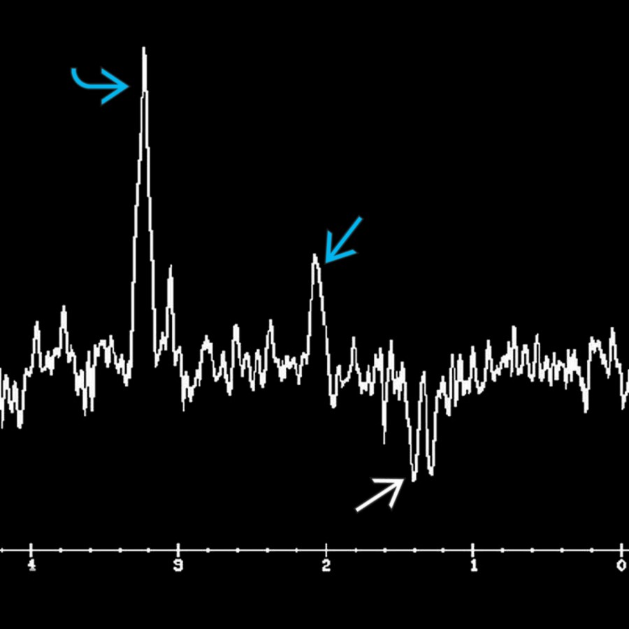
*MRS at long TE in a patient with acute lesions in ADEM demonstrates ↑ choline , ↓ NAA , and the presence of a lactate doublet . Increase in choline with corresponding reductions in NAA normalize as the clinical and conventional neuroimaging abnormalities resolve.*

*MRS at long TE in a patient with acute lesions in ADEM demonstrates ↑ choline , ↓ NAA , and the presence of a lactate doublet . Increase in choline with corresponding reductions in NAA normalize as the clinical and conventional neuroimaging abnormalities resolve.*
### Additional Images

*Axial FLAIR MR shows multiple bilateral, asymmetric, flocculent, hyperintense lesions of acute disseminated encephalomyelitis.*

*Coronal T1 C+ MR demonstrates partial peripheral enhancement around multiple asymmetric, flocculent lesions of acute disseminated encephalomyelitis. Note the supra- and infratentorial lesions.*
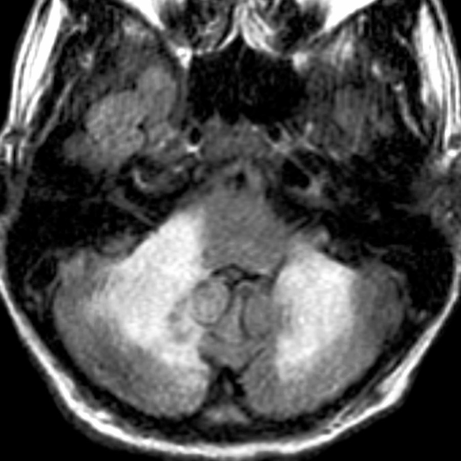
*Axial FLAIR MR shows asymmetric, flocculent, nearly confluent, hyperintense lesions of acute disseminated encephalomyelitis within posterior fossa structures.*

*Axial FLAIR MR shows asymmetric, flocculent, nearly confluent, hyperintense lesions of acute disseminated encephalomyelitis within posterior fossa structures.*
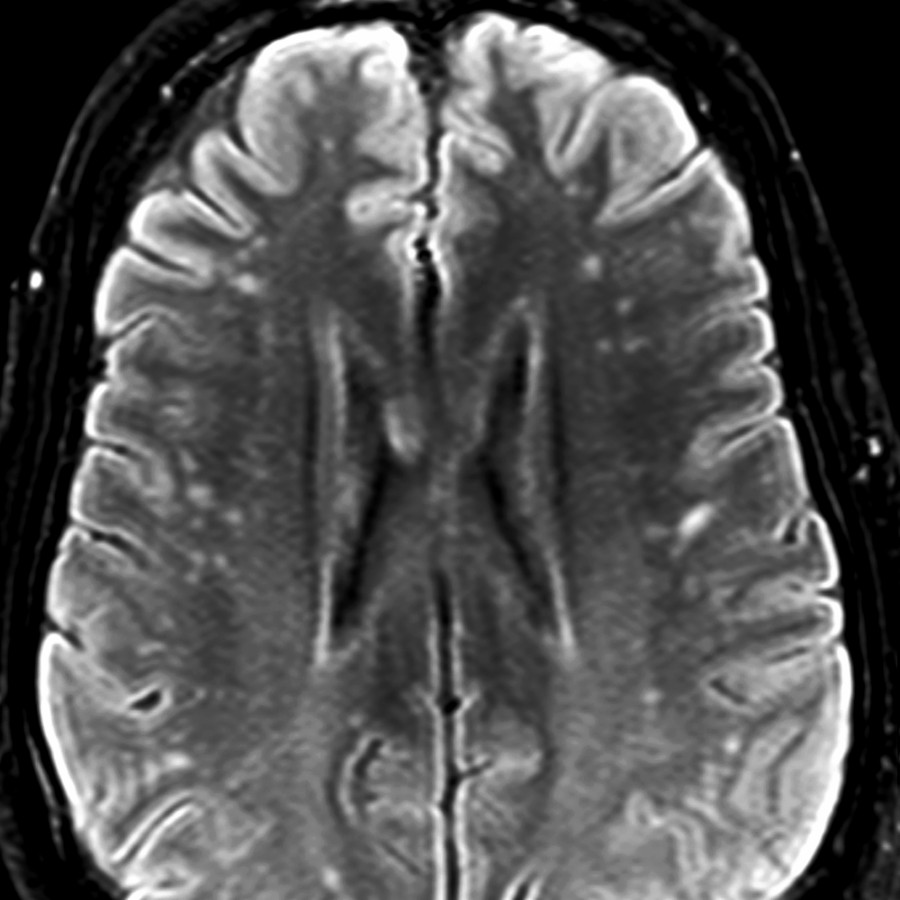
*Axial FLAIR MR reveals multiple asymmetric, primarily punctate, hyperintense lesions of acute disseminated encephalomyelitis.*

*Axial FLAIR MR reveals multiple asymmetric, primarily punctate, hyperintense lesions of acute disseminated encephalomyelitis.*
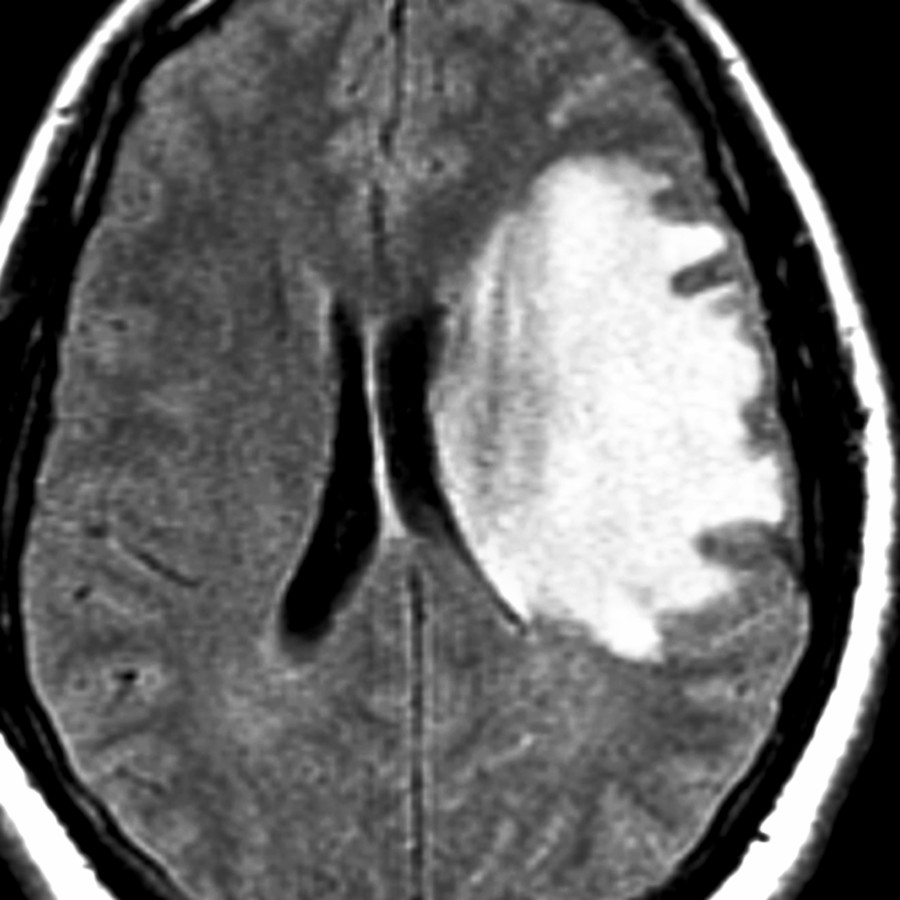
*Axial FLAIR MR demonstrates a large, tumefactive, hyperintense lesion. Less mass effect is present than expected for lesion size. Smaller lesions were also present at other locations.*

*Axial FLAIR MR demonstrates a large, tumefactive, hyperintense lesion. Less mass effect is present than expected for lesion size. Smaller lesions were also present at other locations.*
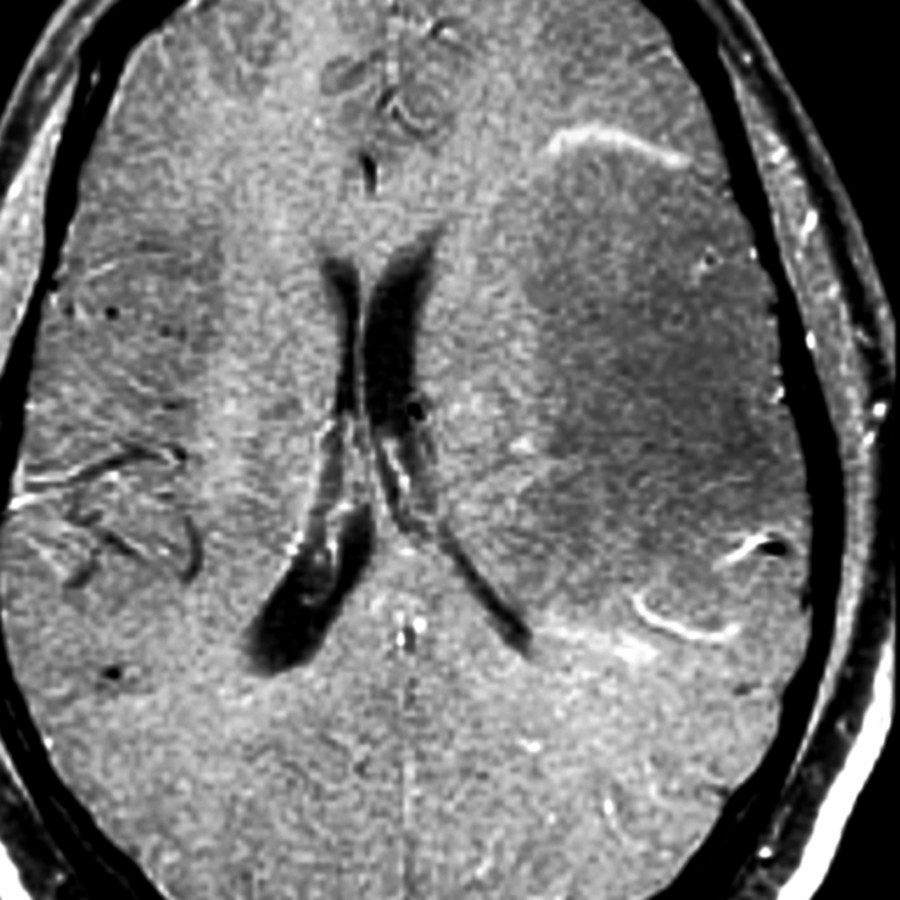
*Axial T1 C+ MR demonstrates a large, tumefactive, hypointense lesion with minimal partial peripheral enhancement. Less mass effect is present than expected for lesion size. More lesions were seen elsewhere.*

*Axial T1 C+ MR demonstrates a large, tumefactive, hypointense lesion with minimal partial peripheral enhancement. Less mass effect is present than expected for lesion size. More lesions were seen elsewhere.*
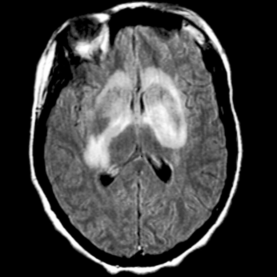
*Axial FLAIR MR demonstrates a rare manifestation of ADEM: Bilateral striatal necrosis, evidenced by asymmetric confluent hyperintensity involving the gray and white matter of bilateral corpus striatum.*

*Axial FLAIR MR demonstrates a rare manifestation of ADEM: Bilateral striatal necrosis, evidenced by asymmetric confluent hyperintensity involving the gray and white matter of bilateral corpus striatum.*
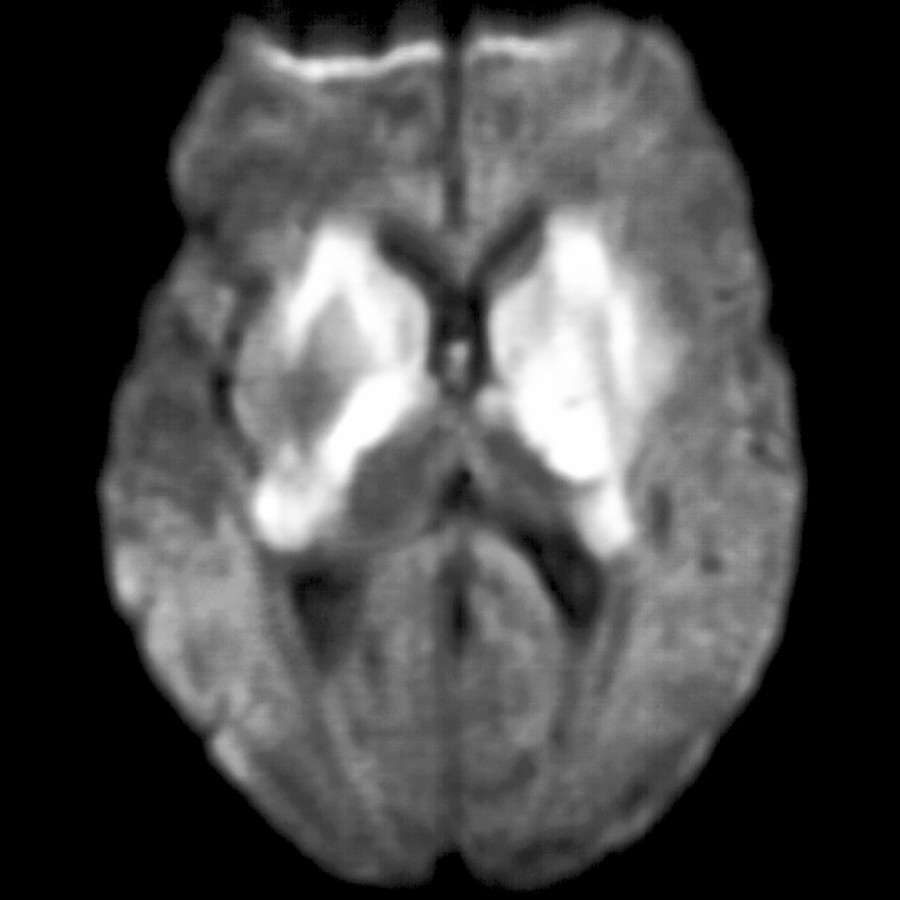
*Axial DWI MR confirms the rare manifestation of ADEM, displaying bilateral striatal necrosis, as evidenced by asymmetric confluent restricted diffusion involving gray and white matter of bilateral corpus striatum.*

*Axial DWI MR confirms the rare manifestation of ADEM, displaying bilateral striatal necrosis, as evidenced by asymmetric confluent restricted diffusion involving gray and white matter of bilateral corpus striatum.*
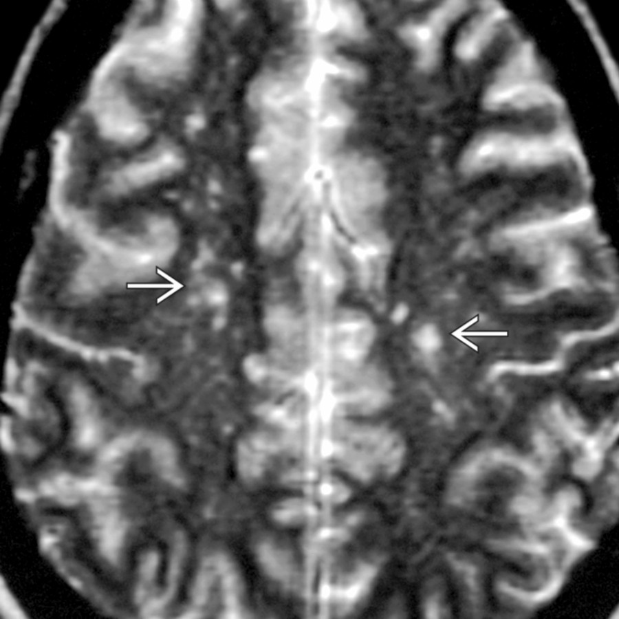
*Axial T2 MR shows multiple bilateral, but asymmetric, T2-hyperintense foci . None of the lesions demonstrate significant mass effect in this adult patient with ADEM. Imaging mimics multiple sclerosis, vasculitis, and microvascular ischemia.*

*Axial T2 MR shows multiple bilateral, but asymmetric, T2-hyperintense foci . None of the lesions demonstrate significant mass effect in this adult patient with ADEM. Imaging mimics multiple sclerosis, vasculitis, and microvascular ischemia.*

*Coronal T2 MR shows large, confluent regions of hyperintense signal in the WM and deep gray nuclei of a child with ADEM. Although ADEM predominantly involves WM, gray matter is often affected.*

*MRS at long TE in a patient with acute lesions in ADEM demonstrates ↑ choline , ↓ NAA , and the presence of a lactate doublet . Increase in choline with corresponding reductions in NAA normalize as the clinical and conventional neuroimaging abnormalities resolve.*

*MRS at long TE in a patient with acute lesions in ADEM demonstrates ↑ choline , ↓ NAA , and the presence of a lactate doublet . Increase in choline with corresponding reductions in NAA normalize as the clinical and conventional neuroimaging abnormalities resolve.*
### Additional Images

*Axial FLAIR MR shows multiple bilateral, asymmetric, flocculent, hyperintense lesions of acute disseminated encephalomyelitis.*

*Coronal T1 C+ MR demonstrates partial peripheral enhancement around multiple asymmetric, flocculent lesions of acute disseminated encephalomyelitis. Note the supra- and infratentorial lesions.*

*Axial FLAIR MR shows asymmetric, flocculent, nearly confluent, hyperintense lesions of acute disseminated encephalomyelitis within posterior fossa structures.*

*Axial FLAIR MR shows asymmetric, flocculent, nearly confluent, hyperintense lesions of acute disseminated encephalomyelitis within posterior fossa structures.*

*Axial FLAIR MR reveals multiple asymmetric, primarily punctate, hyperintense lesions of acute disseminated encephalomyelitis.*

*Axial FLAIR MR reveals multiple asymmetric, primarily punctate, hyperintense lesions of acute disseminated encephalomyelitis.*

*Axial FLAIR MR demonstrates a large, tumefactive, hyperintense lesion. Less mass effect is present than expected for lesion size. Smaller lesions were also present at other locations.*

*Axial FLAIR MR demonstrates a large, tumefactive, hyperintense lesion. Less mass effect is present than expected for lesion size. Smaller lesions were also present at other locations.*

*Axial T1 C+ MR demonstrates a large, tumefactive, hypointense lesion with minimal partial peripheral enhancement. Less mass effect is present than expected for lesion size. More lesions were seen elsewhere.*

*Axial T1 C+ MR demonstrates a large, tumefactive, hypointense lesion with minimal partial peripheral enhancement. Less mass effect is present than expected for lesion size. More lesions were seen elsewhere.*

*Axial FLAIR MR demonstrates a rare manifestation of ADEM: Bilateral striatal necrosis, evidenced by asymmetric confluent hyperintensity involving the gray and white matter of bilateral corpus striatum.*

*Axial FLAIR MR demonstrates a rare manifestation of ADEM: Bilateral striatal necrosis, evidenced by asymmetric confluent hyperintensity involving the gray and white matter of bilateral corpus striatum.*

*Axial DWI MR confirms the rare manifestation of ADEM, displaying bilateral striatal necrosis, as evidenced by asymmetric confluent restricted diffusion involving gray and white matter of bilateral corpus striatum.*

*Axial DWI MR confirms the rare manifestation of ADEM, displaying bilateral striatal necrosis, as evidenced by asymmetric confluent restricted diffusion involving gray and white matter of bilateral corpus striatum.*

*Axial T2 MR shows multiple bilateral, but asymmetric, T2-hyperintense foci . None of the lesions demonstrate significant mass effect in this adult patient with ADEM. Imaging mimics multiple sclerosis, vasculitis, and microvascular ischemia.*

*Axial T2 MR shows multiple bilateral, but asymmetric, T2-hyperintense foci . None of the lesions demonstrate significant mass effect in this adult patient with ADEM. Imaging mimics multiple sclerosis, vasculitis, and microvascular ischemia.*
 .](images/app.statdx.com_image_f2086d11-e747-4b89-99d4-34c16ebd985c_cd1a99bd_20251018T065014Z.jpg)
*Axial FLAIR MR shows a large, tumefactive, hyperintense ADEM lesion with mass effect less than expected for the size of the lesion. Another clue to its nonneoplastic nature is the right-sided lesion .*

*Axial FLAIR MR shows a large, tumefactive, hyperintense ADEM lesion with mass effect less than expected for the size of the lesion. Another clue to its nonneoplastic nature is the right-sided lesion .*
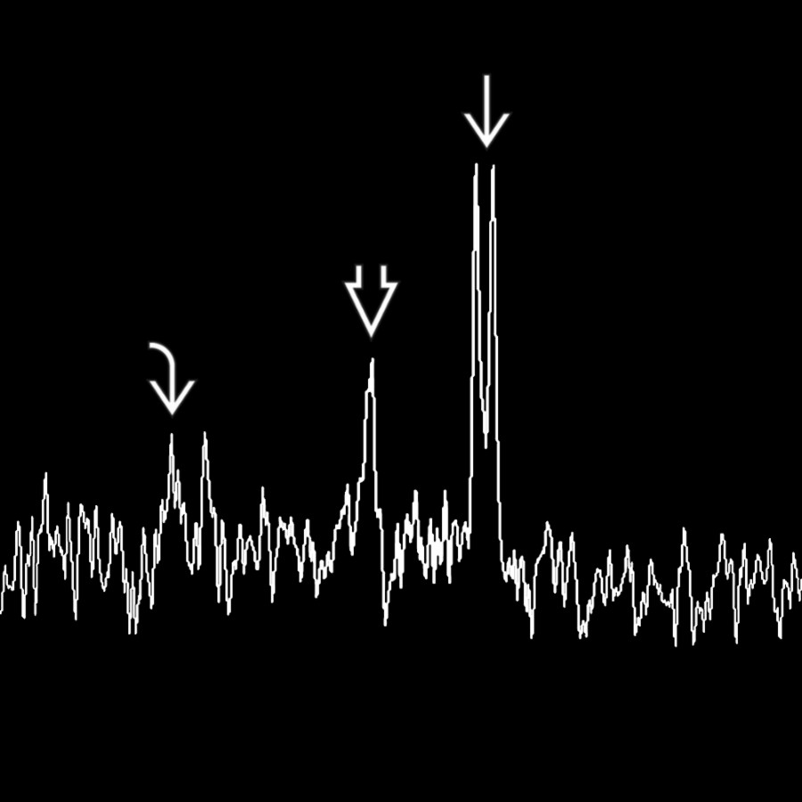
*MRS at a long TE in the same patient shows the tumefactive lesion has a depressed choline and NAA metabolites in the presence of a large lactate doublet . This MRS helps distinguish this lesion from a neoplasm. MRS of ADEM may show elevated choline acutely.*

*MRS at a long TE in the same patient shows the tumefactive lesion has a depressed choline and NAA metabolites in the presence of a large lactate doublet . This MRS helps distinguish this lesion from a neoplasm. MRS of ADEM may show elevated choline acutely.*
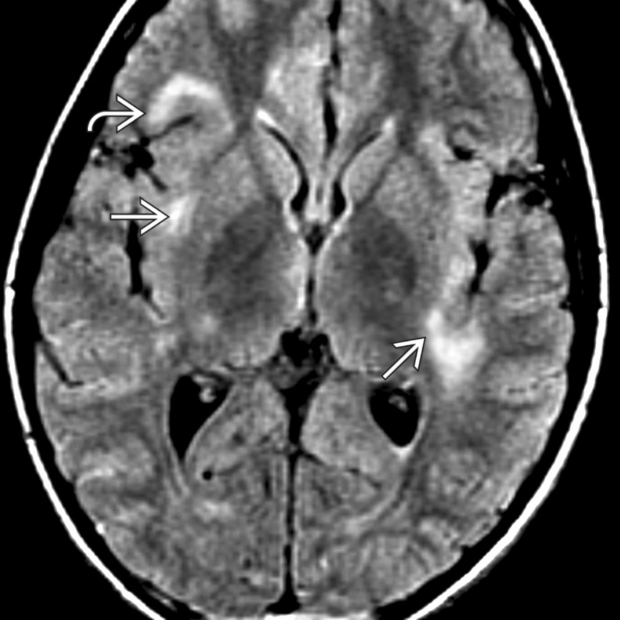
*Axial FLAIR MR shows typical findings of ADEM with peripheral, subcortical hyperintense foci . Bilateral insular involvement is seen . Periventricular and callososeptal lesions, which are typical of multiple sclerosis, are not commonly seen in ADEM.*

*Axial FLAIR MR shows typical findings of ADEM with peripheral, subcortical hyperintense foci . Bilateral insular involvement is seen . Periventricular and callososeptal lesions, which are typical of multiple sclerosis, are not commonly seen in ADEM.*
.](images/app.statdx.com_image_f2086d11-e747-4b89-99d4-34c16ebd985c_cd1a99bd_20251018T065014Z.jpg)
*Axial FLAIR MR shows a large, tumefactive, hyperintense ADEM lesion with mass effect less than expected for the size of the lesion. Another clue to its nonneoplastic nature is the right-sided lesion .*

*Axial FLAIR MR shows a large, tumefactive, hyperintense ADEM lesion with mass effect less than expected for the size of the lesion. Another clue to its nonneoplastic nature is the right-sided lesion .*

*MRS at a long TE in the same patient shows the tumefactive lesion has a depressed choline and NAA metabolites in the presence of a large lactate doublet . This MRS helps distinguish this lesion from a neoplasm. MRS of ADEM may show elevated choline acutely.*

*MRS at a long TE in the same patient shows the tumefactive lesion has a depressed choline and NAA metabolites in the presence of a large lactate doublet . This MRS helps distinguish this lesion from a neoplasm. MRS of ADEM may show elevated choline acutely.*

*Axial FLAIR MR shows typical findings of ADEM with peripheral, subcortical hyperintense foci . Bilateral insular involvement is seen . Periventricular and callososeptal lesions, which are typical of multiple sclerosis, are not commonly seen in ADEM.*

*Axial FLAIR MR shows typical findings of ADEM with peripheral, subcortical hyperintense foci . Bilateral insular involvement is seen . Periventricular and callososeptal lesions, which are typical of multiple sclerosis, are not commonly seen in ADEM.*