---
title: "Adrenal Cyst"
docid: "c5d717a3-3d6e-4e86-9efe-1ad0ec14740f"
authors:
- key: "c3463c5c-31d3-4489-bbfe-6b895abdb86d"
value: "Mitchell Tublin, MD"
breadcrumbs:
-
name: "Genitourinary"
slug: "genitourinary"
treeNodeId: "bd0eb4fe-d465-4faa-a3b7-526e8f01802d"
-
name: "Diagnosis"
slug: "diagnosis"
treeNodeId: "e82a3e55-c0be-4ed1-acd6-b03ae9167c31"
-
name: "Adrenal"
slug: "adrenal"
treeNodeId: "d3b85dea-43cb-4be3-b103-902e38d0336e"
-
name: "Benign Neoplasms"
slug: "benign-neoplasms"
treeNodeId: "eeebc0ba-f71a-4ae6-8daf-525d0d18fa16"
-
name: "Adrenal Cyst"
slug: "adrenal-cyst"
treeNodeId: null
category: "Genitourinary"
documentVersionId: "c0408d4e-b577-4561-89ba-5fd1d7888eaf"
imageCount: 14
lastUpdated: "09/09/21"
pageDescription: "Adrenal Cyst"
pageKeywords: "Genitourinary, Diagnosis, Adrenal, Benign Neoplasms, Adrenal Cyst"
pageTitle: "Adrenal Cyst | STATdx"
enhancedTitle: "Adrenal Cyst"
type: "DX"
references: true
breadcrumbs:
- "Genitourinary"
- "Diagnosis"
- "Adrenal"
- "Benign Neoplasms"
- "Adrenal Cyst"
---
# KEY FACTS
- ## Imaging
- "Adrenal cyst" is descriptive term, not pathological diagnosis
- True adrenal cysts
- Majority are endothelial cysts (lymphangiomas)
- Epithelial cysts exceedingly rare
- Simple, or minimally complex, adrenal cyst, thin rim calcification, no enhancement
- Pseudocysts
- Prior hemorrhage inferred
- Nonenhancing but complex contents and wall calcification
- Relevant history (extraadrenal malignancy, rapid growth), biochemical evaluation (cortisol, metanephrines): Consider underlying adrenal neoplasm
- Enhancing soft tissue components may suggest adrenal mass hemorrhage and pseudocyst formation
- Parasitic (echinococcal) cyst
- Rare outside endemic areas
- Typically in setting of generalized echinococcus
- ## Top Differential Diagnoses
- Adrenal adenoma
- CECT: Enhancing mass without visible wall or peripheral calcifications
- Gastric diverticulum
- Air-, fluid-, or contrast-filled mass with no enhancement of contents
- Adrenal myelolipoma
- Macroscopic fat
- Necrotic adrenal tumor
- Complex wall with heterogeneous contents
- Retroperitoneal bronchogenic cyst
- ## Clinical Issues
- No treatment required usually
- Imaging surveillance performed, although intensity and length of follow-up not defined
- Biochemical evaluation (cortisol, metanephrines) routinely performed to exclude underlying adrenal neoplasm
- Surgical resection for complex cyst with enhancing components, or symptomatic cyst
- ## Diagnostic Checklist
- Complicated cyst has high attenuation, thick enhancing wall, &/or septations
# TERMINOLOGY
- ## Definitions
- "Adrenal cyst" is descriptive term, not pathological diagnosis
- Can mean true cyst, pseudocyst, or cystic mass
# IMAGING
- ## General Features
- ### Best diagnostic clue
- Well-defined, nonenhancing, water-density adrenal mass ± calcifications
- ### Location
- Suprarenal
- Unilateral > bilateral (8-10% of cases)
- ### Size
- < 5 cm (50%), up to 20 cm
- ## CT Findings
- ### NECT
- Unilocular or multilocular mass
- Well-defined, round to oval, homogeneous mass usually with water (0 HU) or near-water density
- Higher- or mixed-attenuation mass (hemorrhage, intracystic debris, crystals)
- Wall usually very thin
- ↑ wall thickness, up to 3 mm for complex cysts
- Calcifications
- Rim-like or nodular (51-69%)
- Centrally in intracystic septation (19%)
- Punctate within intracystic hemorrhage (5%)
- ### CECT
- No central enhancement ± wall enhancement
- Coronal reformats helpful to determine organ of origin if large cyst
- ## MR Findings
- ### T1WI
- Homogeneous, hypointense mass
- Hyperintense mass (hemorrhage)
- ### T2WI
- Hyperintense mass
- ## Ultrasonographic Findings
- Simple or septated suprarenal cyst
- Shadowing from calcification
- Real-time examination helpful to differentiate adrenal cyst from adjacent (renal, pancreatic) cyst
- ## Imaging Recommendations
- ### Best imaging tool
- CECT or MR; US for confirmation
# DIFFERENTIAL DIAGNOSIS
- [Adrenal Adenoma](/document/adrenal-adenoma/e2916d86-5f9f-4dd3-9576-1a7b89d8dda0)
- NECT: Lipid-rich adenoma (< 10 HU) mimics adrenal cyst
- Peripheral or septal calcification favors adrenal cyst
- CECT: **Enhancing mass** without visible wall or peripheral calcifications
- Assess washout kinetics to diagnose lipid-poor adenoma
- MR: Signal suppression at out-of-phase, chemical-shift imaging
- US: Solid adrenal lesion
- [Gastric Diverticulum](/document/gastric-diverticulum/eeb101f0-8bdf-4771-b44a-fe6e73b3a463)
- May simulate left adrenal cyst
- Air-, fluid-, or contrast-filled suprarenal mass
- No enhancement
- Normal adjacent adrenal gland
- [Adrenal Myelolipoma](/document/adrenal-myelolipoma/5813a554-06a4-4696-af71-7ce50693039d)
- Fat (not fluid) attenuation mass
- ## Necrotic Adrenal Tumor
- Primary (pheochromocytoma or carcinoma) or metastatic
- Clinical history, biochemical evaluation, lesion complexity suggest correct diagnosis
- Enhancing soft tissue components
- ## Retroperitoneal Bronchogenic Cyst
- Rare, benign, suprarenal fluid or soft tissue attenuation lesion
- Adjacent to but separate from adrenal gland
- ## Renal Cyst
- Coronal MR/CT or US useful to determine organ of origin of large, retroperitoneal cystic lesions
# PATHOLOGY
- ## General Features
- ### Etiology
- Congenital (endothelial, epithelial) cysts
- Acquired (post hemorrhagic, inflammatory) pseudocysts
- Cystic, hemorrhagic degeneration of underlying adrenal neoplasm
- ## Staging, Grading, & Classification
- Accepted classification scheme
- Pseudocyst
- Most common type of cystic adrenal lesion in surgical series
- No epithelial or endothelial lining: Fibrous cyst wall
- Potentially as complication of prior trauma or hemorrhage though history of such often not elicited
- May be associated with underlying adrenal neoplasm (pheochromocytoma, adrenal carcinoma, myelolipoma)
- Attenuation and complexity at imaging varies depending upon hemorrhagic component
- Wall and septal calcification common
- Endothelial cyst
- Subtypes: Lymphangiomatous and hemangiomatous
- True cyst: Endothelial lining
- Originate from preexisting vascular malformation or obstructed, ectatic lymphatic channels
- Thin rim calcification typical
- Epithelial cyst
- Extremely rare: No acinar structures within normal adrenal gland
- Mesothelial origin suggested (mesothelial cells potentially incorporated within adrenal gland during embryogenesis)
- Parasitic (hydatid) cyst
# CLINICAL ISSUES
- ## Presentation
- ### Most common signs/symptoms
- Typically asymptomatic
- Larger cysts may be symptomatic
- Abdominal pain
- Hemorrhage
- Clinical history (malignancy, hypertension) elicited
- May indicate cystic degeneration of underlying adrenal neoplasm (e.g., metastasis, pheochromocytoma)
- Diagnosis
- Usually incidental finding at imaging
- Endocrine-biochemical evaluation performed to exclude underlying functional adrenal tumor
- ## Demographics
- ### Age
- Any, though patients 20-50 years of age most common
- ### Sex
- M:F = 1:3
- ### Epidemiology
- Uncommon entity: Autopsy incidence 0.064-0.18%
- Accounts for 1% of incidental adrenal lesions in large imaging series
- ## Natural History & Prognosis
- Complications
- Hypertension, infection, rupture, hemorrhage
- Excellent prognosis for vast majority of incidental, benign adrenal cysts
- Prognosis for pseudocysts secondary to adrenal neoplasm depends upon tumor histology
- ## Treatment
- No treatment required usually
- Imaging follow-up typically performed
- Intensity and length of surveillance not defined
- Cysts may enlarge over time
- Endocrine evaluation (cortisol, metanephrine, etc.) performed
- Surgical resection if symptomatic, underlying adrenal neoplasm
- Laparoscopic resection preferred
# DIAGNOSTIC CHECKLIST
- ## Consider
- Complicated cyst may suggest underlying adrenal neoplasm
- Clinical history, biochemical evaluation, and prior imaging helpful
- ## Image Interpretation Pearls
- Simple adrenal cyst: Scant septation, no enhancement, thin rim calcification
- Likely benign endothelial cyst or pseudocyst
- Coronal imaging helpful to determine organ of origin (and exclude exophytic renal or pancreatic cyst)
- Complicated cyst: High attenuation, thick enhancing wall, &/or septations
- Complexity may suggest underlying adrenal neoplasm and secondary pseudocyst
2a5b237c-1238-48d3-9b6c-f76846cba1cb
## References
# Selected References
1. [Wang F et al: CT and MRI of adrenal gland pathologies. Quant Imaging Med Surg. 8(8):853-75, 2018](http://www.ncbi.nlm.nih.gov/pubmed/?term=30306064%5Bpmid%5D)
1. [Lattin GE Jr et al: From the radiologic pathology archives: adrenal tumors and tumor-like conditions in the adult: radiologic-pathologic correlation. Radiographics. 34(3):805-29, 2014](http://www.ncbi.nlm.nih.gov/pubmed/?term=24819798%5Bpmid%5D)
1. [Kyoda Y et al: Adrenal hemorrhagic pseudocyst as the differential diagnosis of pheochromocytoma--a review of the clinical features in cases with radiographically diagnosed pheochromocytoma. J Endocrinol Invest. 36(9):707-11, 2013](http://www.ncbi.nlm.nih.gov/pubmed/?term=23563219%5Bpmid%5D)
1. [Ricci Z et al: Adrenal cysts: natural history by long-term imaging follow-up. AJR Am J Roentgenol. 201(5):1009-16, 2013](http://www.ncbi.nlm.nih.gov/pubmed/?term=24147471%5Bpmid%5D)
1. [Saadai P et al: The pathological features of surgically managed adrenal cysts: a 15-year retrospective review. Am Surg. 79(11):1159-62, 2013](http://www.ncbi.nlm.nih.gov/pubmed/?term=24165250%5Bpmid%5D)
1. [Sebastiano C et al: Cystic lesions of the adrenal gland: our experience over the last 20 years. Hum Pathol. 44(9):1797-803, 2013](http://www.ncbi.nlm.nih.gov/pubmed/?term=23618356%5Bpmid%5D)
1. [El-Hefnawy AS et al: Surgical management of adrenal cysts: single-institution experience. BJU Int. 104(6):847-50, 2009](http://www.ncbi.nlm.nih.gov/pubmed/?term=19389014%5Bpmid%5D)
1. [Chien HP et al: Adrenal cystic lesions: a clinicopathological analysis of 25 cases with proposed histogenesis and review of the literature. Endocr Pathol. 19(4):274-81, 2008](http://www.ncbi.nlm.nih.gov/pubmed/?term=18972224%5Bpmid%5D)
1. [Song JH et al: The incidental adrenal mass on CT: prevalence of adrenal disease in 1,049 consecutive adrenal masses in patients with no known malignancy. AJR Am J Roentgenol. 190(5):1163-8, 2008](http://www.ncbi.nlm.nih.gov/pubmed/?term=18430826%5Bpmid%5D)
1. Elsayes KM et al: Adrenal masses: MR imaging features with pathologic correlation. Radiographics. 24 Suppl 1:S73-86, 2004
1. [Guo YK et al: Uncommon adrenal masses: CT and MRI features with histopathologic correlation. Eur J Radiol. 62(3):359-70, 2007](http://www.ncbi.nlm.nih.gov/pubmed/?term=17532488%5Bpmid%5D)
1. [Sanal HT et al: Imaging features of benign adrenal cysts. Eur J Radiol. 60(3):465-9, 2006](http://www.ncbi.nlm.nih.gov/pubmed/?term=16962278%5Bpmid%5D)
1. [Akçay MN et al: Hydatid cysts of the adrenal gland: review of nine patients. World J Surg. 28(1):97-9, 2004](http://www.ncbi.nlm.nih.gov/pubmed/?term=14639487%5Bpmid%5D)
1. [Elsayes KM et al: Adrenal masses: MR findings with pathologic correlation. RadioGraphics 24: S73-86; 2004](http://www.ncbi.nlm.nih.gov/pubmed/?term=15486251%5Bpmid%5D)
1. [Kawashima A et al: Imaging of nontraumatic hemorrhage of the adrenal gland. Radiographics. 19(4):949-63, 1999](http://www.ncbi.nlm.nih.gov/pubmed/?term=10464802%5Bpmid%5D)
1. [Neri LM et al: Management of adrenal cysts. Am Surg. 65(2):151-63, 1999](http://www.ncbi.nlm.nih.gov/pubmed/?term=9926751%5Bpmid%5D)
1. [Otal P et al: Imaging features of uncommon adrenal masses with histopathologic correlation. Radiographics. 19(3):569-81, 1999](http://www.ncbi.nlm.nih.gov/pubmed/?term=10336189%5Bpmid%5D)
1. [Kawashima A et al: Spectrum of CT findings in nonmalignant disease of the adrenal gland. Radiographics. 18(2):393-412, 1998](http://www.ncbi.nlm.nih.gov/pubmed/?term=9536486%5Bpmid%5D)
1. [Tung GA et al: Adrenal cysts: imaging and percutaneous aspiration. Radiology. 173(1):107-10, 1989](http://www.ncbi.nlm.nih.gov/pubmed/?term=2675177%5Bpmid%5D)
## Images
### Selected Images
 . Note thin cyst septation
  , a finding characteristic of endothelial adrenal cyst.](images/app.statdx.com_image_thumbnail_066f9db3-8b3b-4db5-8b74-5ea109b74746_size_168_quality_85_ff218a4f_20251017T212542Z.jpg)
*Axial CECT in a 28-year-old woman with abdominal pain shows an incidental left adrenal cystic lesion . Note thin cyst septation , a finding characteristic of endothelial adrenal cyst.*

*Axial CECT in a 28-year-old woman with abdominal pain shows an incidental left adrenal cystic lesion . Note thin cyst septation , a finding characteristic of endothelial adrenal cyst.*

*Axial CECT in a 28-year-old woman with abdominal pain shows an incidental left adrenal cystic lesion . Note thin cyst septation , a finding characteristic of endothelial adrenal cyst.*

*Axial CECT in a 28-year-old woman with abdominal pain shows an incidental left adrenal cystic lesion . Note thin cyst septation , a finding characteristic of endothelial adrenal cyst.*
 . The simple appearance of the cyst and the lack of additional relevant clinical history (malignancy, HTN, etc.) prompted surveillance rather than resection for this incidental, benign endothelial cyst.](images/app.statdx.com_image_263c08e3-8cc2-46ee-bbab-eedca9354007_c4146213_20251017T212611Z.jpg)
*Longitudinal US in the same patient confirms an anechoic left suprarenal-adrenal cyst . The simple appearance of the cyst and the lack of additional relevant clinical history (malignancy, HTN, etc.) prompted surveillance rather than resection for this incidental, benign endothelial cyst.*

*Longitudinal US in the same patient confirms an anechoic left suprarenal-adrenal cyst . The simple appearance of the cyst and the lack of additional relevant clinical history (malignancy, HTN, etc.) prompted surveillance rather than resection for this incidental, benign endothelial cyst.*
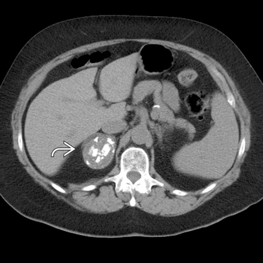
*Axial NECT in a 71-year-old woman shows a 4-cm, complex cystic right adrenal mass containing coarse calcifications. Adrenalectomy (performed given lesion complexity and size) confirmed hemorrhagic pseudocyst.*

*Axial NECT in a 71-year-old woman shows a 4-cm, complex cystic right adrenal mass containing coarse calcifications. Adrenalectomy (performed given lesion complexity and size) confirmed hemorrhagic pseudocyst.*
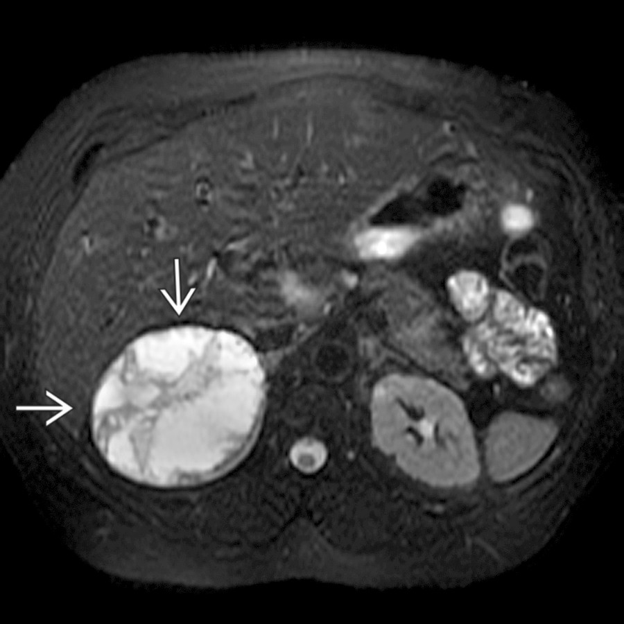
*Axial T2 FS MR in a 54-year-old woman with left flank pain shows a 7-cm, complex cystic right adrenal mass with a low-signal hemosiderin ring . A pseudocyst was resected. Signal intensity of pseudocysts varies depending on the age of hemorrhage.*

*Axial T2 FS MR in a 54-year-old woman with left flank pain shows a 7-cm, complex cystic right adrenal mass with a low-signal hemosiderin ring . A pseudocyst was resected. Signal intensity of pseudocysts varies depending on the age of hemorrhage.*

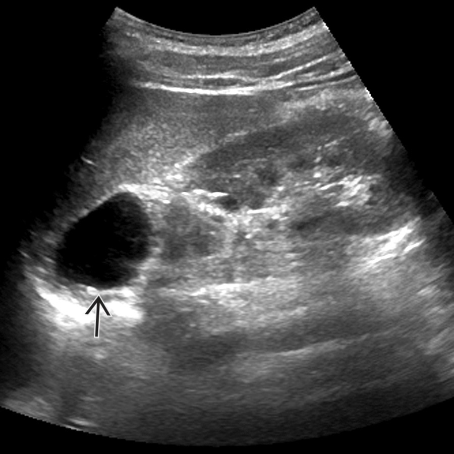
*Sagittal US in a 35-year-old woman shows a > 10-cm right suprarenal cyst . Real-time examination and a follow-up MR (not shown) confirmed simple extrarenal-adrenal cyst that was subsequently resected. An epithelial cyst (a rare subtype of adrenal cysts) was shown at histology.*

*Axial CECT in a 51-year-old woman shows an incidental, peripherally calcified left adrenal cyst . Coarse calcification suggests a pseudocyst. Pseudocysts may be due to prior trauma/hemorrhage, but this history is often absent.*

*Axial CECT in a hypertensive 36-year-old woman shows a peripherally enhancing, septated, 7-cm left adrenal pheochromocytoma . Enhancing soft tissue should prompt testing for an underlying adrenal neoplasm.*

*Axial CECT in a 72-year-old man with pancreatitis shows peripancreatic infiltration and a 3-cm adrenal pseudocyst . The pseudocyst resolved on follow-up CT. Imaging surveillance is advocated for asymptomatic, probable pseudocysts.*

*Staging CECT in a 66-year-old man with metastatic lung carcinoma shows large, bilateral, necrotic adrenal metastases . The clinical history and an enhancing rind prevent an erroneous diagnosis of benign adrenal cysts.*
*Longitudinal US in the same patient confirms an anechoic left suprarenal-adrenal cyst . The simple appearance of the cyst and the lack of additional relevant clinical history (malignancy, HTN, etc.) prompted surveillance rather than resection for this incidental, benign endothelial cyst.*

*Longitudinal US in the same patient confirms an anechoic left suprarenal-adrenal cyst . The simple appearance of the cyst and the lack of additional relevant clinical history (malignancy, HTN, etc.) prompted surveillance rather than resection for this incidental, benign endothelial cyst.*

*Axial NECT in a 71-year-old woman shows a 4-cm, complex cystic right adrenal mass containing coarse calcifications. Adrenalectomy (performed given lesion complexity and size) confirmed hemorrhagic pseudocyst.*

*Axial NECT in a 71-year-old woman shows a 4-cm, complex cystic right adrenal mass containing coarse calcifications. Adrenalectomy (performed given lesion complexity and size) confirmed hemorrhagic pseudocyst.*

*Axial T2 FS MR in a 54-year-old woman with left flank pain shows a 7-cm, complex cystic right adrenal mass with a low-signal hemosiderin ring . A pseudocyst was resected. Signal intensity of pseudocysts varies depending on the age of hemorrhage.*

*Axial T2 FS MR in a 54-year-old woman with left flank pain shows a 7-cm, complex cystic right adrenal mass with a low-signal hemosiderin ring . A pseudocyst was resected. Signal intensity of pseudocysts varies depending on the age of hemorrhage.*

*Sagittal US in a 35-year-old woman shows a > 10-cm right suprarenal cyst . Real-time examination and a follow-up MR (not shown) confirmed simple extrarenal-adrenal cyst that was subsequently resected. An epithelial cyst (a rare subtype of adrenal cysts) was shown at histology.*

*Axial CECT in a 51-year-old woman shows an incidental, peripherally calcified left adrenal cyst . Coarse calcification suggests a pseudocyst. Pseudocysts may be due to prior trauma/hemorrhage, but this history is often absent.*

*Axial CECT in a hypertensive 36-year-old woman shows a peripherally enhancing, septated, 7-cm left adrenal pheochromocytoma . Enhancing soft tissue should prompt testing for an underlying adrenal neoplasm.*

*Axial CECT in a 72-year-old man with pancreatitis shows peripancreatic infiltration and a 3-cm adrenal pseudocyst . The pseudocyst resolved on follow-up CT. Imaging surveillance is advocated for asymptomatic, probable pseudocysts.*

*Staging CECT in a 66-year-old man with metastatic lung carcinoma shows large, bilateral, necrotic adrenal metastases . The clinical history and an enhancing rind prevent an erroneous diagnosis of benign adrenal cysts.*
 and separate from the left adrenal indicates a retroperitoneal bronchogenic cyst, a mimic of adrenal cyst.](images/app.statdx.com_image_thumbnail_fc2010de-959a-4384-8188-96d011832bf5_size_168_quality_85_473f29cf_20251017T212542Z.jpg)
*Axial CECT of a 62-year-old man shows a 2-cm, incidental left suprarenal lesion . Location adjacent to the left crus and separate from the left adrenal indicates a retroperitoneal bronchogenic cyst, a mimic of adrenal cyst.*
### Additional Images

*Sagittal US shows a sonolucent mass above the right kidney that proved to be an adrenal cyst.*

*Axial T2 MR shows a high-signal left adrenal cyst .*

*Axial CECT shows a nonenhancing water density right adrenal cyst.*

*Sagittal reformation of CECT shows a nonenhancing right adrenal cyst .*
and separate from the left adrenal indicates a retroperitoneal bronchogenic cyst, a mimic of adrenal cyst.](images/app.statdx.com_image_thumbnail_fc2010de-959a-4384-8188-96d011832bf5_size_168_quality_85_473f29cf_20251017T212542Z.jpg)
*Axial CECT of a 62-year-old man shows a 2-cm, incidental left suprarenal lesion . Location adjacent to the left crus and separate from the left adrenal indicates a retroperitoneal bronchogenic cyst, a mimic of adrenal cyst.*
### Additional Images

*Sagittal US shows a sonolucent mass above the right kidney that proved to be an adrenal cyst.*

*Axial T2 MR shows a high-signal left adrenal cyst .*

*Axial CECT shows a nonenhancing water density right adrenal cyst.*

*Sagittal reformation of CECT shows a nonenhancing right adrenal cyst .*