---
title: "AHLE"
docid: "0ec0bca6-abee-4931-a6ed-43541b626261"
authors:
- key: "5cff4116-3654-4b3a-bb75-5ebe0b8c9850"
value: "Anne G. Osborn, MD, FACR"
breadcrumbs:
-
name: "Brain"
slug: "brain"
treeNodeId: "6d8829f1-14d7-45af-8675-255189aa526a"
-
name: "Diagnosis"
slug: "diagnosis"
treeNodeId: "51c00394-446e-4a38-94af-d3b1d14d34e8"
-
name: "Pathology-Based Diagnoses"
slug: "pathology-based-diagnoses"
treeNodeId: "d9d3a8ed-f21b-4831-8c77-591a3500ef77"
-
name: "Infectious, Inflammatory, and Demyelinating Disease"
slug: "infectious-inflammatory-and-demyel-"
treeNodeId: "7210f860-fe5f-4a2d-81cc-4fe06c769607"
-
name: "Inflammatory and Demyelinating Disease"
slug: "inflammatory-and-demyelinating-dis-"
treeNodeId: "62ab4dc3-dbf6-45a9-8532-f0e962aa62dc"
-
name: "AHLE"
slug: "ahle"
treeNodeId: null
category: "Brain"
documentVersionId: "05d076c7-6110-4f03-952a-c22726d85e4d"
imageCount: 12
lastUpdated: "08/05/20"
pageDescription: "AHLE"
pageKeywords: "Brain, Diagnosis, Pathology-Based Diagnoses, Infectious, Inflammatory, and Demyelinating Disease, Inflammatory and Demyelinating Disease, AHLE"
pageTitle: "AHLE | STATdx"
enhancedTitle: "AHLE"
type: "DX"
references: true
breadcrumbs:
- "Brain"
- "Diagnosis"
- "Pathology-Based Diagnoses"
- "Infectious, Inflammatory, and Demyelinating Disease"
- "Inflammatory and Demyelinating Disease"
- "AHLE"
---
# KEY FACTS
- ## Terminology
- Hyperacute, fulminant hemorrhagic perivascular demyelinating disorder
- Usually viral or immunization related
- ## Imaging
- Best imaging: MR with T2* GRE, SWI
- CT: May be normal if only microbleeds present
- MR
- Multifocal scattered or confluent WM hyperintensities on T2/FLAIR
- T2* shows multifocal blooming microbleeds in WM (often striking sparing of cortex)
- Most striking in corpus callosum
- Less common: Large, lobar, confluent hemorrhages
- SWI significantly more sensitive than GRE
- ## Top Differential Diagnoses
- Acute disseminated encephalomyelitis (ADEM)
- Multiple sclerosis
- Acute necrotizing encephalopathy
- Other brain "microbleeds"
- Critical illness associated (e.g., ARDS ± on ECMO)
- Trauma (DAI), fat emboli, HUS/TTP
- Sepsis, vasculitis, hemorrhagic viral fevers
- High-altitude cerebral edema
- ## Clinical Issues
- Demographics
- AHLE represents ~ 2% of ADEM cases
- All ages but children, young adults most common
- Presentation and course
- Fever, then rapid neurologic deterioration
- 60-80% mortality without treatment
- ## Diagnostic Checklist
- T2* (GRE or SWI) MR in all febrile CNS illnesses with rapid clinical deterioration
# TERMINOLOGY
- ## Abbreviations
- Acute hemorrhagic leukoencephalitis (AHLE)
- Acute disseminated encephalomyelopathy (ADEM)
- ## Synonyms
- Acute hemorrhagic encephalomyelitis (AHEM)
- Weston-Hurst disease
- ## Definitions
- Hyperacute, fulminant, hemorrhagic perivascular demyelinating disorder
- Usually post viral or immunization related
- May be exceptionally severe, fulminant form of ADEM
# IMAGING
- ## General Features
- ### Best diagnostic clue
- T2* shows multifocal petechial white matter (WM) microhemorrhages with striking sparing of cortex
- Less common: Large, lobar, confluent hemorrhages
- ### Location
- WM
- Corpus callosum, subcortical WM (U fibers)
- Less common: Basal ganglia, midbrain, pons, cerebellum
- ### Size
- Punctate microbleeds > large, lobar hemorrhages
- ## CT Findings
- ### NECT
- May be normal if only microbleeds present
- ± WM edema with hypodensity
- ## MR Findings
- ### T1WI
- Often normal
- ### T2WI
- Multifocal scattered or confluent WM hyperintensities
- Most striking in corpus callosum
- May have reversible corpus callosum splenium lesion
- ### T2* GRE
- Multifocal blooming microbleeds
- SWI significantly more sensitive than GRE
- ## Imaging Recommendations
- ### Best imaging tool
- MR with T2* GRE, SWI
# DIFFERENTIAL DIAGNOSIS
- [Acute Disseminated Encephalomyelitis](/document/adem/a3fafeb7-5861-4364-beb8-c0e30220564e)
- AHLE may be most severe form of ADEM
- ADEM usually less fulminant
- ADEM lacks lobar or perivascular hemorrhages of AHLE
- [Multiple Sclerosis](/document/multiple-sclerosis/7892b2a2-f52a-4d7f-9858-a326f2b7ab04)
- Acute fulminant multiple sclerosis
- Lacks hemorrhage, high fever, marked leukocytosis
- ## Acute Necrotizing Encephalopathy
- Rare complication of acute viral infection, such as influenza A
- Children < 4 years most common
- Bilateral, symmetric lesions in thalami typical
- WM predominance rare
- ## Other Etiologies of Brain Microbleeds
- Diffuse, traumatic vascular injury
- Critical illness-associated (e.g., ARDS ± on ECMO)
- Sepsis, vasculitis
- Fat emboli
- HUS/TTP
- Hemorrhagic infections
- Viral (H1N1, coronavirus), malaria, rickettsia
- High-altitude cerebral edema
# PATHOLOGY
- ## Gross Pathologic & Surgical Features
- Predominantly involves WM of brain ± spinal cord
- May affect basal ganglia but usually spares cortical gray matter
- Focal confluent &/or multifocal petechial WM hemorrhages
- ## Microscopic Features
- Fibrinoid necrosis of vessel walls
- Perivascular demyelination, hemorrhages
# CLINICAL ISSUES
- ## Presentation
- ### Most common signs/symptoms
- Fever, then rapid neurologic deterioration
- Somnolence, impaired consciousness
- ### Other signs/symptoms
- Long-tract signs
- ## Demographics
- AHLE represents ~ 2% of ADEM cases
- All ages but children, young adults most common
- ## Natural History & Prognosis
- Rapid clinical deterioration; death within days typical course
- Mortality: 60-80%
- ## Treatment
- Aggressive IV steroids, immunoglobulin, plasmapheresis
# DIAGNOSTIC CHECKLIST
- ## Consider
- T2* (GRE or SWI) MR in all febrile CNS illnesses with rapid clinical deterioration
- ## Image Interpretation Pearls
- Corpus callosum, subcortical WM microbleeds with striking sparing of overlying cortex typical for AHLE
8f325204-5ce7-4da8-b816-cb545fa5b054
## References
# Selected References
1. [Mondia MWL et al: Acute hemorrhagic leukoencephalitis of Weston Hurst secondary to herpes encephalitis presenting as status epilepticus: a case report and review of literature. J Clin Neurosci. 67:265-70, 2019](http://www.ncbi.nlm.nih.gov/pubmed/?term=31239199%5Bpmid%5D)
1. [Yae Y et al: Fulminant acute disseminated encephalomyelitis in children. Brain Dev. 41(4):373-7, 2019](http://www.ncbi.nlm.nih.gov/pubmed/?term=30522797%5Bpmid%5D)
1. [Bonduelle T et al: Weston-Hurst syndrome with acute hemorrhagic cerebellitis. Clin Neurol Neurosurg. 173:118-9, 2018](http://www.ncbi.nlm.nih.gov/pubmed/?term=30121019%5Bpmid%5D)
1. [Fanou EM et al: Critical illness-associated cerebral microbleeds. Stroke. 48(4):1085-7, 2017](http://www.ncbi.nlm.nih.gov/pubmed/?term=28235962%5Bpmid%5D)
1. [Nabi S et al: Weston-Hurst syndrome: a rare fulminant form of acute disseminated encephalomyelitis (ADEM). BMJ Case Rep. 2016, 2016](http://www.ncbi.nlm.nih.gov/pubmed/?term=27797801%5Bpmid%5D)
1. [Jeganathan N et al: Acute hemorrhagic leukoencephalopathy associated with influenza A (H1N1) virus. Neurocrit Care. 19(2):218-21, 2013](http://www.ncbi.nlm.nih.gov/pubmed/?term=23943349%5Bpmid%5D)
## Images
### Selected Images
 and corpus callosum
  . Note the overlying cortex is almost completely spared
  . (Courtesy E. Rushing, MD.)](images/app.statdx.com_image_93f46c03-6a13-429c-9a24-57e552795482_70fac743_20251018T070801Z.jpg)
*Close-up view of autopsied brain in a patient with acute hemorrhagic leukoencephalitis (AHLE) shows innumerable tiny microbleeds in the subcortical and deep white matter (WM) and corpus callosum . Note the overlying cortex is almost completely spared . (Courtesy E. Rushing, MD.)*

*Close-up view of autopsied brain in a patient with acute hemorrhagic leukoencephalitis (AHLE) shows innumerable tiny microbleeds in the subcortical and deep white matter (WM) and corpus callosum . Note the overlying cortex is almost completely spared . (Courtesy E. Rushing, MD.)*

*Close-up view of autopsied brain in a patient with acute hemorrhagic leukoencephalitis (AHLE) shows innumerable tiny microbleeds in the subcortical and deep white matter (WM) and corpus callosum . Note the overlying cortex is almost completely spared . (Courtesy E. Rushing, MD.)*
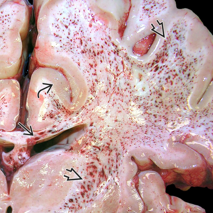
*Close-up view of autopsied brain in a patient with acute hemorrhagic leukoencephalitis (AHLE) shows innumerable tiny microbleeds in the subcortical and deep white matter (WM) and corpus callosum . Note the overlying cortex is almost completely spared . (Courtesy E. Rushing, MD.)*

*Close-up view of autopsied brain in a patient with acute hemorrhagic leukoencephalitis (AHLE) shows innumerable tiny microbleeds in the subcortical and deep white matter (WM) and corpus callosum . Note the overlying cortex is almost completely spared . (Courtesy E. Rushing, MD.)*

*Close-up view of autopsied brain in a patient with acute hemorrhagic leukoencephalitis (AHLE) shows innumerable tiny microbleeds in the subcortical and deep white matter (WM) and corpus callosum . Note the overlying cortex is almost completely spared . (Courtesy E. Rushing, MD.)*
 and deep/subcortical WM
  .](images/app.statdx.com_image_bc85bdf7-9d59-41a2-aa1c-4936434aeb8a_bb2e3243_20251018T070813Z.jpg)
*Axial FLAIR MR in a 28-year-old man with rapidly declining mental status after a flu-like illness shows patchy hyperintensities in the corpus callosum and deep/subcortical WM .*
*Axial FLAIR MR in a 28-year-old man with rapidly declining mental status after a flu-like illness shows patchy hyperintensities in the corpus callosum and deep/subcortical WM .*
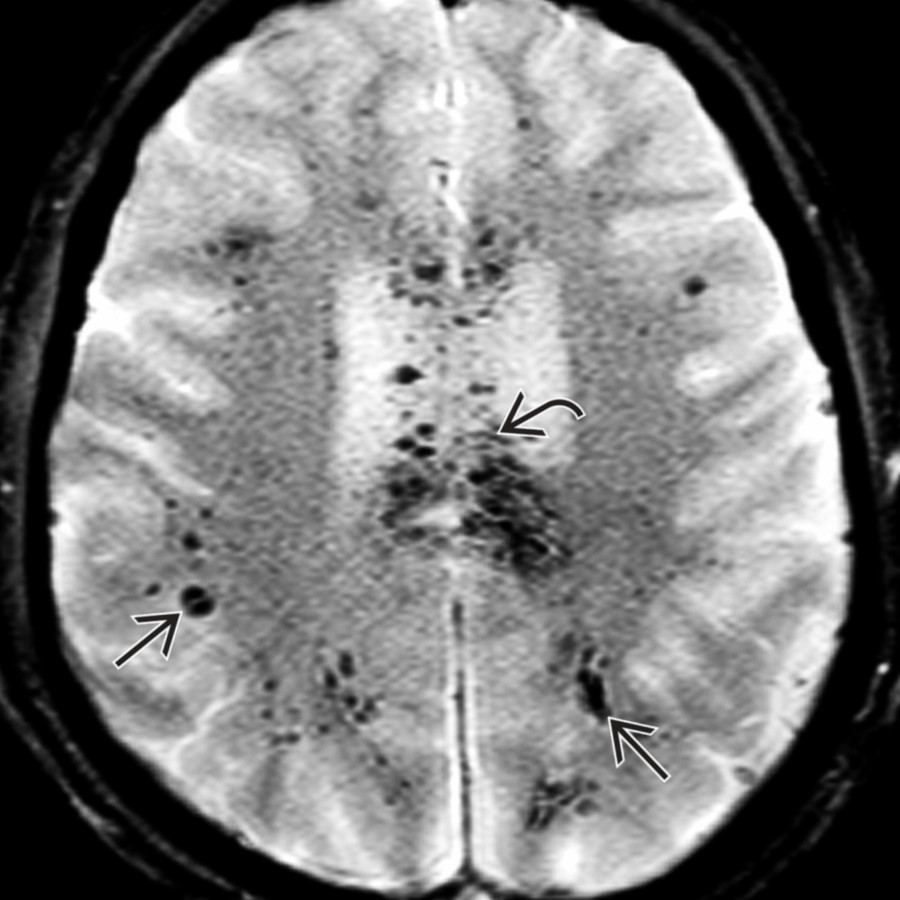
*Axial T2* GRE MR in the same patient shows multiple tiny hypointensities in the corpus callosum and deep/subcortical WM . The cortex is largely spared.*

*Axial T2* GRE MR in the same patient shows multiple tiny hypointensities in the corpus callosum and deep/subcortical WM . The cortex is largely spared.*
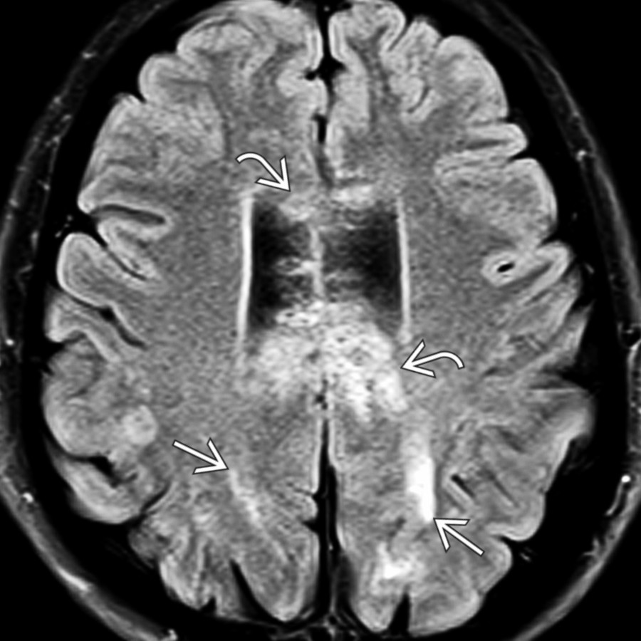
*Axial FLAIR MR in a 28-year-old man with rapidly declining mental status after a flu-like illness shows patchy hyperintensities in the corpus callosum and deep/subcortical WM .*

*Axial FLAIR MR in a 28-year-old man with rapidly declining mental status after a flu-like illness shows patchy hyperintensities in the corpus callosum and deep/subcortical WM .*

*Axial T2* GRE MR in the same patient shows multiple tiny hypointensities in the corpus callosum and deep/subcortical WM . The cortex is largely spared.*

*Axial T2* GRE MR in the same patient shows multiple tiny hypointensities in the corpus callosum and deep/subcortical WM . The cortex is largely spared.*
 with diffuse involvement of the hemispheric WM
  in this patient with AHLE.](images/app.statdx.com_image_9228cf7b-7628-4416-aff0-05b17a828cbc_2992bc89_20251018T070821Z.jpg)
*Axial T2* SWI MR MIP shows the innumerable microbleeds in the corpus callosum with diffuse involvement of the hemispheric WM in this patient with AHLE.*

*Axial T2* SWI MR MIP shows the innumerable microbleeds in the corpus callosum with diffuse involvement of the hemispheric WM in this patient with AHLE.*
### Additional Images
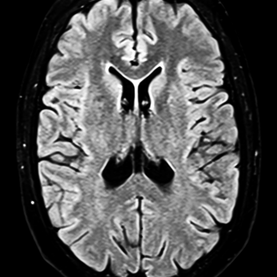
*Axial FLAIR MR in a 25-year-old man with fever and rapidly decreasing mental status shows no definite abnormalities.*

*Axial FLAIR MR in a 25-year-old man with fever and rapidly decreasing mental status shows no definite abnormalities.*
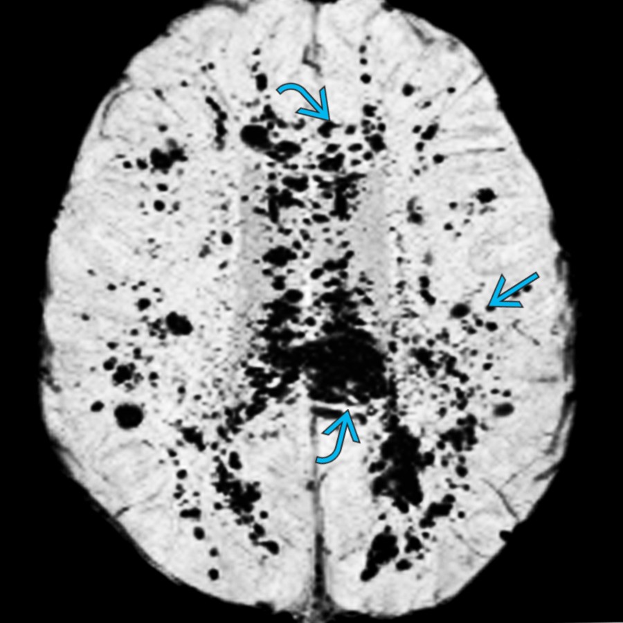
*Axial T2* SWI MR MIP shows the innumerable microbleeds in the corpus callosum with diffuse involvement of the hemispheric WM in this patient with AHLE.*

*Axial T2* SWI MR MIP shows the innumerable microbleeds in the corpus callosum with diffuse involvement of the hemispheric WM in this patient with AHLE.*
### Additional Images

*Axial FLAIR MR in a 25-year-old man with fever and rapidly decreasing mental status shows no definite abnormalities.*

*Axial FLAIR MR in a 25-year-old man with fever and rapidly decreasing mental status shows no definite abnormalities.*
 and splenium . The remainder of the WM appears normal.](images/app.statdx.com_image_cb9da6f4-8eb1-4e0f-8f9b-b3bc86522b86_9f11d750_20251018T070825Z.jpg)
*Axial T2* GRE MR in the same patient shows a few punctate "blooming" foci in the corpus callosum genu and splenium . The remainder of the WM appears normal.*

*Axial T2* GRE MR in the same patient shows a few punctate "blooming" foci in the corpus callosum genu and splenium . The remainder of the WM appears normal.*

*Axial SWI MR n the same patient shows innumerable tiny "blooming" microbleeds in the corpus callosum and subcortical and deep WM . The cortex is largely spared.*

*More cephalad axial T2* SWI MR shows innumerable tiny "blooming" foci in the WM, especially in the corpus callosum . These imaging findings are characteristic of AHLE.*

*Autopsy shows 2 areas of gross hemorrhagic necrosis . These findings and clinical history of prior flu-like illness with rapidly progressive fatal clinical course are characteristic of AHLE. (Courtesy R. Hewlett, MD).*

*Axial T2* GRE MR in a patient with rapid decline after a flu-like illness shows a large left frontal hemorrhage with numerous "blooming" foci in multiple WM lesions . Diagnosis was AHLE. (Courtesy R. Ramakantan, MD).*

*Axial NECT in a patient with AHLE shows patchy hemorrhages in the corpus callosum splenium .*

*Axial T2* SWI MR MIP in the same patient shows the microbleeds are heavily concentrated in the corpus callosum and hemispheric WM . Note involvement of the internal capsules and relative sparing of the basal ganglia and cortex.*
and splenium . The remainder of the WM appears normal.](images/app.statdx.com_image_cb9da6f4-8eb1-4e0f-8f9b-b3bc86522b86_9f11d750_20251018T070825Z.jpg)
*Axial T2* GRE MR in the same patient shows a few punctate "blooming" foci in the corpus callosum genu and splenium . The remainder of the WM appears normal.*

*Axial T2* GRE MR in the same patient shows a few punctate "blooming" foci in the corpus callosum genu and splenium . The remainder of the WM appears normal.*

*Axial SWI MR n the same patient shows innumerable tiny "blooming" microbleeds in the corpus callosum and subcortical and deep WM . The cortex is largely spared.*

*More cephalad axial T2* SWI MR shows innumerable tiny "blooming" foci in the WM, especially in the corpus callosum . These imaging findings are characteristic of AHLE.*

*Autopsy shows 2 areas of gross hemorrhagic necrosis . These findings and clinical history of prior flu-like illness with rapidly progressive fatal clinical course are characteristic of AHLE. (Courtesy R. Hewlett, MD).*

*Axial T2* GRE MR in a patient with rapid decline after a flu-like illness shows a large left frontal hemorrhage with numerous "blooming" foci in multiple WM lesions . Diagnosis was AHLE. (Courtesy R. Ramakantan, MD).*

*Axial NECT in a patient with AHLE shows patchy hemorrhages in the corpus callosum splenium .*

*Axial T2* SWI MR MIP in the same patient shows the microbleeds are heavily concentrated in the corpus callosum and hemispheric WM . Note involvement of the internal capsules and relative sparing of the basal ganglia and cortex.*