---
title: "Aortic Intramural Abnormality"
docid: "75d7b37f-bc37-493b-8961-8b2a9001fb94"
authors:
- key: "df804626-c042-4296-96e3-836a6da50fd6"
value: "Gregory Kicska, MD, PhD"
breadcrumbs:
-
name: "Cardiac"
slug: "cardiac"
treeNodeId: "fa90100b-619c-430e-8074-b5b9789bab39"
-
name: "Differential Diagnosis"
slug: "differential-diagnosis"
treeNodeId: "952326a0-b3ea-4a21-aa7a-d796cc9325ed"
-
name: "Aortic Intramural Abnormality"
slug: "aortic-intramural-abnormality"
treeNodeId: null
category: "Cardiac"
documentVersionId: "8ad4b88e-7101-4be0-a839-eb819f6dfe08"
imageCount: 9
lastUpdated: "03/17/22"
pageDescription: "Aortic Intramural Abnormality"
pageKeywords: "Cardiac, Differential Diagnosis, Aortic Intramural Abnormality"
pageTitle: "Aortic Intramural Abnormality | STATdx"
enhancedTitle: "Aortic Intramural Abnormality"
type: "DDX"
references: true
breadcrumbs:
- "Cardiac"
- "Differential Diagnosis"
- "Aortic Intramural Abnormality"
---
# ESSENTIAL INFORMATION
- ## Key Differential Diagnosis Issues
- Aortic wall should measure < 4 mm
- Aortic wall should be isointense to lumen
- ## Helpful Clues for Common Diagnoses
- **Atherosclerosis/Adherent Thrombus**
- Concentric diffuse involvement vs. spiral involvement of intramural hematoma
- Aorta often tortuous with atherosclerotic disease in branch vessels
- **Aortic Dissection**
- Intimal flap readily seen on CECT as unenhanced line through lumen
- Intraluminal calcifications on NECT suggest diagnosis and represent displaced intimal calcifications
- "Beak" sign: False lumen side of dissection flap meets outer wall with acute angle
- "Cobweb" sign: False lumen traversed by media fibers
- Confusion with pulsation artifact at aortic root avoided by inspecting coronal images
- ## Helpful Clues for Less Common Diagnoses
- **Aortic Intramural Hematoma**
- Hyperdense aortic wall compared to lumen when acute, isodense when old
- Check LV chamber for hypodense blood to avoid pitfall of confusion anemia
- Patient more likely to progress to dissection with coexistence of ulcer-like projections
- Most commonly in descending aorta
- **Penetrating Atherosclerotic Ulcer**
- Luminal irregularity
- Must extend beyond expected contour of intima
- Outer aortic wall thickening indicates acuity
- ## Helpful Clues for Rare Diagnoses
- **Takayasu/Giant Cell Arteritis**
- Radiographically indistinguishable, differentiated based on age (Takayasu < 50 years, giant cell > 50 years)
- FDG PET can determine active disease
- Aortic caliber will be reduced
- Subclavian stenosis is hallmark finding
- Pulmonary artery strictures and mesenteric vessel stenosis are common
- **Radiation**
- Vascular calcifications confined to radiation field
- Radiation history will be present
## References
# Selected References
1. [Ko JP et al: Chest CT Angiography for acute aortic pathologic conditions: pearls and pitfalls. Radiographics. 41(2):399-424, 2021](http://www.ncbi.nlm.nih.gov/pubmed/?term=33646903%5Bpmid%5D)
1. [Murillo H et al: Aortic dissection and other acute aortic syndromes: diagnostic imaging findings from acute to chronic longitudinal progression. Radiographics. 41(2):425-46, 2021](http://www.ncbi.nlm.nih.gov/pubmed/?term=33646901%5Bpmid%5D)
1. [Carroll BJ et al: Imaging for acute aortic syndromes. Heart. 106(3):182-9, 2019](http://www.ncbi.nlm.nih.gov/pubmed/?term=31822571%5Bpmid%5D)
1. [Abbara S et al: Thoracic aortic disease: spectrum of multidetector computed tomography imaging findings. J Cardiovasc Comput Tomogr. 1(1):40-54, 2007](http://www.ncbi.nlm.nih.gov/pubmed/?term=19083876%5Bpmid%5D)
## Images
### Selected Images
 .](images/app.statdx.com_image_thumbnail_6f26598e-b01e-456b-bf06-9708e5f5b758_annotated_true_size_900_quality_90_22dbe83187bf10aa89cc47d04f43d3121dc35b9c.jpg)
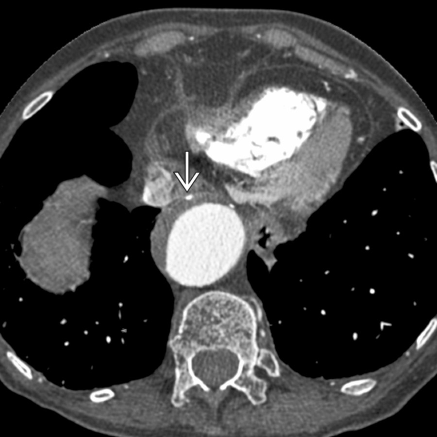
**Atherosclerosis/Adherent Thrombus**
*Axial CECT shows a mural thrombus in an otherwise dilated aorta. Note that intimal calcifications are on the outer edge of the thrombus .*
**Atherosclerosis/Adherent Thrombus**
*Axial CECT shows a mural thrombus in an otherwise dilated aorta. Note that intimal calcifications are on the outer edge of the thrombus .*
**Atherosclerosis/Adherent Thrombus**
*Axial CECT shows a mural thrombus in an otherwise dilated aorta. Note that intimal calcifications are on the outer edge of the thrombus .*

**Atherosclerosis/Adherent Thrombus**
*Axial CECT shows a mural thrombus in an otherwise dilated aorta. Note that intimal calcifications are on the outer edge of the thrombus .*
 . Note true lumen and false lumen
  . This was treated with fenestration.](images/app.statdx.com_image_thumbnail_ab9ba180-d18f-4a83-b390-3c1cb9228182_annotated_true_size_900_quality_90_932d5640e98dc34e85af08affa4ea2735e281ac2.jpg)
**Aortic Dissection**
*Axial CECT shows a severely displaced dissection flap, compressing the true lumen and occluding the SMA . Note true lumen and false lumen . This was treated with fenestration.*
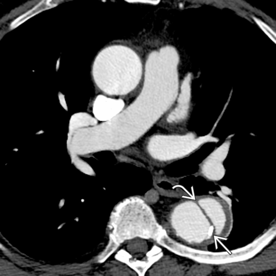
**Aortic Dissection**
*Axial CECT shows a dissection flap in the descending aorta with a displaced intimal calcification . The dissection did not involve the arch and was managed medically.*
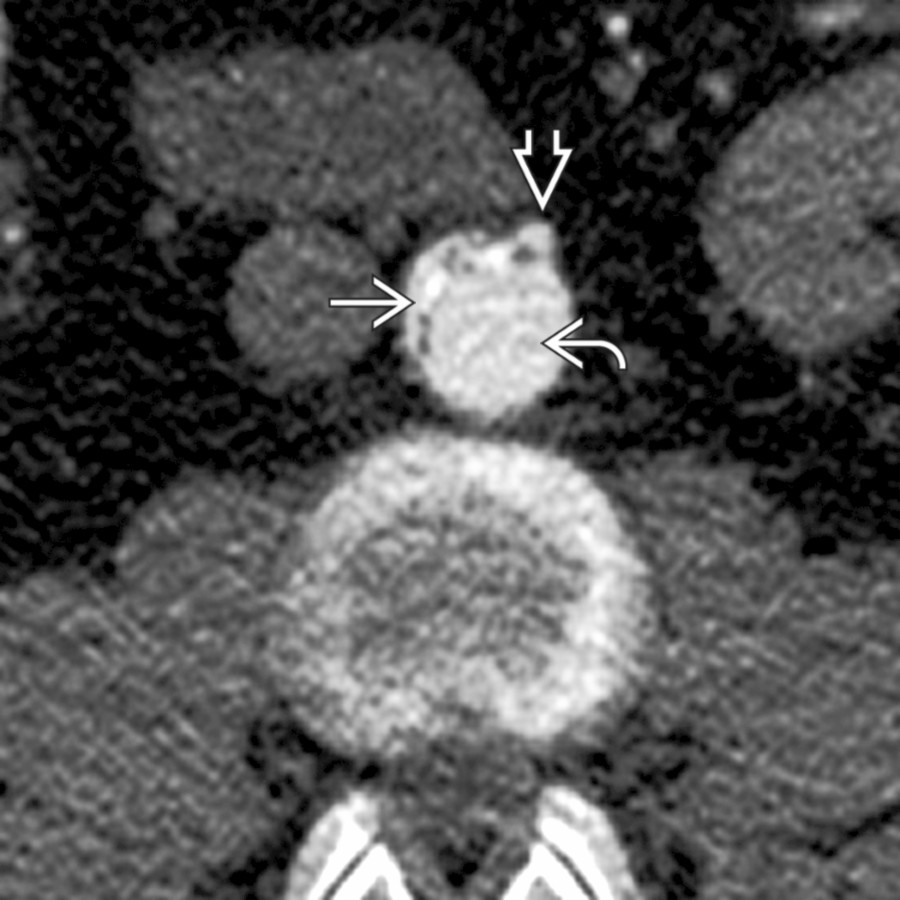
**Aortic Dissection**
*Axial CECT shows a severely displaced dissection flap, compressing the true lumen and occluding the SMA . Note true lumen and false lumen . This was treated with fenestration.*

**Aortic Dissection**
*Axial CECT shows a dissection flap in the descending aorta with a displaced intimal calcification . The dissection did not involve the arch and was managed medically.*
 aortic wall, representing the hematoma. Note the displaced intimal calcification .](images/app.statdx.com_image_thumbnail_3d098690-8549-4fc3-b311-bdffbe9f7019_annotated_true_size_900_quality_90_7d86fbf457c3890658e9cd35ccd020cb6f994314.jpg)
**Aortic Intramural Hematoma**
*Coronal NECT shows asymmetric thickening and hyperdense aortic wall, representing the hematoma. Note the displaced intimal calcification .*
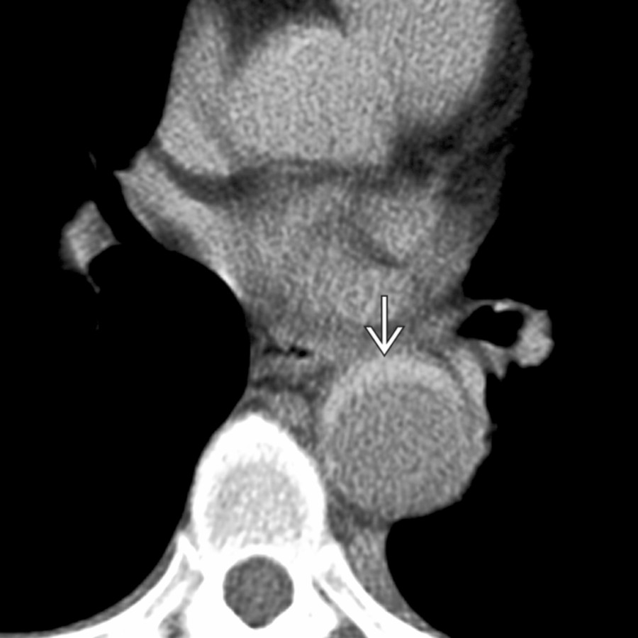
**Aortic Intramural Hematoma**
*Axial CECT shows hyperdense, thickened aortic wall in a hypertensive patient who presented to the ER with back pain. This finding can be missed on an enhanced exam.*
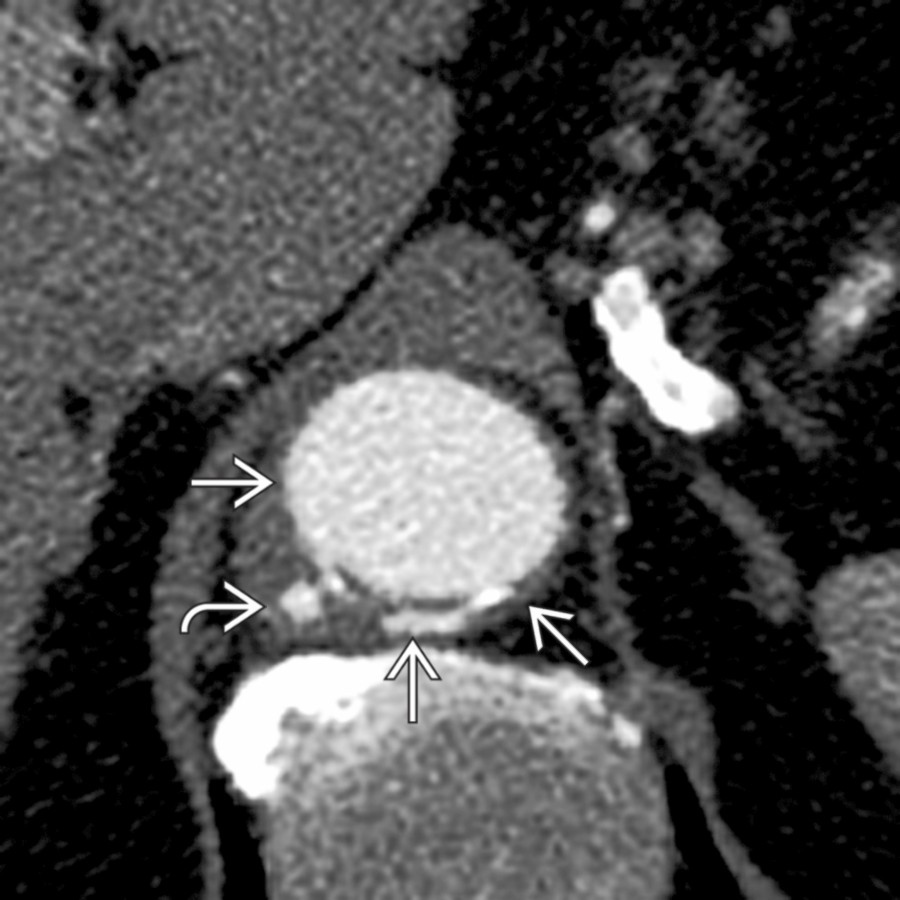
**Penetrating Atherosclerotic Ulcer**
*Axial CECT shows a small focus of contrast extending beyond the expected aortic wall . Adjacent wall thickening suggested it is likely acute and explains the patient's back pain.*

**Takayasu/Giant Cell Arteritis**
*Axial CECT shows thickened aortic wall with inner intimal calcifications. Although causing aortic narrowing, this may progress to aneurysmal dilation.*
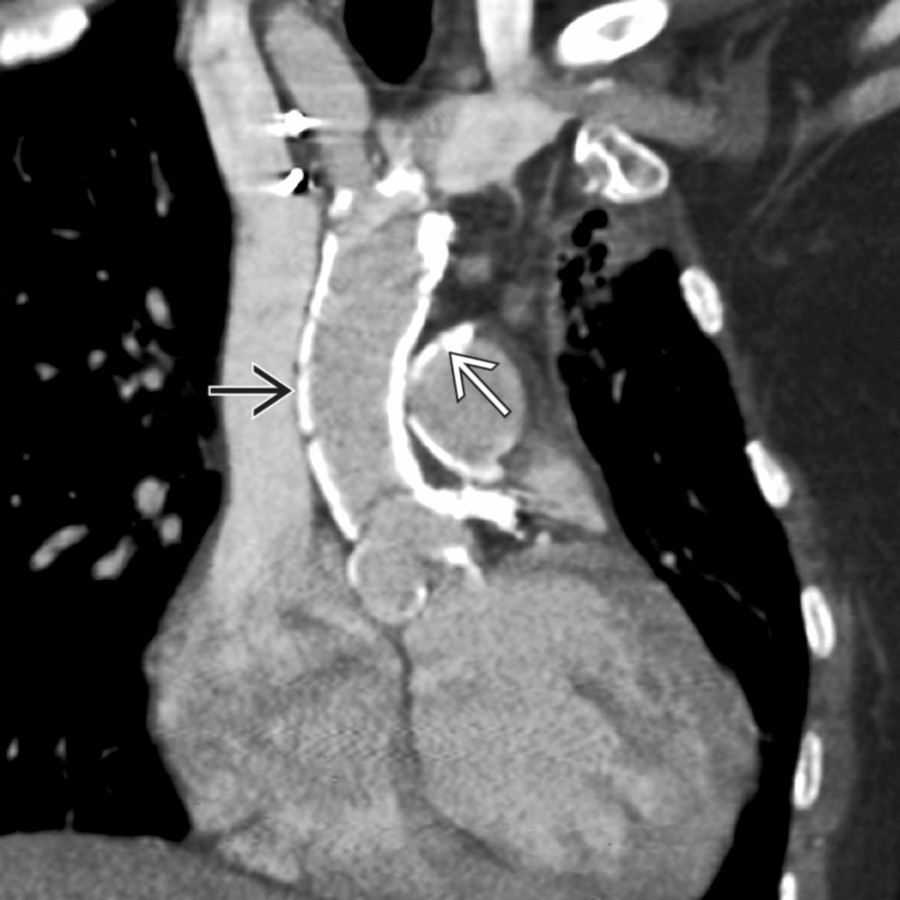
**Radiation**
*Double oblique CECT shows dense aortic and pulmonary artery calcifications in a patient who received prior mediastinal radiation. Vascular calcifications were not present elsewhere. Note that calcifications are spatially confined to the radiation field.*
### Additional Images
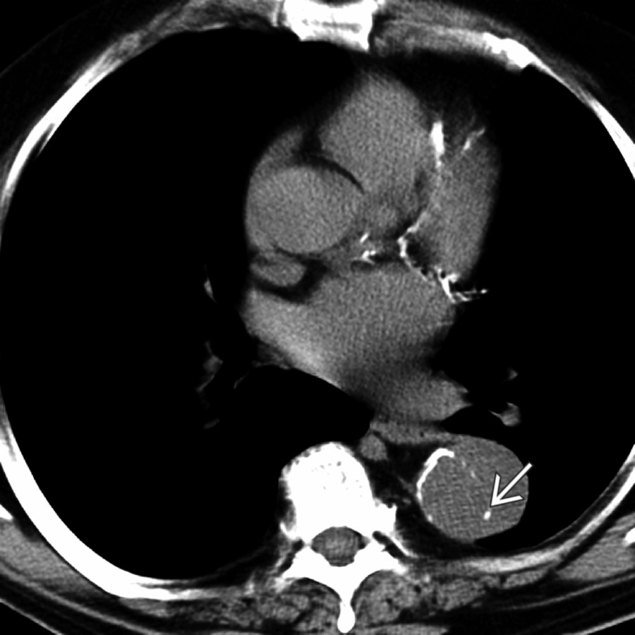
**Aortic Dissection**
*Axial NECT shows displaced intimal calcifications representing aortic dissection .*
aortic wall, representing the hematoma. Note the displaced intimal calcification .](images/app.statdx.com_image_thumbnail_3d098690-8549-4fc3-b311-bdffbe9f7019_annotated_true_size_900_quality_90_7d86fbf457c3890658e9cd35ccd020cb6f994314.jpg)
**Aortic Intramural Hematoma**
*Coronal NECT shows asymmetric thickening and hyperdense aortic wall, representing the hematoma. Note the displaced intimal calcification .*

**Aortic Intramural Hematoma**
*Axial CECT shows hyperdense, thickened aortic wall in a hypertensive patient who presented to the ER with back pain. This finding can be missed on an enhanced exam.*

**Penetrating Atherosclerotic Ulcer**
*Axial CECT shows a small focus of contrast extending beyond the expected aortic wall . Adjacent wall thickening suggested it is likely acute and explains the patient's back pain.*

**Takayasu/Giant Cell Arteritis**
*Axial CECT shows thickened aortic wall with inner intimal calcifications. Although causing aortic narrowing, this may progress to aneurysmal dilation.*

**Radiation**
*Double oblique CECT shows dense aortic and pulmonary artery calcifications in a patient who received prior mediastinal radiation. Vascular calcifications were not present elsewhere. Note that calcifications are spatially confined to the radiation field.*
### Additional Images

**Aortic Dissection**
*Axial NECT shows displaced intimal calcifications representing aortic dissection .*