---
title: "Asymmetric Lateral Ventricles"
docid: "87387f0d-9b20-4288-a250-aa3ec83520c4"
authors:
- key: "1fa14dfd-71ea-4960-908e-e720313bc63a"
value: "Santhosh Gaddikeri, MD"
- key: "30ce27b2-237f-4aff-a88f-65ead356335b"
value: "Marinos Kontzialis, MD"
breadcrumbs:
-
name: "Brain"
slug: "brain"
treeNodeId: "6d8829f1-14d7-45af-8675-255189aa526a"
-
name: "Differential Diagnosis"
slug: "differential-diagnosis"
treeNodeId: "a7fdd139-664e-4bb8-8d18-400e4733ff60"
-
name: "Ventricles, Periventricular Regions"
slug: "ventricles-periventricular-regions"
treeNodeId: "353c434a-a6fc-4ef1-8786-d30a1988a4dc"
-
name: "Generic Imaging Patterns"
slug: "generic-imaging-patterns"
treeNodeId: "969c31a2-ef56-4fc3-9125-05857cf9aac3"
-
name: "Asymmetric Lateral Ventricles"
slug: "asymmetric-lateral-ventricles"
treeNodeId: null
category: "Brain"
cmeTopicId: "5536eb32-54f9-4eb3-83c6-15950dc4efe6"
documentVersionId: "7b24399c-3ad6-47c3-9521-b6125b27d26d"
imageCount: 54
lastUpdated: "01/25/23"
pageDescription: "Asymmetric Lateral Ventricles"
pageKeywords: "Brain, Differential Diagnosis, Ventricles, Periventricular Regions, Generic Imaging Patterns, Asymmetric Lateral Ventricles"
pageTitle: "Asymmetric Lateral Ventricles | STATdx"
enhancedTitle: "Asymmetric Lateral Ventricles"
type: "DDX"
references: true
breadcrumbs:
- "Brain"
- "Differential Diagnosis"
- "Ventricles, Periventricular Regions"
- "Generic Imaging Patterns"
- "Asymmetric Lateral Ventricles"
---
# ESSENTIAL INFORMATION
- ## Key Differential Diagnosis Issues
- Asymmetric lateral ventricles are most commonly seen as normal variant
- ## Helpful Clues for Common Diagnoses
- **Normal Variant**
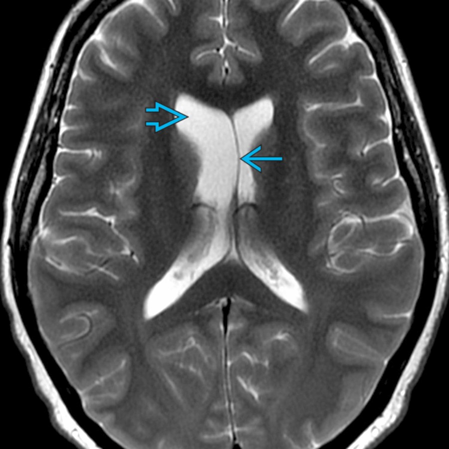
- Asymmetric lateral ventricles seen in 5-10% of normal population
- Asymmetry mild to moderate, left > right
- Septum may be displaced across midline
- No associated mass effect, herniation, or parenchymal atrophy
- Must exclude parenchymal or intraventricular abnormality
- **Extrinsic Mass Effect**
- Etiologies include mass, hemorrhage, infarct, infection
- Mass can cause ventricular deformity, subfalcine herniation
- **Encephalomalacia, General**
- Parenchymal loss results in compensatory ventricular enlargement
- Common etiologies include chronic infarct, trauma, surgery
- **Intraventricular Hemorrhage**
- Involved ventricle may dilate early from mass effect
- Chronic dilation may be due to scarring and adhesions
- Etiologies include trauma, arteriovenous malformation (AVM), basal ganglia hemorrhage
- **Herniation Syndromes, Intracranial**
- Subfalcine herniation: Cingulate gyrus herniates under falx
- Ipsilateral lateral ventricle compressed
- Foramen of Monro obstructs and causes contralateral lateral ventricle enlargement
- Unilateral descending transtentorial herniation (uncal): Herniation of medial temporal lobe inferiorly
- Contralateral temporal horn becomes entrapped and enlarges
- Entrapped ventricle: Typically temporal horn, by extrinsic mass effect
- **Surgical Defects**
- Look for calvarial defect or "tract"
- Typically related to resection of mass
- Ventricle enlarged unilateral to defect
- **Obstructive Hydrocephalus**
- Typically acquired and bilateral
- May be unilateral if shunt complication or obstructing tumor is cause
- Rare: Colloid cyst may obstruct unilateral foramen of Monro and cause unilateral ventriculomegaly
- **Choroid Plexus Cyst**
- a.k.a. choroid plexus xanthogranuloma
- Most common choroid plexus mass in adults
- Nonneoplastic, noninflammatory cyst of choroid plexus
- Common incidental finding in older patients (40% prevalence)
- Typically bilateral; may be unilateral and enlarge lateral ventricle
- Most in lateral ventricular atria
- 60-80% bright on DWI
- ## Helpful Clues for Less Common Diagnoses
- **Ventriculitis**
- Ventriculomegaly with debris level, enhancing ependyma, periventricular T2/FLAIR hyperintensities
- May affect lateral ventricles asymmetrically, particularly if related to shunt placement or abscess rupture
- Restricted diffusion of layering debris is characteristic
- Bacterial ventriculitis may occur in healthy individuals after trauma or neurosurgical procedure
- Fungal or viral ventriculitis occurs most commonly in immunosuppressed patients
- **CSF Shunts and Complications**
- Common complications include shunt obstruction or breakage, infection, overdrainage
- Asymmetric ventricles may result from overdrainage or underdrainage of "isolated" ventricle
- **Meningioma**
- Although rare, still one of more common intraventricular neoplasms in adults
- More common in females (F:M = 2:1)
- Most common location is ventricular atrium with slight majority on left
- Smooth, avidly enhancing intraventricular mass
- 50% calcified; cystic changes may be present
- **Choroid Plexus Papilloma**
- Enhancing, lobulated intraventricular mass in child
- 50% in lateral ventricle atrium, left > right
- 40% in 4th ventricle
- Increased CSF production in most cases, CSF obstruction in some cases
- May have CSF spread of tumor
- **Neurocytoma, Central**
- Neurocytoma arising from ventricular system, usually septum pellucidum or lateral ventricle
- Well-circumscribed, lobulated, "bubbly" lesions
- 50% calcifications, cyst-like areas, enhancement, prominent associated flow voids
- MRS: Glycine peak (3.55 ppm) may help differentiation from other intraventricular neoplasms
- **Neurocysticercosis**
- May involve cisterns > parenchyma > ventricles
- Intraventricular disease anywhere from 0.7-33% of cases
- May lead to obstructive hydrocephalus, ventriculitis, adhesions
- Intraventricular cyst signal intensity may differ slightly from CSF on T1, T2, FLAIR
- ## Helpful Clues for Rare Diagnoses
- **Intraventricular Synechiae/Adhesions**
- May be congenital or acquired (prior bleed, infection, tumor)
- Look for enhancing septa, intraventricular cysts within ventricle
- **Choroid Plexus Carcinoma**
- Enhancing intraventricular mass and ependymal invasion in young child
- CSF seeding common
- Imaging does not allow reliable distinction between choroid plexus papilloma and carcinoma
- May be more heterogeneous than choroid plexus papilloma in part reflecting areas of necrosis
- May demonstrate elevation of lactate level
- **Ependymal Cyst**
- Nonenhancing, thin-walled cyst with CSF density/intensity
- Lateral ventricle most common location
- Most are incidental
- Best diagnostic clue: Nonenhancing, thin-walled CSF density/intensity cyst in lateral ventricle
- **Dyke-Davidoff-Masson**
- Antenatal unilateral hemispheric insult causes cerebral hemiatrophy
- Compensatory ipsilateral calvarial thickening, hyperpneumatized frontal sinuses and temporal bones due to longstanding cerebral hemiatrophy
- Dilated ventricle from volume loss is ipsilateral to small hemisphere
- **Hemimegalencephaly**
- Unilateral hemispheric cortical thickening
- Dilated, usually dysmorphic ventricle ipsilateral to enlarged hemisphere
- Ipsilateral extracalvarial soft tissues may be larger
- Pachygyria, polymicrogyria, heterotopias, abnormal white matter signal, blurring of gray-white matter junction
- **Rasmussen Encephalitis**
- Chronic unilateral brain inflammation of uncertain etiology leading to progressive hemispheric atrophy
- Early focal swelling of gyri
- Unilateral, predominantly frontoinsular cortical and subcortical T2/FLAIR hyperintensity progress to atrophy
- ## Other Essential Information
- High-resolution MR cisternography: CISS, balanced FFE, FIESTA
- May detect small septations or arachnoid membranes causing obstruction
- Cine CSF flow study may help detect physiologic flow obstruction from arachnoid webs or membranes
- May assess adequacy of drainage procedures
## References
# Selected References
1. [Balasubramaniam C: Shunt complications - staying out of trouble. Neurol India. 69(Supplement):S495-501, 2021](http://www.ncbi.nlm.nih.gov/pubmed/?term=35103008%5Bpmid%5D)
1. [Lanska DJ: Cruveilhier's unrecognized case (c1831) of Dyke-Davidoff-Masson syndrome. Eur Neurol. 84(4):300-6, 2021](http://www.ncbi.nlm.nih.gov/pubmed/?term=33965957%5Bpmid%5D)
1. [Cay-Martinez KC et al: Rasmussen encephalitis: an update. Semin Neurol. 40(2):201-10, 2020](http://www.ncbi.nlm.nih.gov/pubmed/?term=32185790%5Bpmid%5D)
1. [Crawford JR et al: Perinatal (fetal and neonatal) choroid plexus tumors: a review. Childs Nerv Syst. 35(6):937-44, 2019](http://www.ncbi.nlm.nih.gov/pubmed/?term=30953158%5Bpmid%5D)
1. [Tan LA et al: Obstructive hydrocephalus due to intraventricular hemorrhage after incidental durotomy during lumbar spine surgery. Spine (Phila Pa 1976). 43(5):E316-9, 2018](http://www.ncbi.nlm.nih.gov/pubmed/?term=26208226%5Bpmid%5D)
1. [Zamora CA et al: Teaching neuroImages: Dyke-Davidoff-Masson in Sturge-Weber syndrome. Neurology. 85(16):e128, 2015](http://www.ncbi.nlm.nih.gov/pubmed/?term=26481933%5Bpmid%5D)
1. [Smith AB et al: From the radiologic pathology archives: intraventricular neoplasms: radiologic-pathologic correlation. Radiographics. 33(1):21-43, 2013](http://www.ncbi.nlm.nih.gov/pubmed/?term=23322825%5Bpmid%5D)
1. [Kimura-Hayama ET et al: Neurocysticercosis: radiologic-pathologic correlation. Radiographics. 30(6):1705-19, 2010](http://www.ncbi.nlm.nih.gov/pubmed/?term=21071384%5Bpmid%5D)
1. [Kiroğlu Y et al: Cerebral lateral ventricular asymmetry on CT: how much asymmetry is representing pathology? Surg Radiol Anat. 30(3):249-55, 2008](http://www.ncbi.nlm.nih.gov/pubmed/?term=18253688%5Bpmid%5D)
1. [Rastogi S et al: Neuroimaging in pediatric epilepsy: a multimodality approach. Radiographics. 28(4):1079-95, 2008](http://www.ncbi.nlm.nih.gov/pubmed/?term=18635630%5Bpmid%5D)
1. [Osborn AG, Preece MT. Intracranial cysts: radiologic-pathologic correlation and imaging approach. Radiology. 2006 Jun;239(3):650-64.](http://www.ncbi.nlm.nih.gov/pubmed/?term=16714456%5Bpmid%5D)
1. [Koeller KK et al: Cerebral intraventricular neoplasms: radiologic-pathologic correlation. Radiographics. 22(6):1473-505, 2002](http://www.ncbi.nlm.nih.gov/pubmed/?term=12432118%5Bpmid%5D)
## Images
### Selected Images
 when compared to the left. Note the septum is slightly deviated to the left
  .](images/app.statdx.com_image_thumbnail_74e87ea4-3c2a-400b-b714-f9fa2db4e66a_annotated_true_size_900_quality_90_2466c18c_20251018T165124Z.jpg)
**Normal Variant**
*Axial T2 MR demonstrates normal variant anatomy with mild asymmetric prominence of the right lateral ventricle when compared to the left. Note the septum is slightly deviated to the left .*
**Normal Variant**
*Axial T2 MR demonstrates normal variant anatomy with mild asymmetric prominence of the right lateral ventricle when compared to the left. Note the septum is slightly deviated to the left .*
**Normal Variant**
*Axial T2 MR demonstrates normal variant anatomy with mild asymmetric prominence of the right lateral ventricle when compared to the left. Note the septum is slightly deviated to the left .*

**Normal Variant**
*Axial T2 MR demonstrates normal variant anatomy with mild asymmetric prominence of the right lateral ventricle when compared to the left. Note the septum is slightly deviated to the left .*
 .](images/app.statdx.com_image_thumbnail_ddc74acb-a5fe-4147-bc17-3e3862305da7_annotated_true_size_900_quality_90_d599964a_20251018T165124Z.jpg)
**Extrinsic Mass Effect**
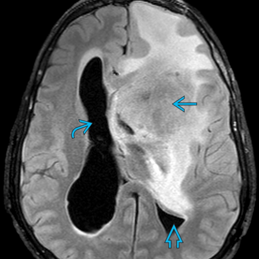
*Axial FLAIR MR demonstrates a large, heterogeneous mass in the left frontal lobe with surrounding edema and mass effect with compression of the left lateral ventricle and moderate dilation of the right lateral ventricle .*
**Encephalomalacia, General**
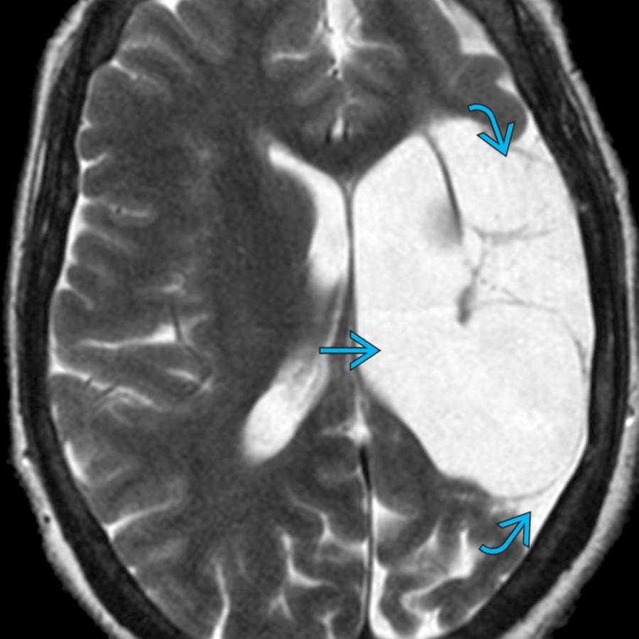
*Axial T2 MR demonstrates a large, cystic encephalomalacia in the left frontal and parietal lobes due to chronic infarction with ex-vacuo dilation of the left lateral ventricle .*

**Intraventricular Hemorrhage**
*Axial T2 MR demonstrates a large amount of acute intraventricular hemorrhage with mild dilation of the right lateral ventricle . Note CSF seepage in the peritrigonal region .*
**Extrinsic Mass Effect**
*Axial FLAIR MR demonstrates a large, heterogeneous mass in the left frontal lobe with surrounding edema and mass effect with compression of the left lateral ventricle and moderate dilation of the right lateral ventricle .*

**Encephalomalacia, General**
*Axial T2 MR demonstrates a large, cystic encephalomalacia in the left frontal and parietal lobes due to chronic infarction with ex-vacuo dilation of the left lateral ventricle .*

**Intraventricular Hemorrhage**
*Axial T2 MR demonstrates a large amount of acute intraventricular hemorrhage with mild dilation of the right lateral ventricle . Note CSF seepage in the peritrigonal region .*
 and a small parenchymal hemorrhage
  , resulting in rightward subfalcine herniation , compression of the left lateral ventricle , and mild dilation of the right lateral ventricle .](b9992ffd-4f31-44e2-8c92-83ab8fad30c1)
**Herniation Syndromes, Intracranial**
*Axial NECT demonstrates a large left subdural hematoma and a small parenchymal hemorrhage , resulting in rightward subfalcine herniation , compression of the left lateral ventricle , and mild dilation of the right lateral ventricle .*

**Surgical Defects**
*Axial FLAIR MR demonstrates a large surgical defect in the left frontal lobe due to a tumor resection with resultant mild prominence of the left lateral ventricle .*

**Obstructive Hydrocephalus**
*Axial T1 C+ MR demonstrates enhancing intraventricular mass with obstructive dilation of the left temporal horn .*

**Herniation Syndromes, Intracranial**
*Axial NECT demonstrates a large left subdural hematoma and a small parenchymal hemorrhage , resulting in rightward subfalcine herniation , compression of the left lateral ventricle , and mild dilation of the right lateral ventricle .*

**Surgical Defects**
*Axial FLAIR MR demonstrates a large surgical defect in the left frontal lobe due to a tumor resection with resultant mild prominence of the left lateral ventricle .*

**Obstructive Hydrocephalus**
*Axial T1 C+ MR demonstrates enhancing intraventricular mass with obstructive dilation of the left temporal horn .*
 due to the shunt reservoir.](6a5a42a2-43a2-4fe6-8dd5-b2b1fc3e83fd)
**CSF Shunts and Complications**
*Axial T2 MR demonstrates a shunt catheter in the right lateral ventricle with asymmetric lateral ventricles due to overdrainage of the right and mild underdrainage of the left . Note susceptibility artifact due to the shunt reservoir.*

**Meningioma**
*Axial T2 MR demonstrates a left lateral intraventricular isointense mass in the trigone with dilatation of the left trigone . Note periventricular edema . Biopsy revealed meningioma.*

**Neurocytoma, Central**
*Axial T2 MR demonstrates a well-circumscribed, isointense, left lateral ventricular mass attached to the septum pellucidum with peripheral tiny cysts . Note moderate dilation of the left lateral ventricle . Biopsy revealed neurocytoma.*

**Intraventricular Synechiae/Adhesions**
*Coronal T2 MR demonstrates asymmetric mild enlargement of the left lateral ventricle due to synechiae obstructing the foramen of Monro.*

**Choroid Plexus Carcinoma**
*Axial T2 MR demonstrates a poorly circumscribed heterogeneous left trigonal mass with internal cysts/necrosis. Note extensive periventricular edema . Biopsy revealed choroid plexus carcinoma.*

**Dyke-Davidoff-Masson**
*Axial NECT demonstrates atrophy of the left cerebral hemisphere with encephalomalacia due to antenatal vascular insult. Note the dilated left lateral ventricle and mild overlying calvarial thickening .*

**Rasmussen Encephalitis**
*Axial T2 MR in a patient with refractory epilepsy due to Rasmussen encephalitis demonstrates left cerebral hemispheric atrophy and mild ex-vacuo dilation of the left lateral ventricle .*
### Additional Images

**CSF Shunts and Complications**
*Axial T2 MR demonstrates a shunt catheter in the right lateral ventricle with asymmetric lateral ventricles due to overdrainage of the right and mild underdrainage of the left . Note susceptibility artifact due to the shunt reservoir.*

**Meningioma**
*Axial T2 MR demonstrates a left lateral intraventricular isointense mass in the trigone with dilatation of the left trigone . Note periventricular edema . Biopsy revealed meningioma.*

**Neurocytoma, Central**
*Axial T2 MR demonstrates a well-circumscribed, isointense, left lateral ventricular mass attached to the septum pellucidum with peripheral tiny cysts . Note moderate dilation of the left lateral ventricle . Biopsy revealed neurocytoma.*

**Intraventricular Synechiae/Adhesions**
*Coronal T2 MR demonstrates asymmetric mild enlargement of the left lateral ventricle due to synechiae obstructing the foramen of Monro.*

**Choroid Plexus Carcinoma**
*Axial T2 MR demonstrates a poorly circumscribed heterogeneous left trigonal mass with internal cysts/necrosis. Note extensive periventricular edema . Biopsy revealed choroid plexus carcinoma.*

**Dyke-Davidoff-Masson**
*Axial NECT demonstrates atrophy of the left cerebral hemisphere with encephalomalacia due to antenatal vascular insult. Note the dilated left lateral ventricle and mild overlying calvarial thickening .*

**Rasmussen Encephalitis**
*Axial T2 MR in a patient with refractory epilepsy due to Rasmussen encephalitis demonstrates left cerebral hemispheric atrophy and mild ex-vacuo dilation of the left lateral ventricle .*
### Additional Images
 , primary CNS lymphoma. Extrinsic mass effect is a common cause of ventricular asymmetry.](images/app.statdx.com_image_thumbnail_9c7a2c27-fa74-474a-a33e-3cfbf2329f76_annotated_true_size_900_quality_90_8cafe5b4_20251018T165124Z.jpg)
**Extrinsic Mass Effect**
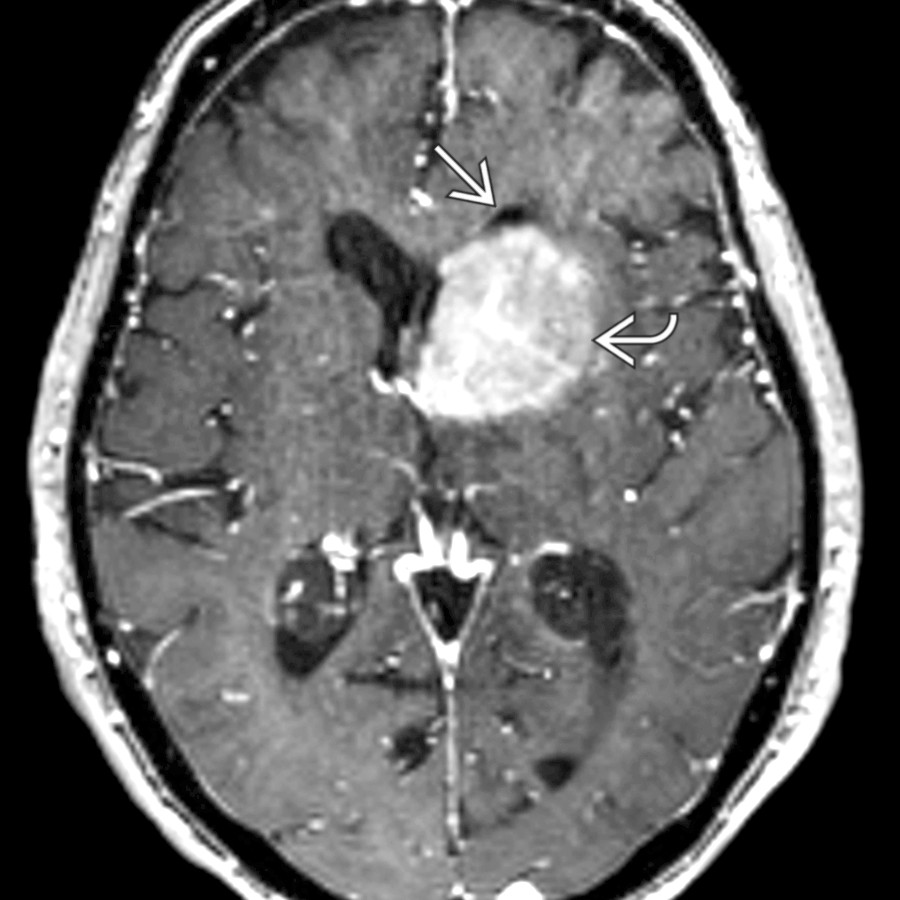
*Axial T1WI C+ MR shows compression of the left frontal horn by a large, periventricular, enhancing mass , primary CNS lymphoma. Extrinsic mass effect is a common cause of ventricular asymmetry.*
**Extrinsic Mass Effect**
*Axial T1WI C+ MR shows compression of the left frontal horn by a large, periventricular, enhancing mass , primary CNS lymphoma. Extrinsic mass effect is a common cause of ventricular asymmetry.*
 .](images/app.statdx.com_image_thumbnail_d875dfa4-5133-4dd6-9471-a019de504b4a_annotated_true_size_900_quality_90_e15566aa_20251018T165124Z.jpg)
**Encephalomalacia, General**
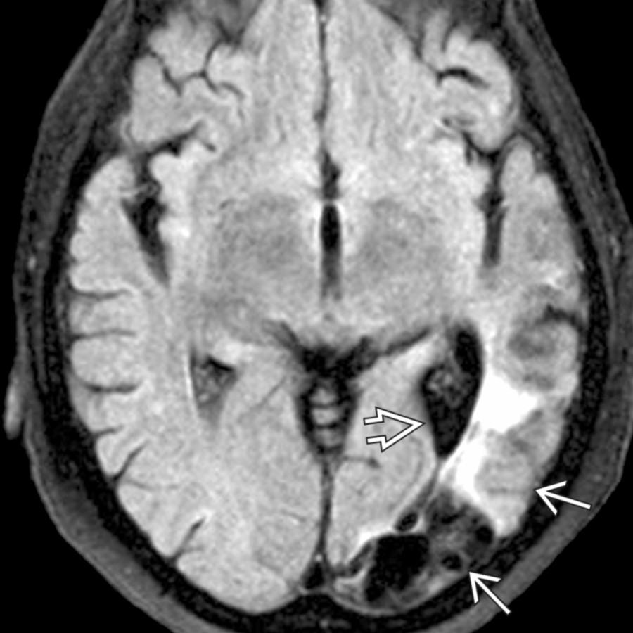
*Axial FLAIR MR demonstrates a left posterior MCA encephalomalacia resulting in mild ex-vacuo dilatation of the left atrium .*
**Encephalomalacia, General**
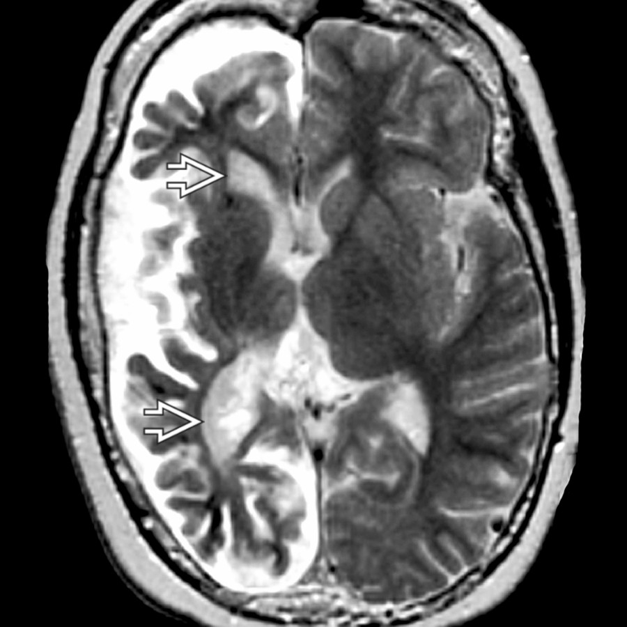
*Axial T2 MR shows right hemiatrophy in Sturge-Weber syndrome. Chronic venous ischemia leads to progressive hemiatrophy. Note the ipsilateral large right ventricle due to volume loss.*

**Intraventricular Hemorrhage**
*Axial NECT shows a basal ganglia hypertensive hemorrhage with intraventricular extension . Associated midline shift results in dilation of the contralateral ventricles from foramen of Monro obstruction.*

**Herniation Syndromes, Intracranial**
*Coronal FLAIR MR shows a diffusely enlarged hyperintense supratentorial cortex compared to the cerebellum in this patient in longstanding status epilepticus. Disproportionate left hemisphere involvement has resulted in left ventricular compression , right foramen of Monro outlet obstruction, and dilation of the right ventricular system .*

**Herniation Syndromes, Intracranial**
*Coronal T1WI C+ MR shows a right hemispheric, subacute, subdural hematoma causing subfalcine and uncal herniation. Mass effect compresses the right frontal horn. The left ventricle is enlarged from foramen of Monro obstruction.*

**Herniation Syndromes, Intracranial**
*Coronal T2WI MR shows right temporal viral encephalitis causing local mass effect . The left lateral ventricle is larger from foramen of Monro obstruction due to midline shift.*

**Surgical Defects**
*Axial T2WI MR shows widening of right foramen of Monro , septum pellucidum deviation , and an enlarged right lateral ventricle in this tuberous sclerosis patient with remote tumor resection.*

**Obstructive Hydrocephalus**
*Axial NECT shows marked enlargement of the left lateral ventricle with bowing of septum pellucidum across midline and transependymal CSF migration indicating acute obstruction. Findings were related to a small atrial diverticulum.*

**Obstructive Hydrocephalus**
*Axial T1WI C+ MR shows a colloid cyst believed to be complicated by inflammatory changes. Obstruction of the left foramen of Monro causes unilateral left lateral ventricle dilation .*

**Obstructive Hydrocephalus**
*Axial T2WI MR shows a medial atrial diverticulum , a rare complication of severe hydrocephalus. CSF pouch herniates inferomedially through tentorial incisura.*

**Obstructive Hydrocephalus**
*Coronal T1WI C+ MR shows typical case of coccidioidomycosis meningitis. Note marked enhancement of basal cisterns and asymmetric ventricular enlargement from CSF obstruction.*

**Choroid Plexus Cyst**
*Axial T1WI C+ MR shows a lobulated, nonenhancing mass in the lateral ventricle atrium, a choroid plexus xanthogranuloma. This degenerative cyst of the choroid plexus is often found incidentally in older patients.*

**Ventriculitis**
*Axial T1WI C+ MR shows ventriculitis with asymmetric lateral ventricles related to a temporal lobe abscess rupture and meningitis . Note ventriculomegaly and ventricular wall enhancement characteristic of ventriculitis.*

**Ventriculitis**
*Axial CECT shows marked ventriculomegaly and ependymal enhancement . A dependent debris level is noted in both lateral ventricles. Ventriculitis has resulted from abscess rupture.*

**CSF Shunts and Complications**
*Axial NECT shows an infant with hydrocephalus after placement of a right frontal ventricular drain . The shunt did not cross midline. The left lateral ventricle remained enlarged, and the right became slit-like.*

**CSF Shunts and Complications**
*Axial T2WI MR shows marked enlargement of the isolated right lateral ventricle with transependymal flow of CSF indicating acute obstruction. Note left shunt and completely decompressed left lateral ventricle.*

**CSF Shunts and Complications**
*Axial NECT shows asymmetric left ventricular dilation post shunting . Shunt tip may be occluded from clot or from imperforate septum, preventing drainage of the left ventricular system.*

**Meningioma**
*Axial T2WI FS MR shows a hypointense choroid plexus mass in the atrium of the left lateral ventricle that enhanced intensely (not shown). Note striking surrounding vasogenic edema in adjacent brain parenchyma, thought due to locally obstructed CSF.*

**Neurocytoma, Central**
*Axial T1WI C+ MR demonstrates a mildly enhancing and heterogeneous mass arising from the septum pellucidum . Note asymmetric dilatation of the right lateral ventricle .*

**Choroid Plexus Papilloma**
*Axial T1WI C+ MR shows enhancement within the mass in the atrium of the lateral ventricle with an encysted asymmetrically larger left lateral ventricle.*

**Neurocysticercosis**
*Axial T1WI C+ MR shows asymmetric lateral ventricles caused by a giant neurocysticercosis cyst in the body of the left lateral ventricle.*

**Intraventricular Synechiae/Adhesions**
*Axial T1WI C+ FS MR shows a large mass in the atrium of the right lateral ventricle with a trapped, encysted occipital horn . Ependymal enhancement represents tumor spread from choroidal metastasis.*
**Encephalomalacia, General**
*Axial FLAIR MR demonstrates a left posterior MCA encephalomalacia resulting in mild ex-vacuo dilatation of the left atrium .*

**Encephalomalacia, General**
*Axial T2 MR shows right hemiatrophy in Sturge-Weber syndrome. Chronic venous ischemia leads to progressive hemiatrophy. Note the ipsilateral large right ventricle due to volume loss.*

**Intraventricular Hemorrhage**
*Axial NECT shows a basal ganglia hypertensive hemorrhage with intraventricular extension . Associated midline shift results in dilation of the contralateral ventricles from foramen of Monro obstruction.*

**Herniation Syndromes, Intracranial**
*Coronal FLAIR MR shows a diffusely enlarged hyperintense supratentorial cortex compared to the cerebellum in this patient in longstanding status epilepticus. Disproportionate left hemisphere involvement has resulted in left ventricular compression , right foramen of Monro outlet obstruction, and dilation of the right ventricular system .*

**Herniation Syndromes, Intracranial**
*Coronal T1WI C+ MR shows a right hemispheric, subacute, subdural hematoma causing subfalcine and uncal herniation. Mass effect compresses the right frontal horn. The left ventricle is enlarged from foramen of Monro obstruction.*

**Herniation Syndromes, Intracranial**
*Coronal T2WI MR shows right temporal viral encephalitis causing local mass effect . The left lateral ventricle is larger from foramen of Monro obstruction due to midline shift.*

**Surgical Defects**
*Axial T2WI MR shows widening of right foramen of Monro , septum pellucidum deviation , and an enlarged right lateral ventricle in this tuberous sclerosis patient with remote tumor resection.*

**Obstructive Hydrocephalus**
*Axial NECT shows marked enlargement of the left lateral ventricle with bowing of septum pellucidum across midline and transependymal CSF migration indicating acute obstruction. Findings were related to a small atrial diverticulum.*

**Obstructive Hydrocephalus**
*Axial T1WI C+ MR shows a colloid cyst believed to be complicated by inflammatory changes. Obstruction of the left foramen of Monro causes unilateral left lateral ventricle dilation .*

**Obstructive Hydrocephalus**
*Axial T2WI MR shows a medial atrial diverticulum , a rare complication of severe hydrocephalus. CSF pouch herniates inferomedially through tentorial incisura.*

**Obstructive Hydrocephalus**
*Coronal T1WI C+ MR shows typical case of coccidioidomycosis meningitis. Note marked enhancement of basal cisterns and asymmetric ventricular enlargement from CSF obstruction.*

**Choroid Plexus Cyst**
*Axial T1WI C+ MR shows a lobulated, nonenhancing mass in the lateral ventricle atrium, a choroid plexus xanthogranuloma. This degenerative cyst of the choroid plexus is often found incidentally in older patients.*

**Ventriculitis**
*Axial T1WI C+ MR shows ventriculitis with asymmetric lateral ventricles related to a temporal lobe abscess rupture and meningitis . Note ventriculomegaly and ventricular wall enhancement characteristic of ventriculitis.*

**Ventriculitis**
*Axial CECT shows marked ventriculomegaly and ependymal enhancement . A dependent debris level is noted in both lateral ventricles. Ventriculitis has resulted from abscess rupture.*

**CSF Shunts and Complications**
*Axial NECT shows an infant with hydrocephalus after placement of a right frontal ventricular drain . The shunt did not cross midline. The left lateral ventricle remained enlarged, and the right became slit-like.*

**CSF Shunts and Complications**
*Axial T2WI MR shows marked enlargement of the isolated right lateral ventricle with transependymal flow of CSF indicating acute obstruction. Note left shunt and completely decompressed left lateral ventricle.*

**CSF Shunts and Complications**
*Axial NECT shows asymmetric left ventricular dilation post shunting . Shunt tip may be occluded from clot or from imperforate septum, preventing drainage of the left ventricular system.*

**Meningioma**
*Axial T2WI FS MR shows a hypointense choroid plexus mass in the atrium of the left lateral ventricle that enhanced intensely (not shown). Note striking surrounding vasogenic edema in adjacent brain parenchyma, thought due to locally obstructed CSF.*

**Neurocytoma, Central**
*Axial T1WI C+ MR demonstrates a mildly enhancing and heterogeneous mass arising from the septum pellucidum . Note asymmetric dilatation of the right lateral ventricle .*

**Choroid Plexus Papilloma**
*Axial T1WI C+ MR shows enhancement within the mass in the atrium of the lateral ventricle with an encysted asymmetrically larger left lateral ventricle.*

**Neurocysticercosis**
*Axial T1WI C+ MR shows asymmetric lateral ventricles caused by a giant neurocysticercosis cyst in the body of the left lateral ventricle.*

**Intraventricular Synechiae/Adhesions**
*Axial T1WI C+ FS MR shows a large mass in the atrium of the right lateral ventricle with a trapped, encysted occipital horn . Ependymal enhancement represents tumor spread from choroidal metastasis.*
 .](29370a1b-3c42-4629-8032-765ef4ce7c56)
**Dyke-Davidoff-Masson**
*Axial T1WI C+ MR shows an asymmetrically larger right atrium in this patient with Sturge-Weber syndrome. Note associated ipsilateral enlarged frontal sinus and calvarial thickening .*

**Hemimegalencephaly**
*Axial CECT shows enlargement of the right hemisphere and lateral ventricle compared to the left side. Expansion of the hemisphere is mostly due to increased white matter. An enlarged, often deformed, lateral ventricle on the abnormal side is typical.*

**Ependymal Cyst**
*Axial FLAIR MR shows a cyst enlarging the left lateral ventricle with signal intensity isointense to CSF . There was no enhancement of the cyst wall, typical of ependymal cyst.*

**Ependymal Cyst**
*Axial FLAIR MR shows a cystic lesion arising from the ependymal lining of the left temporal horn , consistent with an ependymal cyst. The lesion followed CSF signal on every sequence.*

**CSF Shunts and Complications**
*Axial T1WI MR demonstrates a right parietal shunt catheter with its tip in the right frontal horn in a patient with congenital aqueductal stenosis. The right lateral ventricle is collapsed, while the 3rd and left lateral ventricles are moderately dilated.*

**Ventriculitis**
*Axial T1WI C+ MR shows ependymal enhancement and mild asymmetric dilatation of the left occipital horn in ventriculitis. There is also asymmetric enhancement of the adjacent choroid plexus consistent with choroid plexitis .*

**Choroid Plexus Cyst**
*Axial T2WI MR shows a large, hyperintense choroid plexus cyst in a newborn. The lesion increased in size on sequential imaging, requiring endoscopic fenestration.*

**Surgical Defects**
*Axial FLAIR MR shows asymmetric dilatation of the left frontal horn and surrounding gliosis following the resection of an intraaxial metastasis. Part of the surgical tract is seen . Note multiple additional metastatic lesions in the right cerebral hemisphere.*

**Herniation Syndromes, Intracranial**
*Axial NECT demonstrates a large left frontal intraparenchymal hematoma causing subfalcine herniation to the right . The left lateral ventricle is effaced. The posterior right lateral ventricle is mildly dilated .*

**Intraventricular Hemorrhage**
*Axial NECT shows a large amount of intraventricular hemorrhage resulting in moderate asymmetric expansion of the left lateral ventricle. A smaller amount of layering blood products is seen in the right lateral ventricle .*

**Rasmussen Encephalitis**
*Axial T2WI MR shows mild, asymmetric, right hemispheric volume loss with prominent sulci and mild, asymmetric, right lateral ventricle dilatation in a pediatric patient with Rasmussen encephalitis.*
**Extrinsic Mass Effect**
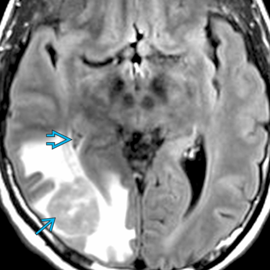
*Axial FLAIR MR shows a round mass in the right occipital lobe (primary CNS lymphoma) that effaces the adjacent right occipital horn and atrium . Extrinsic mass effect is a common cause of ventricular asymmetry and compression.*

**Neurocytoma, Central**
*Axial T1WI C+ MR demonstrates a heterogeneous enhancing mass arising from the septum pellucidum, a pathologically proven central neurocytoma. Note moderate asymmetric dilatation of the right lateral ventricle .*

**Obstructive Hydrocephalus**
*Axial FLAIR MR shows a left thalamic expansile mass , a glioblastoma, which demonstrated heterogeneous enhancement (not shown). It protrudes into and obstructs the left ventricular atrium, which is asymmetrically dilated .*
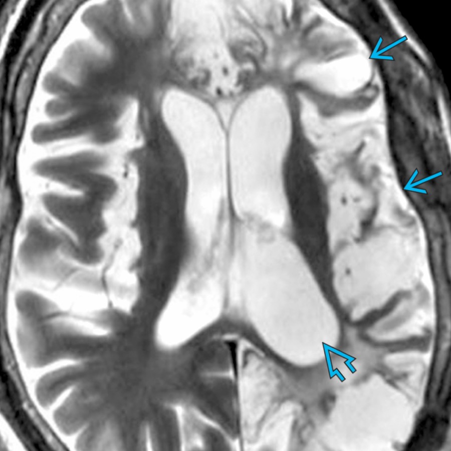
**Encephalomalacia, General**
*Axial T2WI MR demonstrates generalized left hemispheric encephalomalacia following necrotizing encephalitis of unknown origin. There is resultant mild ex-vacuo dilatation of the left lateral ventricle .*

**Meningioma**
*Axial T1WI C+ MR shows an avidly enhancing, lobulated mass arising from the right ventricular atrium , a meningioma. Note the mild asymmetric dilatation of the left lateral ventricle .*
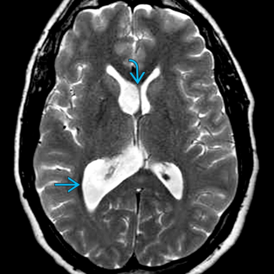
**Normal Variant**
*Axial T2WI MR shows lateral ventricles with the right being larger than the left, representing a normal variant. Note mild bowing of the septum pellucidum across the midline .*
.](29370a1b-3c42-4629-8032-765ef4ce7c56)
**Dyke-Davidoff-Masson**
*Axial T1WI C+ MR shows an asymmetrically larger right atrium in this patient with Sturge-Weber syndrome. Note associated ipsilateral enlarged frontal sinus and calvarial thickening .*

**Hemimegalencephaly**
*Axial CECT shows enlargement of the right hemisphere and lateral ventricle compared to the left side. Expansion of the hemisphere is mostly due to increased white matter. An enlarged, often deformed, lateral ventricle on the abnormal side is typical.*

**Ependymal Cyst**
*Axial FLAIR MR shows a cyst enlarging the left lateral ventricle with signal intensity isointense to CSF . There was no enhancement of the cyst wall, typical of ependymal cyst.*

**Ependymal Cyst**
*Axial FLAIR MR shows a cystic lesion arising from the ependymal lining of the left temporal horn , consistent with an ependymal cyst. The lesion followed CSF signal on every sequence.*

**CSF Shunts and Complications**
*Axial T1WI MR demonstrates a right parietal shunt catheter with its tip in the right frontal horn in a patient with congenital aqueductal stenosis. The right lateral ventricle is collapsed, while the 3rd and left lateral ventricles are moderately dilated.*

**Ventriculitis**
*Axial T1WI C+ MR shows ependymal enhancement and mild asymmetric dilatation of the left occipital horn in ventriculitis. There is also asymmetric enhancement of the adjacent choroid plexus consistent with choroid plexitis .*

**Choroid Plexus Cyst**
*Axial T2WI MR shows a large, hyperintense choroid plexus cyst in a newborn. The lesion increased in size on sequential imaging, requiring endoscopic fenestration.*

**Surgical Defects**
*Axial FLAIR MR shows asymmetric dilatation of the left frontal horn and surrounding gliosis following the resection of an intraaxial metastasis. Part of the surgical tract is seen . Note multiple additional metastatic lesions in the right cerebral hemisphere.*

**Herniation Syndromes, Intracranial**
*Axial NECT demonstrates a large left frontal intraparenchymal hematoma causing subfalcine herniation to the right . The left lateral ventricle is effaced. The posterior right lateral ventricle is mildly dilated .*

**Intraventricular Hemorrhage**
*Axial NECT shows a large amount of intraventricular hemorrhage resulting in moderate asymmetric expansion of the left lateral ventricle. A smaller amount of layering blood products is seen in the right lateral ventricle .*

**Rasmussen Encephalitis**
*Axial T2WI MR shows mild, asymmetric, right hemispheric volume loss with prominent sulci and mild, asymmetric, right lateral ventricle dilatation in a pediatric patient with Rasmussen encephalitis.*

**Extrinsic Mass Effect**
*Axial FLAIR MR shows a round mass in the right occipital lobe (primary CNS lymphoma) that effaces the adjacent right occipital horn and atrium . Extrinsic mass effect is a common cause of ventricular asymmetry and compression.*

**Neurocytoma, Central**
*Axial T1WI C+ MR demonstrates a heterogeneous enhancing mass arising from the septum pellucidum, a pathologically proven central neurocytoma. Note moderate asymmetric dilatation of the right lateral ventricle .*

**Obstructive Hydrocephalus**
*Axial FLAIR MR shows a left thalamic expansile mass , a glioblastoma, which demonstrated heterogeneous enhancement (not shown). It protrudes into and obstructs the left ventricular atrium, which is asymmetrically dilated .*

**Encephalomalacia, General**
*Axial T2WI MR demonstrates generalized left hemispheric encephalomalacia following necrotizing encephalitis of unknown origin. There is resultant mild ex-vacuo dilatation of the left lateral ventricle .*

**Meningioma**
*Axial T1WI C+ MR shows an avidly enhancing, lobulated mass arising from the right ventricular atrium , a meningioma. Note the mild asymmetric dilatation of the left lateral ventricle .*

**Normal Variant**
*Axial T2WI MR shows lateral ventricles with the right being larger than the left, representing a normal variant. Note mild bowing of the septum pellucidum across the midline .*