---
title: "Calcified Suprasellar Mass"
docid: "b61b0b70-de36-45f9-88a2-162a9758b729"
authors:
- key: "e0282a62-994d-4550-a127-1eb773b1e920"
value: "Blair A. Winegar, MD"
- key: "5cff4116-3654-4b3a-bb75-5ebe0b8c9850"
value: "Anne G. Osborn, MD, FACR"
- key: "8d5254e9-8dda-478b-8f08-bdee97a32c79"
value: "Karen L. Salzman, MD, FACR"
breadcrumbs:
-
name: "Brain"
slug: "brain"
treeNodeId: "6d8829f1-14d7-45af-8675-255189aa526a"
-
name: "Differential Diagnosis"
slug: "differential-diagnosis"
treeNodeId: "a7fdd139-664e-4bb8-8d18-400e4733ff60"
-
name: "Sella/Juxtasellar, Pineal Region"
slug: "sellajuxtasellar-pineal-region"
treeNodeId: "5e38b9c1-3137-47e3-aa83-1fc82cb4099a"
-
name: "Anatomically Based Differentials"
slug: "anatomically-based-differentials"
treeNodeId: "7a51b2ca-8fee-4c16-aff3-b7189f68ea60"
-
name: "Calcified Suprasellar Mass"
slug: "calcified-suprasellar-mass"
treeNodeId: null
category: "Brain"
documentVersionId: "9a1f2879-7179-44a8-abaa-f97284492f8d"
imageCount: 15
lastUpdated: "02/01/23"
pageDescription: "Calcified Suprasellar Mass"
pageKeywords: "Brain, Differential Diagnosis, Sella/Juxtasellar, Pineal Region, Anatomically Based Differentials, Calcified Suprasellar Mass"
pageTitle: "Calcified Suprasellar Mass | STATdx"
enhancedTitle: "Calcified Suprasellar Mass"
type: "DDX"
references: true
breadcrumbs:
- "Brain"
- "Differential Diagnosis"
- "Sella/Juxtasellar, Pineal Region"
- "Anatomically Based Differentials"
- "Calcified Suprasellar Mass"
---
# ESSENTIAL INFORMATION
- ## Key Differential Diagnosis Issues
- Is Ca⁺⁺ curvilinear, punctate, globular, etc.?
- Does lesion enhance?
- ## Helpful Clues for Common Diagnoses
- **Atherosclerosis****, Intracranial**
- Curvilinear Ca⁺⁺ along vessel walls
- Usually bilateral, often multifocal
- Older patients
- **Craniopharyngioma**
- Globular, punctate, &/or ring Ca⁺⁺
- **Adamantinomatous**: Bimodal age distribution (peaks 5-15 years and 45-60 years)
- **Papillary**: Older adult tumors more often solid, Ca⁺⁺ less frequent
- **Meningioma**
- Psammomatous (sand-like) Ca⁺⁺
- Solid > rim enhancement
- Middle-aged, older patients (unless NF2)
- **Aneurysm**
- **Saccular****a****neurysm**
- Calcification less common than with fusiform aneurysm, ASVD
- Curvilinear (peripheral arcs, rings) pattern
- **Fusiform****a****neurysm, atherosclerotic**
- Linear ± rim Ca⁺⁺
- Ca⁺⁺ often present in other vessels
- ## Helpful Clues for Less Common Diagnoses
- **Neurocysticercosis**
- Nodular calcified stage
- Usually parenchymal > > cisternal Ca⁺⁺
- **Pilocytic Astrocytoma**
- Common in children, young adults
- Ca⁺⁺ uncommon in hypothalamic PA
- **Dermoid Cyst**
- 20% have capsular Ca⁺⁺
- Contain lipid
- Look for evidence of rupture (fatty droplets in subarachnoid spaces, cisterns)
- No enhancement unless chemical meningitis
- ## Helpful Clues for Rare Diagnoses
- **Pituitary Macroadenoma**
- Only 1-2% of macroadenomas calcify
- Adenomas usually isodense with gray matter
- Majority result in smooth enlargement of sella turcica
- **Rathke Cleft Cyst**
- Vast majority have no calcification (> 85%)
- May rarely see thin, curvilinear rim calcification (10-15%)
- Typically hypodense to brain parenchyma
- **Tuberculosis**
- TB, healing/healed granulomatous infections cause parenchymal > > cisternal Ca⁺⁺
- **Chondroid Tumor**
- Chondromas, enchondromas arise from central skull base
- Chondroid matrix on CT
- May extend to suprasellar region
## References
# Selected References
1. [Alali AA et al: Do we need gadolinium-based contrast agents for routine MRI surveillance of unoperated pituitary macroadenoma? AJNR Am J Neuroradiol. 43(7):1024-8, 2022](http://www.ncbi.nlm.nih.gov/pubmed/?term=35738673%5Bpmid%5D)
1. [Perret CM et al: A giant internal carotid bifurcation aneurysm as a rare and dangerous differential diagnosis of a craniopharyngioma. Cureus. 14(1):e21588, 2022](http://www.ncbi.nlm.nih.gov/pubmed/?term=35228946%5Bpmid%5D)
1. [Jipa A et al: Imaging of the sellar and parasellar regions. Clin Imaging. 77:254-75, 2021](http://www.ncbi.nlm.nih.gov/pubmed/?term=34153590%5Bpmid%5D)
1. [Chapman PR et al: Neuroimaging of the pituitary gland: practical anatomy and pathology. Radiol Clin North Am. 58(6):1115-33, 2020](http://www.ncbi.nlm.nih.gov/pubmed/?term=33040852%5Bpmid%5D)
1. [Snyder MH et al: Neurocysticercosis presenting as an isolated suprasellar lesion. World Neurosurg. 141:352-6, 2020](http://www.ncbi.nlm.nih.gov/pubmed/?term=32522639%5Bpmid%5D)
1. [Xin WQ et al: Patient with epilepsy caused by the spontaneous rupture of an intracerebral dermoid cyst. World Neurosurg. 136:140-5, 2020](http://www.ncbi.nlm.nih.gov/pubmed/?term=31954900%5Bpmid%5D)
1. [Jacków J et al: Ruptured intracranial dermoid cysts: a pictorial review. Pol J Radiol. 83:e465-70, 2018](http://www.ncbi.nlm.nih.gov/pubmed/?term=30655926%5Bpmid%5D)
1. [Seeburg DP et al: Imaging of the sella and parasellar region in the pediatric population. Neuroimaging Clin N Am. 27(1):99-121, 2017](http://www.ncbi.nlm.nih.gov/pubmed/?term=27889026%5Bpmid%5D)
1. [Hoffmann A et al: Fusiform dilatation of the internal carotid artery in childhood-onset craniopharyngioma: multicenter study on incidence and long-term outcome. Pituitary. 19(4):422-8, 2016](http://www.ncbi.nlm.nih.gov/pubmed/?term=27125511%5Bpmid%5D)
1. [Pan J et al: Growth patterns of craniopharyngiomas: clinical analysis of 226 patients. J Neurosurg Pediatr. 17(4):418-33, 2016](http://www.ncbi.nlm.nih.gov/pubmed/?term=26636252%5Bpmid%5D)
1. [Sekiguchi K et al: Osteochondroma presenting as a calcified mass in the sellar region and review of the literature. J Neurol Surg A Cent Eur Neurosurg. 78(4):380-5, 2016](http://www.ncbi.nlm.nih.gov/pubmed/?term=27903019%5Bpmid%5D)
1. [Yildiz AE et al: Suprasellar masses in children: characteristic MR imaging features. J Neuroradiol. 43(4):246-59, 2016](http://www.ncbi.nlm.nih.gov/pubmed/?term=27131616%5Bpmid%5D)
1. [Ben Nsir A et al: Calcified suprasellar xanthogranuloma presenting with primary amenorrhea in a 17-year-old girl: case report and literature review. World Neurosurg. 84(3):866.e11-4, 2015](http://www.ncbi.nlm.nih.gov/pubmed/?term=25916181%5Bpmid%5D)
## Images
### Selected Images
 that represents an ectatic, supraclinoid, internal carotid artery (ICA) with calcified atherosclerotic plaque. Coronal and sagittal reformatted images make this diagnosis straightforward.](images/app.statdx.com_image_thumbnail_eecea3f1-ae69-42be-a896-9b64a38071b4_annotated_true_size_900_quality_90_b311b11e.jpg)
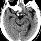
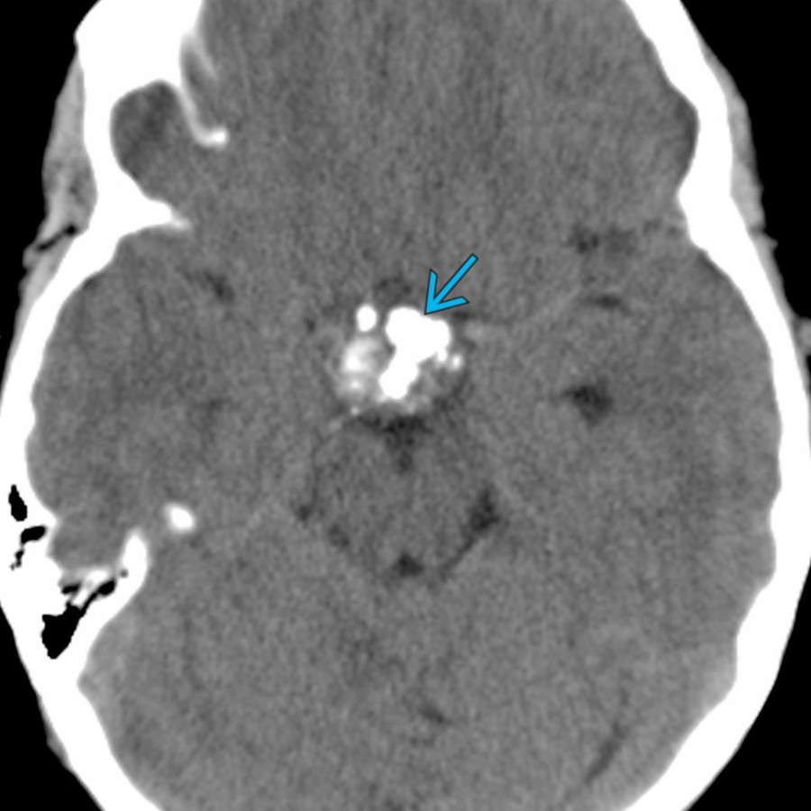
**Atherosclerosis, Intracranial**
*Axial NECT shows a fusiform, partially calcified mass in the suprasellar cistern that represents an ectatic, supraclinoid, internal carotid artery (ICA) with calcified atherosclerotic plaque. Coronal and sagittal reformatted images make this diagnosis straightforward.*
**Atherosclerosis, Intracranial**
*Axial NECT shows a fusiform, partially calcified mass in the suprasellar cistern that represents an ectatic, supraclinoid, internal carotid artery (ICA) with calcified atherosclerotic plaque. Coronal and sagittal reformatted images make this diagnosis straightforward.*
**Craniopharyngioma**
*Axial NECT shows a cystic suprasellar mass with globular calcifications typical of an adamantinomatous craniopharyngioma. These tumors are WHO grade 1 and are typically treated with surgical resection.*
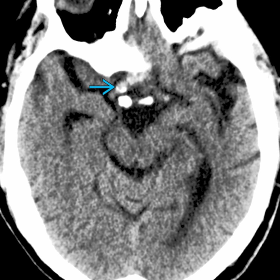
**Atherosclerosis, Intracranial**
*Axial NECT shows a fusiform, partially calcified mass in the suprasellar cistern that represents an ectatic, supraclinoid, internal carotid artery (ICA) with calcified atherosclerotic plaque. Coronal and sagittal reformatted images make this diagnosis straightforward.*

**Atherosclerosis, Intracranial**
*Axial NECT shows a fusiform, partially calcified mass in the suprasellar cistern that represents an ectatic, supraclinoid, internal carotid artery (ICA) with calcified atherosclerotic plaque. Coronal and sagittal reformatted images make this diagnosis straightforward.*

**Craniopharyngioma**
*Axial NECT shows a cystic suprasellar mass with globular calcifications typical of an adamantinomatous craniopharyngioma. These tumors are WHO grade 1 and are typically treated with surgical resection.*
 within a normal-sized sella turcica. Meningiomas in this location often involve the planum sphenoidale and have a dural tail on contrast-enhanced imaging.](images/app.statdx.com_image_thumbnail_ae28f3d5-00a6-4764-9a86-47904bee32be_annotated_true_size_900_quality_90_173c54ee.jpg)
**Meningioma**
*Sagittal NECT shows a partially calcified extraaxial mass projecting from the tuberculum sella , related to a meningioma. This is separate from the adjacent pituitary gland within a normal-sized sella turcica. Meningiomas in this location often involve the planum sphenoidale and have a dural tail on contrast-enhanced imaging.*
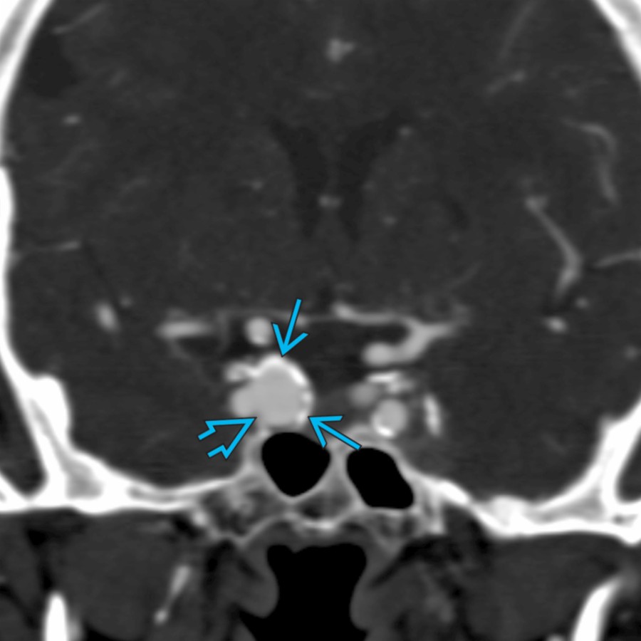
**Saccular Aneurysm**
*Coronal arterial-phase CTA shows a saccular aneurysm projecting medially from the right cavernous ICA with internal arterial enhancement and rim calcifications .*
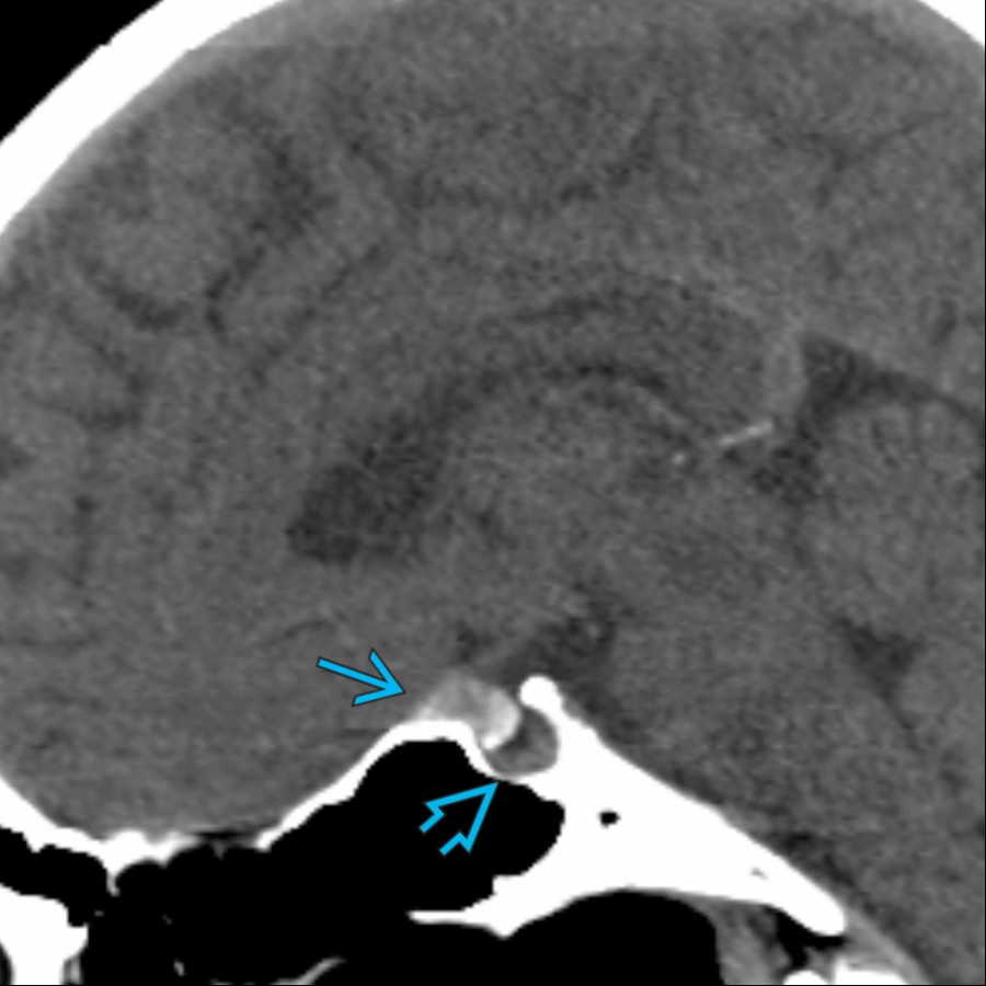
**Meningioma**
*Sagittal NECT shows a partially calcified extraaxial mass projecting from the tuberculum sella , related to a meningioma. This is separate from the adjacent pituitary gland within a normal-sized sella turcica. Meningiomas in this location often involve the planum sphenoidale and have a dural tail on contrast-enhanced imaging.*

**Saccular Aneurysm**
*Coronal arterial-phase CTA shows a saccular aneurysm projecting medially from the right cavernous ICA with internal arterial enhancement and rim calcifications .*
 . Fusiform aneurysms are often related to atherosclerotic disease, as in this patient.](images/app.statdx.com_image_thumbnail_c4bf6e23-0c1b-480d-98f2-364c5dd61993_annotated_true_size_900_quality_90_f5e9fda8.jpg)
**Fusiform Aneurysm, Atherosclerotic**
*Axial NECT shows a hyperdense, fusiform basilar artery aneurysm and calcified, fusiform aneurysmal ectasias of both ICAs . Fusiform aneurysms are often related to atherosclerotic disease, as in this patient.*

**Pilocytic Astrocytoma**
*Axial NECT shows a low-density, suprasellar mass with rim and globular calcification . Biopsy disclosed a pilomyxoid subtype of pilocytic astrocytoma. This variant often occurs in the hypothalamic region in an infant or young child. These tumors rarely calcify.*
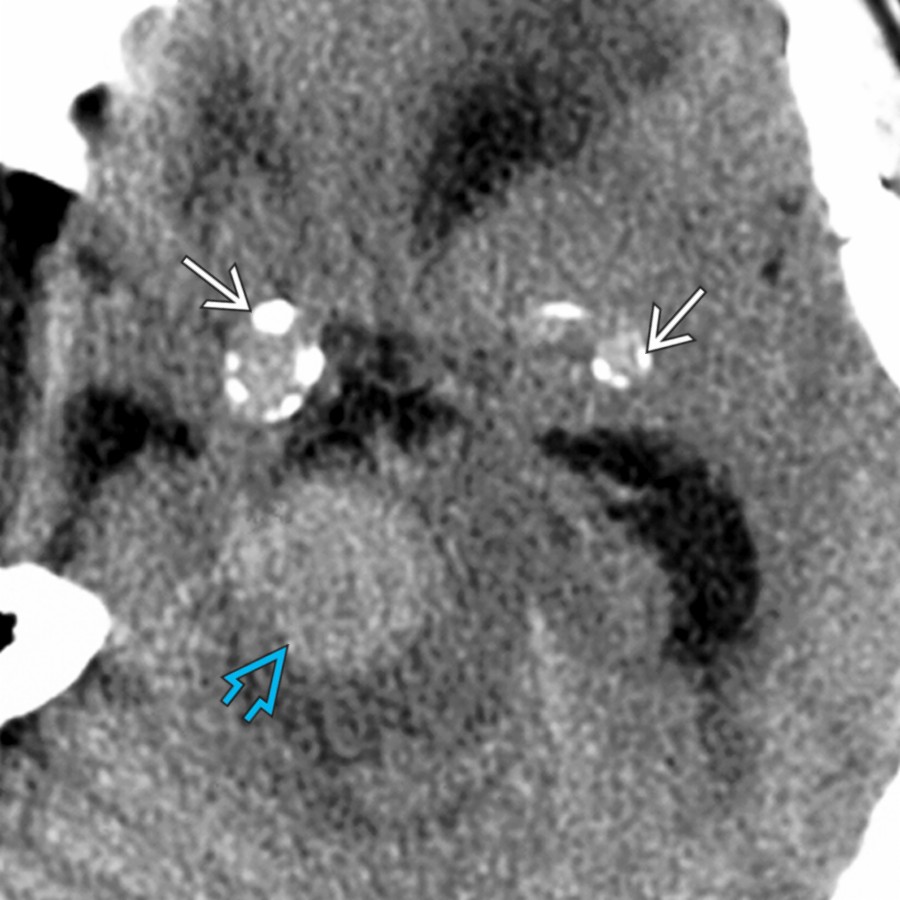
**Fusiform Aneurysm, Atherosclerotic**
*Axial NECT shows a hyperdense, fusiform basilar artery aneurysm and calcified, fusiform aneurysmal ectasias of both ICAs . Fusiform aneurysms are often related to atherosclerotic disease, as in this patient.*

**Pilocytic Astrocytoma**
*Axial NECT shows a low-density, suprasellar mass with rim and globular calcification . Biopsy disclosed a pilomyxoid subtype of pilocytic astrocytoma. This variant often occurs in the hypothalamic region in an infant or young child. These tumors rarely calcify.*
 in the suprasellar cistern. There are scattered fat droplets in the subarachnoid spaces in this case of a ruptured dermoid cyst.](b1cf55ae-d0d7-4a2d-92f4-3cedacf35c4c)
**Dermoid Cyst**
*Sagittal NECT shows a fat density lesion with rim calcification in the suprasellar cistern. There are scattered fat droplets in the subarachnoid spaces in this case of a ruptured dermoid cyst.*

**Rathke Cleft Cyst**
*Axial NECT shows curvilinear calcification along the anterior aspect of a cystic suprasellar mass in a young adult. This mass was T1 hyperintense and did not enhance. A Rathke cleft cyst was diagnosed at resection. These benign cysts are usually not calcified, which helps to differentiate them from craniopharyngiomas.*
### Additional Images

**Dermoid Cyst**
*Sagittal NECT shows a fat density lesion with rim calcification in the suprasellar cistern. There are scattered fat droplets in the subarachnoid spaces in this case of a ruptured dermoid cyst.*

**Rathke Cleft Cyst**
*Axial NECT shows curvilinear calcification along the anterior aspect of a cystic suprasellar mass in a young adult. This mass was T1 hyperintense and did not enhance. A Rathke cleft cyst was diagnosed at resection. These benign cysts are usually not calcified, which helps to differentiate them from craniopharyngiomas.*
### Additional Images
 within the mass, an uncommon finding in macroadenomas.](95b8c653-566b-4169-8af8-5fe85a13bdfb)
**Pituitary Macroadenoma**
*Axial CECT shows a very large intra- and suprasellar enhancing mass invading the central skull base. Note floccular calcifications within the mass, an uncommon finding in macroadenomas.*

**Chondroid Tumor**
*Sagittal bone CT shows globular calcification in a suprasellar mass in this typical osteochondroma of the central skull base with calcification in the cartilaginous cap.*
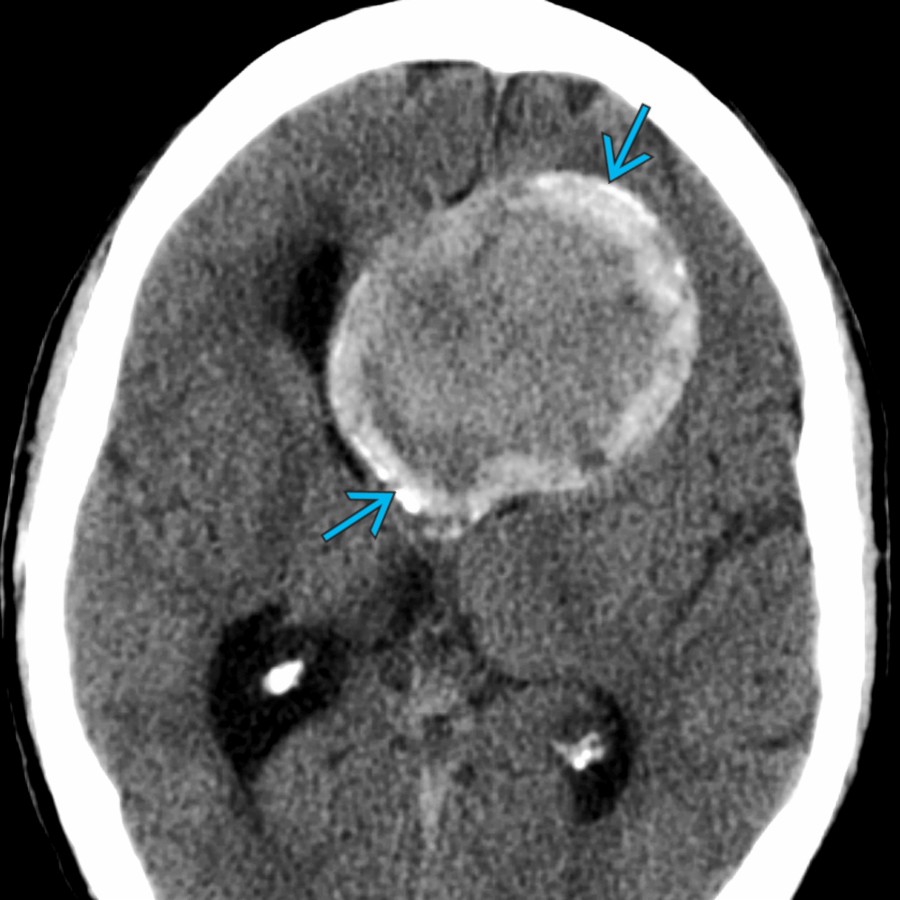
**Meningioma**
*Axial NECT shows a hyperdense suprasellar mass with focal calcifications in this patient with a classic meningioma arising from the central skull base. Note the associated hydrocephalus related to ventricular obstruction.*
**Saccular Aneurysm**
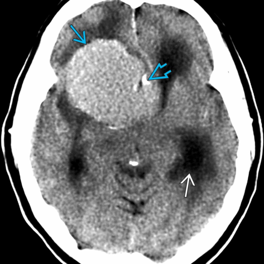
*Axial NECT shows a huge, well-delineated, hyperdense suprasellar mass with rim calcifications . A giant, mostly thrombosed, saccular aneurysm was diagnosed and treated surgically. MR may show a laminated appearance with pulsation artifact if the aneurysm is patent.*

**Pituitary Macroadenoma**
*Axial CECT shows a very large intra- and suprasellar enhancing mass invading the central skull base. Note floccular calcifications within the mass, an uncommon finding in macroadenomas.*

**Chondroid Tumor**
*Sagittal bone CT shows globular calcification in a suprasellar mass in this typical osteochondroma of the central skull base with calcification in the cartilaginous cap.*

**Meningioma**
*Axial NECT shows a hyperdense suprasellar mass with focal calcifications in this patient with a classic meningioma arising from the central skull base. Note the associated hydrocephalus related to ventricular obstruction.*

**Saccular Aneurysm**
*Axial NECT shows a huge, well-delineated, hyperdense suprasellar mass with rim calcifications . A giant, mostly thrombosed, saccular aneurysm was diagnosed and treated surgically. MR may show a laminated appearance with pulsation artifact if the aneurysm is patent.*
 in the subarachnoid space related to a ruptured dermoid cyst.](aaeaa251-a331-4ca5-b854-b42d581c1782)
**Dermoid Cyst**
*Axial NECT shows a large, hypodense, calcified suprasellar mass . Note fat density droplets in the subarachnoid space related to a ruptured dermoid cyst.*

**Pituitary Macroadenoma**
*Coronal NECT shows a complex cystic expansile mass in the sella and suprasellar cistern with peripheral calcification in this case of pituitary macroadenoma with cystic degeneration.*

**Chondroid Tumor**
*Coronal NECT shows a destructive low- density mass with internal chondroid calcifications extending from the central skull base into the suprasellar cistern in this case of chondrosarcoma.*
in the subarachnoid space related to a ruptured dermoid cyst.](aaeaa251-a331-4ca5-b854-b42d581c1782)
**Dermoid Cyst**
*Axial NECT shows a large, hypodense, calcified suprasellar mass . Note fat density droplets in the subarachnoid space related to a ruptured dermoid cyst.*

**Pituitary Macroadenoma**
*Coronal NECT shows a complex cystic expansile mass in the sella and suprasellar cistern with peripheral calcification in this case of pituitary macroadenoma with cystic degeneration.*

**Chondroid Tumor**
*Coronal NECT shows a destructive low- density mass with internal chondroid calcifications extending from the central skull base into the suprasellar cistern in this case of chondrosarcoma.*