---
title: "Cavum Velum Interpositum (CVI)"
docid: "849ee468-35c4-46e3-9297-96196109cdb8"
authors:
- key: "a25c450b-3d34-4f64-bba3-cc0834813df6"
value: "Miral D. Jhaveri, MD, MBA"
- key: "5cff4116-3654-4b3a-bb75-5ebe0b8c9850"
value: "Anne G. Osborn, MD, FACR"
breadcrumbs:
-
name: "Brain"
slug: "brain"
treeNodeId: "6d8829f1-14d7-45af-8675-255189aa526a"
-
name: "Diagnosis"
slug: "diagnosis"
treeNodeId: "51c00394-446e-4a38-94af-d3b1d14d34e8"
-
name: "Anatomy-Based Diagnoses"
slug: "anatomy-based-diagnoses"
treeNodeId: "529d3e33-f508-498c-bc70-cf962e81e629"
-
name: "Ventricles and Cisterns"
slug: "ventricles-and-cisterns"
treeNodeId: "33b267f0-908c-4c77-81f8-f6135d1bc592"
-
name: "Normal Variants"
slug: "normal-variants"
treeNodeId: "bf92256f-cdff-4bcd-8420-d876b9e4031a"
-
name: "Cavum Velum Interpositum (CVI)"
slug: "cavum-velum-interpositum-cvi"
treeNodeId: null
category: "Brain"
documentVersionId: "75312185-3ddd-4300-a4fb-d824b606effc"
imageCount: 12
lastUpdated: "05/08/20"
pageDescription: "Cavum Velum Interpositum (CVI)"
pageKeywords: "Brain, Diagnosis, Anatomy-Based Diagnoses, Ventricles and Cisterns, Normal Variants, Cavum Velum Interpositum (CVI)"
pageTitle: "Cavum Velum Interpositum (CVI) | STATdx"
enhancedTitle: "Cavum Velum Interpositum (CVI)"
type: "DX"
references: true
breadcrumbs:
- "Brain"
- "Diagnosis"
- "Anatomy-Based Diagnoses"
- "Ventricles and Cisterns"
- "Normal Variants"
- "Cavum Velum Interpositum (CVI)"
---
# KEY FACTS
- ## Terminology
- Cavum velum interpositum (CVI), cyst of velum interpositum (VI)
- ## Imaging
- Triangular CSF space
- Between lateral ventricles, over thalami
- Apex points toward foramen of Monro
- Elevates, splays fornices
- Flattens, displaces internal cerebral veins inferiorly
- Size varies from slit-like linear to triangular to round/ovoid CSF collection
- Isodense/isointense with CSF
- Suppresses completely on FLAIR
- Does not restrict on DWI
- Does not enhance
- US shows hypoechoic midline interhemispheric cyst
- ## Top Differential Diagnoses
- Normal cistern of velum interpositum
- Cavum septi pellucidi, cavum vergae
- Arachnoid cyst
- Epidermoid cyst
- ## Clinical Issues
- Can be found at any age
- Common in infants, rare in adults
- Symptoms
- Usually asymptomatic, found incidentally
- Headache (relationship to cyst unclear)
- Large CVI can obstruct normal CSF flow; treated by endoscopic fenestration
- ## Diagnostic Checklist
- CSF-like "cyst" could be epidermoid
- Include FLAIR and DWI to distinguish between CVI, epidermoid cyst
# TERMINOLOGY
- ## Abbreviations
- Cavum velum interpositum (CVI), cyst of velum interpositum (VI)
- ## Definitions
- Cystic dilation of VI
# IMAGING
- ## General Features
- ### Best diagnostic clue
- Triangular-shaped CSF space
- Between lateral ventricles, over thalami
- Apex points toward but does not extend anteriorly beyond foramen of Monro
- Base contiguous with quadrigeminal cistern
- ### Location
- Midline between lateral ventricles, below fornices
- Continuous with choroid plexus subependymally
- ### Size
- Varies (few mm to several cm)
- ## CT Findings
- ### NECT
- Triangle of CSF between lateral ventricles
- ### CECT
- Does not enhance
- ## MR Findings
- ### T1WI
- Sagittal: Varies from slit-like linear to round/ovoid
- Elevates fornices
- Flattens, displaces internal cerebral veins inferiorly
- Axial: Triangle of CSF between lateral ventricles
- ### T2WI
- Isointense with CSF
- ### FLAIR
- Suppresses completely
- ### DWI
- Does not restrict
- ## Ultrasonographic Findings
- ### Color Doppler
- Hypoechoic midline interhemispheric cyst
- ## Imaging Recommendations
- ### Best imaging tool
- MR ± contrast
- ### Protocol advice
- FLAIR/DWI (distinguish between CVI and epidermoid)
# DIFFERENTIAL DIAGNOSIS
- [Normal Cistern of Velum Interpositum](/document/pineal-cyst/eda99e5c-c992-4798-aa35-21437eb505ea)
- Usually small (slit-like or oval)
- Does not elevate fornices or depress internal cerebral veins
- ## Cavum Septi Pellucidi, Vergae
- CSP + CV elongated, finger-like CSF space
- [Arachnoid Cyst](/document/arachnoid-cyst/7d63f0d3-6999-4d8c-a41d-953f738c43a6)
- Lined with arachnoid (may be indistinguishable)
- [Epidermoid Cyst](/document/epidermoid-cyst/704c5ddf-e1f7-4a5d-a1b8-5b0e603170d9)
- Lobulated, insinuating mass
- Does not suppress with FLAIR; DWI shows restriction
# PATHOLOGY
- ## General Features
- ### Etiology
- Pia infolds along transverse fissure, forms CSF cistern (VI)
- Cystic dilatation of VI may occur (precise etiology unknown)
- ### Associated abnormalities
- Usually none (large CVIs may cause hydrocephalus)
- ## Gross Pathologic & Surgical Features
- Normal cistern of VI is small to inapparent at autopsy
- Pial-lined CSF-filled space
- ## Microscopic Features
- Occasionally cysts of midline CSF spaces contain glial cells, scattered neurons
# CLINICAL ISSUES
- ## Presentation
- ### Most common signs/symptoms
- Usually asymptomatic, found incidentally
- Headache (relationship to cyst unclear)
- ## Demographics
- ### Age
- Can be found at any age
- Common in infants, rare in adults
- ### Gender
- M = F
- ## Treatment
- Usually none
# DIAGNOSTIC CHECKLIST
- ## Consider
- CSF-like "cyst" could be epidermoid
- Use DWI, FLAIR to differentiate from other cysts
806ab3b4-9c9a-4887-a52d-d5809cd5a417
## References
# Selected References
1. [Krejčí T et al: Symptomatic cysts of the cavum septi pellucidi, cavum vergae and cavum veli interpositi: A retrospective duocentric study of 10 patients. Clin Neurol Neurosurg. 185:105494, 2019](http://www.ncbi.nlm.nih.gov/pubmed/?term=31472394%5Bpmid%5D)
1. [Akinola RA et al: Caval variations in neurologically diseased patients. Acta Radiol Short Rep. 3(5):2047981614530288, 2014](http://www.ncbi.nlm.nih.gov/pubmed/?term=25298867%5Bpmid%5D)
1. [Tong CK et al: Endoscopic fenestration of cavum velum interpositum cysts: a case study of two symptomatic patients. Childs Nerv Syst. 28(8):1261-4, 2012](http://www.ncbi.nlm.nih.gov/pubmed/?term=22543434%5Bpmid%5D)
1. [Tubbs RS et al: Cavum velum interpositum, cavum septum pellucidum, and cavum vergae: a review. Childs Nerv Syst. 27(11):1927-30, 2011](http://www.ncbi.nlm.nih.gov/pubmed/?term=21687999%5Bpmid%5D)
1. [Tubbs RS et al: The velum interpositum revisited and redefined. Surg Radiol Anat. 30(2):131-5, 2008](http://www.ncbi.nlm.nih.gov/pubmed/?term=18094919%5Bpmid%5D)
1. [Osborn AG et al: Intracranial cysts: radiologic-pathologic correlation and imaging approach. Radiology. 239(3):650-64, 2006](http://www.ncbi.nlm.nih.gov/pubmed/?term=16714456%5Bpmid%5D)
1. [Eisenberg VH et al: Prenatal diagnosis of cavum velum interpositum cysts: significance and outcome. Prenat Diagn. 23(10):779-83, 2003](http://www.ncbi.nlm.nih.gov/pubmed/?term=14558018%5Bpmid%5D)
1. [Vergani P et al: Ultrasonographic differential diagnosis of fetal intracranial interhemispheric cysts. Am J Obstet Gynecol. 180(2 Pt 1):423-8, 1999](http://www.ncbi.nlm.nih.gov/pubmed/?term=9988813%5Bpmid%5D)
1. [Chen CY et al: Sonographic characteristics of the cavum velum interpositum. AJNR Am J Neuroradiol. 19(9):1631-5, 1998](http://www.ncbi.nlm.nih.gov/pubmed/?term=9802483%5Bpmid%5D)
## Images
### Selected Images
 . Also noted is the inferior displacement of the internal cerebral veins and 3rd ventricle
  .](images/app.statdx.com_image_thumbnail_2df127e7-04ef-413c-b33e-46703bf26fc3_annotated_true_size_900_quality_90_43c5eb31_20251018T164844Z.jpg)
*Sagittal graphic with axial insert shows a cavum velum interpositum (CVI). Note the elevation and splaying of the fornices . Also noted is the inferior displacement of the internal cerebral veins and 3rd ventricle .*

*Sagittal graphic with axial insert shows a cavum velum interpositum (CVI). Note the elevation and splaying of the fornices . Also noted is the inferior displacement of the internal cerebral veins and 3rd ventricle .*

*Sagittal graphic with axial insert shows a cavum velum interpositum (CVI). Note the elevation and splaying of the fornices . Also noted is the inferior displacement of the internal cerebral veins and 3rd ventricle .*

*Sagittal graphic with axial insert shows a cavum velum interpositum (CVI). Note the elevation and splaying of the fornices . Also noted is the inferior displacement of the internal cerebral veins and 3rd ventricle .*
 as a CSF-like enlargement that elevates the fornix
  and flattens and displaces the internal cerebral vein
  inferiorly. These are usually asymptomatic; however, large ones can cause CSF obstruction and can be treated by fenestration.](images/app.statdx.com_image_thumbnail_bc8c67a9-f3e5-4673-a4f6-3febba3eccd9_annotated_true_size_900_quality_90_39cc402c_20251018T164845Z.jpg)
*Sagittal T1 MR shows a classic CVI as a CSF-like enlargement that elevates the fornix and flattens and displaces the internal cerebral vein inferiorly. These are usually asymptomatic; however, large ones can cause CSF obstruction and can be treated by fenestration.*
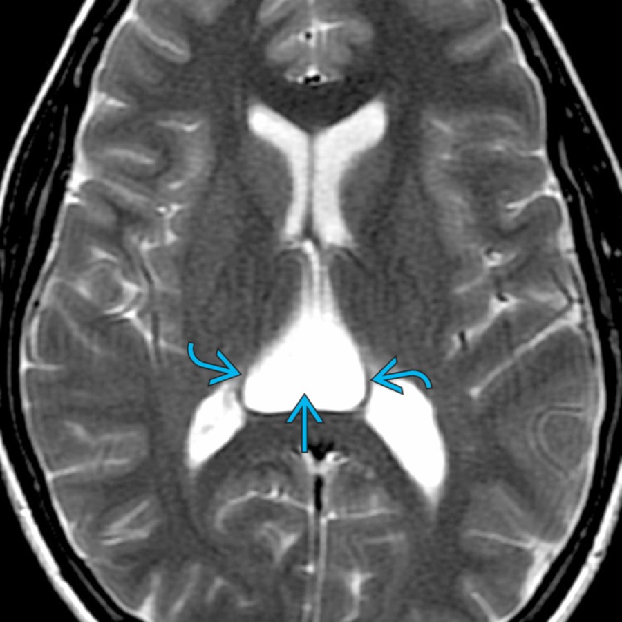
*Axial T2 MR in a 37-year-old man with headaches shows a triangular-shaped CSF collection between the lateral ventricles, spreading the fornices laterally .*

*Axial FLAIR MR in the same patient shows complete suppression of the CSF signal within the cyst similar to the lateral ventricles. Findings are consistent with a classic cavum velum interpositum. FLAIR and DWI distinguish between cavum velum interpositum and an epidermoid cyst.*
### Additional Images

*Sagittal T1 MR shows a classic CVI as a CSF-like enlargement that elevates the fornix and flattens and displaces the internal cerebral vein inferiorly. These are usually asymptomatic; however, large ones can cause CSF obstruction and can be treated by fenestration.*

*Axial T2 MR in a 37-year-old man with headaches shows a triangular-shaped CSF collection between the lateral ventricles, spreading the fornices laterally .*

*Axial FLAIR MR in the same patient shows complete suppression of the CSF signal within the cyst similar to the lateral ventricles. Findings are consistent with a classic cavum velum interpositum. FLAIR and DWI distinguish between cavum velum interpositum and an epidermoid cyst.*
### Additional Images
 along with a very small cavum velum interpositum
  . Note that these 2 unrelated lesions do not communicate with each other. It is common to see a CSP with a cavum vergae; it is rare to see a CSP and a CVI in the same patient.](images/app.statdx.com_image_thumbnail_8f807265-867d-45c4-a0e0-d4b5ed77ec48_annotated_true_size_900_quality_90_14754121_20251018T164844Z.jpg)
*Axial T2 MR shows a cavum septi pellucidi (CSP) along with a very small cavum velum interpositum . Note that these 2 unrelated lesions do not communicate with each other. It is common to see a CSP with a cavum vergae; it is rare to see a CSP and a CVI in the same patient.*
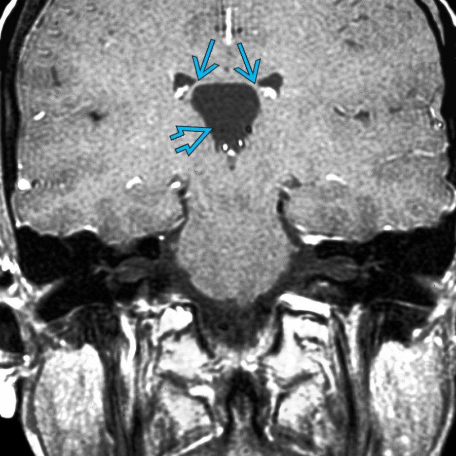
*Coronal T1 C+ MR shows classic cavum velum interpositum that spreads the fornices apart.*

*Axial T2 MR shows a small, triangular-shaped CSF space. Note the cavum velum interpositum interposed between the fornices and lateral ventricles. The CVI ends at the foramen of Monro.*

*Axial T1 MR shows a very large cavum velum interpositum. Note the splaying of fornices and anterior displacement of the septum pellucidum . Mild enlargement of the lateral ventricles is seen.*

*Sagittal T1 MR in the same patient shows anterior/superior displacement of the fornix and inferior displacement of the 3rd ventricle . The corpus callosum is elevated and thinned.*

*Axial T2 MR shows a cavum septi pellucidi (CSP) along with a very small cavum velum interpositum . Note that these 2 unrelated lesions do not communicate with each other. It is common to see a CSP with a cavum vergae; it is rare to see a CSP and a CVI in the same patient.*

*Coronal T1 C+ MR shows classic cavum velum interpositum that spreads the fornices apart.*

*Axial T2 MR shows a small, triangular-shaped CSF space. Note the cavum velum interpositum interposed between the fornices and lateral ventricles. The CVI ends at the foramen of Monro.*

*Axial T1 MR shows a very large cavum velum interpositum. Note the splaying of fornices and anterior displacement of the septum pellucidum . Mild enlargement of the lateral ventricles is seen.*

*Sagittal T1 MR in the same patient shows anterior/superior displacement of the fornix and inferior displacement of the 3rd ventricle . The corpus callosum is elevated and thinned.*
 that elevates the fornix and flattens and displaces the internal cerebral vein inferiorly . This large CVI is probably unrelated to the patient's symptoms.](15050721-daf0-49a6-a3f3-45a4a2b17b85)
*Sagittal T1 MR in a 40-year-old woman with headaches shows CSF-like enlargement of the velum interpositum that elevates the fornix and flattens and displaces the internal cerebral vein inferiorly . This large CVI is probably unrelated to the patient's symptoms.*

*Axial T2 MR in a 46-year-old woman with headaches shows a classic CVI with a triangular-shaped CSF collection , spreading the fornices laterally . The posterior location between the lateral ventricles is typical.*

*Sagittal T1 MR shows a variant CVI that elevates the fornix , flattens the internal cerebral vein , and extends into the quadrigeminal and suprasellar cisterns . This case probably represents an arachnoid cyst of the cavum velum interpositum.*
that elevates the fornix and flattens and displaces the internal cerebral vein inferiorly . This large CVI is probably unrelated to the patient's symptoms.](15050721-daf0-49a6-a3f3-45a4a2b17b85)
*Sagittal T1 MR in a 40-year-old woman with headaches shows CSF-like enlargement of the velum interpositum that elevates the fornix and flattens and displaces the internal cerebral vein inferiorly . This large CVI is probably unrelated to the patient's symptoms.*

*Axial T2 MR in a 46-year-old woman with headaches shows a classic CVI with a triangular-shaped CSF collection , spreading the fornices laterally . The posterior location between the lateral ventricles is typical.*

*Sagittal T1 MR shows a variant CVI that elevates the fornix , flattens the internal cerebral vein , and extends into the quadrigeminal and suprasellar cisterns . This case probably represents an arachnoid cyst of the cavum velum interpositum.*