---
title: "Cisterna Magna Mass"
docid: "047add0c-7e4f-40a0-9933-8d6fa00a24f7"
authors:
- key: "d19354f3-7ff2-495a-ad3f-064122e45602"
value: "Bernadette L. Koch, MD"
- key: "20008105-51da-4bbf-aba6-e8253b2d9c21"
value: "Daniel E. Meltzer, MD"
breadcrumbs:
-
name: "Head and Neck"
slug: "head-and-neck"
treeNodeId: "ed24ed8c-5d57-4629-879b-447b82d2973d"
-
name: "Differential Diagnosis"
slug: "differential-diagnosis"
treeNodeId: "40d68862-8975-4dde-ac2b-ebc43ab0fb5c"
-
name: "CPA-IAC and Posterior Fossa"
slug: "cpa-iac-and-posterior-fossa"
treeNodeId: "c590eedb-4a3b-4158-a04f-ad880564c992"
-
name: "Anatomically Based Differentials"
slug: "anatomically-based-differentials"
treeNodeId: "debfb06c-8656-4f5d-92c1-eaa468185d78"
-
name: "Cisterna Magna Mass"
slug: "cisterna-magna-mass"
treeNodeId: null
category: "Head and Neck"
documentVersionId: "86b48ea6-d9ea-428a-b5cb-32a58aaf84b2"
imageCount: 37
lastUpdated: "07/16/24"
pageDescription: "Cisterna Magna Mass"
pageKeywords: "Head and Neck, Differential Diagnosis, CPA-IAC and Posterior Fossa, Anatomically Based Differentials, Cisterna Magna Mass"
pageTitle: "Cisterna Magna Mass | STATdx"
enhancedTitle: "Cisterna Magna Mass"
type: "DDX"
references: true
breadcrumbs:
- "Head and Neck"
- "Differential Diagnosis"
- "CPA-IAC and Posterior Fossa"
- "Anatomically Based Differentials"
- "Cisterna Magna Mass"
---
# ESSENTIAL INFORMATION
- ## Key Differential Diagnosis Issues
- Cisterna magna (CM) between medulla (anterior), occiput (posterior) (a.k.a. cerebellomedullary cistern)
- Below/behind inferior vermis
- Large medullary cistern masses may extend laterally, posteriorly into CM
- Most common adult lesions are tonsillar associated
- Indirect (secondary effect on tonsil) > direct (lesion in tonsil)
- MR and clinical information helps DDx
- ## Helpful Clues for Common Diagnoses
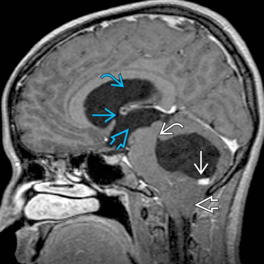
- **Herniation Syndromes, Intracranial**
- Tonsillar herniation most often 2° to posterior fossa (PF) mass effect
- Tonsils pushed down into CM
- Peg-like configuration of tonsils
- Tonsil folia usually oriented horizontally → become vertically oriented when herniated
- 4th ventricle may obstruct → obstructive hydrocephalus
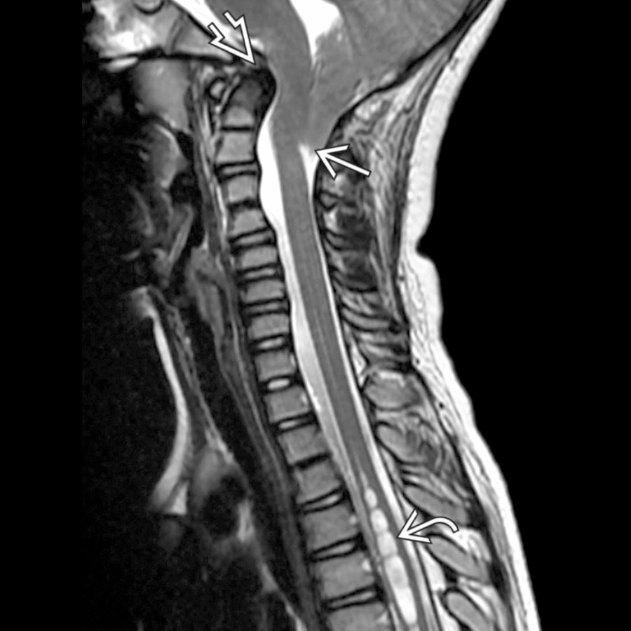
- **Chiari 1**
- Pointed cerebellar tonsils ≥ 5 mm below foramen magnum (FM)
- Crowded FM + effaced CSF
- ± brainstem compression
- ± retroflexed odontoid, horizontal clivus, scoliosis
- PF usually normal size
- Syrinx in 30-70%
- Treatment aim = restore normal CSF flow at FM
- **Chiari 2**
- Small PF → contents herniates inferiorly through FM and superiorly through widened incisura
- Cascade of tissue (vermis, not tonsil) herniates downward through FM
- Caudal brainstem herniation with cervicomedullary kink, tectal beaking, falx insufficiency
- ± ventriculomegaly, subependymal gray matter heterotopia, &/or polymicrogyria
- ~ 100% secondary to open spinal dysraphism, most often myelomeningocele
- **Dandy-Walker Continuum**
- Dandy-Walker continuum is broad spectrum of cystic PF malformations
- 2/3 have associated CNS &/or extracranial anomalies
- All may have inner table occipital bone scalloping
- Dandy-Walker malformation classic triad
- Vermian agenesis/hypogenesis
- Cystic dilatation 4th ventricle
- Large PF with lambdoid-torcular inversion
- ± hydrocephalus
- Vermian hypoplasia
- Varying degrees of vermian hypoplasia
- ± rotation of vermis
- No enlargement of PF
- Blake pouch cyst
- Open 4th ventricle
- Elevated tegmentovermian angle
- Normal size and morphology of vermis
- Mega CM
- Enlarged retrocerebellar cistern communicates with normal 4th ventricle
- Normal vermis
- ## Helpful Clues for Less Common Diagnoses
- **Arachnoid Cyst**
- Sharply demarcated extraaxial cyst that follows CSF attenuation/signal
- FLAIR suppresses; no diffusion restriction
- Size varies from few millimeters to giant
- Often asymptomatic, found incidentally
- Cerebellopontine angle (CPA) location > CM
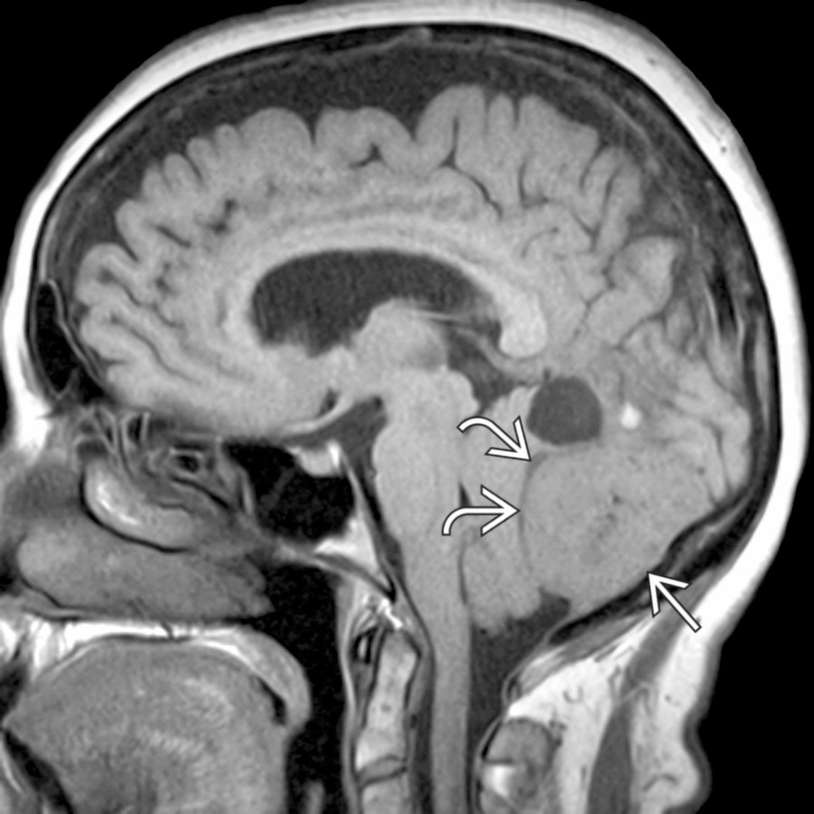
- **Ependymoma**
- Anywhere in neuraxis: 2/3 in PF, 1/3 in supratentorial
- 3rd most common malignant pediatric brain tumor arising in PF
- Heterogeneous T1/T2 signal with mild to moderate enhancement
- Soft or "plastic" tumor squeezes out of 4th ventricle foramina into cisterns
- Calcification common (50%)
- ± cysts, hemorrhage
- Sagittal imaging can distinguish origin as floor vs. roof of 4th ventricle
- CSF seeding may occur → image entire neuraxis
- **Meningioma**
- CM rare PF location
- CPA, medullary cisterns more common than CM
- CM meningiomas usually arise from occipital squamosa
- Well-demarcated, lobulated/rounded enhancing mass with dural attachment
- Hyperostosis, tumoral calcifications, ↑ vascular markings
- **Metastasis**
- Linear or nodular meningeal enhancement
- MR CSF flow may be helpful establishing location and degree of CSF obstruction
- Primary tumors include breast, lung, melanoma, prostate
- Lymphoproliferative malignancy = lymphoma and leukemia
- Primary CNS tumor seed basal cisterns (drop metastases)
- **Image entire neuraxis**
- **Intracranial Hypotension**
- Sagittal shows brain descent in 40-50%
- Caudal displacement of cerebellar tonsils in 25-75%
- Diffusely and intensely enhancing dura in 85%
- Bilateral subdural fluid collections in 15%
- Dural venous sinus may be distended (convex margins)
- Enlarged pituitary
- Frequently misdiagnosed syndrome of headache caused by ↓ intracranial CSF pressure from spontaneous spinal CSF leak
- ## Helpful Clues for Rare Diagnoses
- **Subependymoma**
- T2-hyperintense lobular, nonenhancing intraventricular mass
- When arises from 4th ventricle floor, may extend posteroinferiorly into CM
- More common in middle-aged, older adults
- 0.7% of intracranial neoplasms
- **Epidermoid Cyst**
- Congenital inclusion cysts
- Lobulated, irregular, CSF-like mass with "fronds" insinuate cistern
- FLAIR usually does not completely null; diffusion yields high-signal restriction
- 0.2-1.8% of all primary intracranial tumors
- CPA cistern (40-50%) > 4th ventricle (17%) > sellar/parasellar (10-15%)
- Rare malignant degeneration into squamous cell carcinoma
- **Dermoid Cyst**
- Fat appearance: Use fat suppression sequence to confirm
- With rupture, fat droplets in cisterns, sulci, and ventricles with extensive MR enhancement possible from chemical meningitis
- Rare: < 0.5% of primary intracranial tumors
- Rupture can cause significant morbidity/mortality
- Rare malignant degeneration into squamous cell carcinoma
- **Hemangioblastoma**
- Intraaxial PF mass with cyst, enhancing mural nodule abutting pia
- Classified as 1 of mesenchymal, nonmeningothelial tumors
- Familial = von Hippel-Lindau
- ≈ 30% of patients with hemangioblastoma have von Hippel-Lindau
- 7-10% of PF tumors
- **Neurenteric Cyst**
- Round/lobulated, nonenhancing, slightly hyperintense to CSF mass
- Most intracranial NECs found in PF
- Benign malformative endodermal CNS cyst
- Part of split spinal cord malformation spectrum; persistent neurenteric canal
- Location
- Thoracic (42%), cervical (32%)
- Others: Lumbar spine, basilar cisterns, brain parenchyma
- Anterior medullary, CPA cisterns > CM
## References
# Selected References
1. [Pindrik J et al: Imaging in Chiari I malformation. Neurosurg Clin N Am. 34(1):67-79, 2023](http://www.ncbi.nlm.nih.gov/pubmed/?term=36424066%5Bpmid%5D)
1. [Paun L et al: Foramen magnum meningiomas: a systematic review and meta-analysis. Neurosurg Rev. 44(5):2583-96, 2021](http://www.ncbi.nlm.nih.gov/pubmed/?term=33507444%5Bpmid%5D)
1. [Menezes AH et al: Neurenteric cysts at foramen magnum in children: presentation, imaging characteristics, and surgical management-case series and literature review. Childs Nerv Syst. 36(7):1379-84, 2020](http://www.ncbi.nlm.nih.gov/pubmed/?term=32322975%5Bpmid%5D)
1. [Abdel Razek AA et al: Magnetic resonance imaging of malformations of midbrain-hindbrain. J Comput Assist Tomogr. 40(1):14-25, 2016](http://www.ncbi.nlm.nih.gov/pubmed/?term=26599961%5Bpmid%5D)
1. [Dorfer C et al: Ependymoma: a heterogeneous tumor of uncertain origin and limited therapeutic options. Handb Clin Neurol. 134:417-31, 2016](http://www.ncbi.nlm.nih.gov/pubmed/?term=26948369%5Bpmid%5D)
1. [Louis DN et al: The 2016 World Health Organization Classification of Tumors of the Central Nervous System: a summary. Acta Neuropathol. 131(6):803-20, 2016](http://www.ncbi.nlm.nih.gov/pubmed/?term=27157931%5Bpmid%5D)
1. [Tao C et al: Prognostic factors affecting the surgical outcome of anterolateral benign tumors in the foramen magnum region. Int J Surg. 33 Pt A:172-6, 2016](http://www.ncbi.nlm.nih.gov/pubmed/?term=27528437%5Bpmid%5D)
1. [Tian W et al: A quantitative study of intracranial hypotensive syndrome by magnetic resonance. Clin Neurol Neurosurg. 141:71-6, 2016](http://www.ncbi.nlm.nih.gov/pubmed/?term=26745515%5Bpmid%5D)
1. [Yamahata H et al: Exploitation of simple classification and space created by the tumor for the treatment of foramen magnum meningiomas. World Neurosurg. 87:1-7, 2016](http://www.ncbi.nlm.nih.gov/pubmed/?term=26409077%5Bpmid%5D)
1. [Cesmebasi A et al: The Chiari malformations: a review with emphasis on anatomical traits. Clin Anat. 28(2):184-94, 2015](http://www.ncbi.nlm.nih.gov/pubmed/?term=25065525%5Bpmid%5D)
1. [Liao CC et al: Clinical features and surgical outcomes of sporadic cerebellar hemangioblastomas. Clin Neurol Neurosurg. 125:160-5, 2014](http://www.ncbi.nlm.nih.gov/pubmed/?term=25156409%5Bpmid%5D)
## Images
### Selected Images
 , consistent with hemangioblastoma, pushing the tonsils inferiorly through the foramen magnum
  . Note effaced 4th ventricle
  , enlarged foramen of Monro
  , and enlarged 3rd
  and lateral
  ventricles, consistent with obstructive hydrocephalus.](images/app.statdx.com_image_thumbnail_d93e03ab-ee6a-4de8-b332-8660df2448e1_annotated_true_size_900_quality_90_11e9ec7f.jpg)
**Herniation Syndromes, Intracranial**
*Sagittal T1 C+ MR shows a cystic mass with an enhancing nodule , consistent with hemangioblastoma, pushing the tonsils inferiorly through the foramen magnum . Note effaced 4th ventricle , enlarged foramen of Monro , and enlarged 3rd and lateral ventricles, consistent with obstructive hydrocephalus.*
**Herniation Syndromes, Intracranial**
*Sagittal T1 C+ MR shows a cystic mass with an enhancing nodule , consistent with hemangioblastoma, pushing the tonsils inferiorly through the foramen magnum . Note effaced 4th ventricle , enlarged foramen of Monro , and enlarged 3rd and lateral ventricles, consistent with obstructive hydrocephalus.*

**Herniation Syndromes, Intracranial**
*Sagittal T1 C+ MR shows a cystic mass with an enhancing nodule , consistent with hemangioblastoma, pushing the tonsils inferiorly through the foramen magnum . Note effaced 4th ventricle , enlarged foramen of Monro , and enlarged 3rd and lateral ventricles, consistent with obstructive hydrocephalus.*
**Herniation Syndromes, Intracranial**
*Sagittal T1 C+ MR shows a cystic mass with an enhancing nodule , consistent with hemangioblastoma, pushing the tonsils inferiorly through the foramen magnum . Note effaced 4th ventricle , enlarged foramen of Monro , and enlarged 3rd and lateral ventricles, consistent with obstructive hydrocephalus.*

**Chiari 1**
*Sagittal T2 MR in a patient with occipital headaches shows a significantly pointed configuration of a low-lying cerebellar tonsil to the level of the midposterior ring of C2. Notice also the dorsally tilted dens , effacement of CSF at the foramen magnum, and associated cervicothoracic syrinx .*
**Chiari 1**
*Sagittal T2 MR in a patient with occipital headaches shows a significantly pointed configuration of a low-lying cerebellar tonsil to the level of the midposterior ring of C2. Notice also the dorsally tilted dens , effacement of CSF at the foramen magnum, and associated cervicothoracic syrinx .*

**Herniation Syndromes, Intracranial**
*Sagittal T1 C+ MR shows a cystic mass with an enhancing nodule , consistent with hemangioblastoma, pushing the tonsils inferiorly through the foramen magnum . Note effaced 4th ventricle , enlarged foramen of Monro , and enlarged 3rd and lateral ventricles, consistent with obstructive hydrocephalus.*

**Herniation Syndromes, Intracranial**
*Sagittal T1 C+ MR shows a cystic mass with an enhancing nodule , consistent with hemangioblastoma, pushing the tonsils inferiorly through the foramen magnum . Note effaced 4th ventricle , enlarged foramen of Monro , and enlarged 3rd and lateral ventricles, consistent with obstructive hydrocephalus.*

**Herniation Syndromes, Intracranial**
*Sagittal T1 C+ MR shows a cystic mass with an enhancing nodule , consistent with hemangioblastoma, pushing the tonsils inferiorly through the foramen magnum . Note effaced 4th ventricle , enlarged foramen of Monro , and enlarged 3rd and lateral ventricles, consistent with obstructive hydrocephalus.*

**Herniation Syndromes, Intracranial**
*Sagittal T1 C+ MR shows a cystic mass with an enhancing nodule , consistent with hemangioblastoma, pushing the tonsils inferiorly through the foramen magnum . Note effaced 4th ventricle , enlarged foramen of Monro , and enlarged 3rd and lateral ventricles, consistent with obstructive hydrocephalus.*

**Chiari 1**
*Sagittal T2 MR in a patient with occipital headaches shows a significantly pointed configuration of a low-lying cerebellar tonsil to the level of the midposterior ring of C2. Notice also the dorsally tilted dens , effacement of CSF at the foramen magnum, and associated cervicothoracic syrinx .*

**Chiari 1**
*Sagittal T2 MR in a patient with occipital headaches shows a significantly pointed configuration of a low-lying cerebellar tonsil to the level of the midposterior ring of C2. Notice also the dorsally tilted dens , effacement of CSF at the foramen magnum, and associated cervicothoracic syrinx .*
 .](images/app.statdx.com_image_thumbnail_3c1182ce-603f-414b-9ecd-728488f8ea13_annotated_true_size_900_quality_90_73ce0e43.jpg)
**Chiari 2**
*Sagittal T2 MR shows a small posterior fossa with extension of the cerebellar peg through the foramen magnum to the level of C6. The cervicomedullary junction and 4th ventricle are low-lying and there is mild tectal beaking and moderate prominence of the massa intermedia .*

**Dandy-Walker Continuum**
*Sagittal T2 MR demonstrates a markedly enlarged posterior fossa with cystic dilatation of the 4th ventricle , upward rotation of severely hypoplastic vermis , markedly enlarged posterior fossa, and elevated torcula.*

**Arachnoid Cyst**
*Sagittal T1WI MR shows a CSF isointense arachnoid cyst filling the lower posterior fossa, effacing the cisterna magna, flattening the cervicomedullary junction , and extending caudally into the upper cervical canal.*

**Ependymoma**
*Sagittal T1WI C+ MR shows enhancing tissue extruding through the foramen of Magendie, filling the cisterna magna . Resultant obstruction has caused enlargement of the cerebral aqueduct and dilated 3rd ventricle .*
**Meningioma**
*Sagittal T1WI MR demonstrates dural-based tumor with significant mass effect compressing and displacing the cerebellum. Note the trapped CSF clefts . The tumor encroaches on the cisterna magna.*

**Metastasis**
*Axial T1WI C+ MR shows a typical case of primary CNS lymphoma with subependymal tumor spread. Note a posterior fossa mass near the foramen of Luschka as well as a 2nd dural-based mass .*

**Chiari 2**
*Sagittal T2 MR shows a small posterior fossa with extension of the cerebellar peg through the foramen magnum to the level of C6. The cervicomedullary junction and 4th ventricle are low-lying and there is mild tectal beaking and moderate prominence of the massa intermedia .*

**Dandy-Walker Continuum**
*Sagittal T2 MR demonstrates a markedly enlarged posterior fossa with cystic dilatation of the 4th ventricle , upward rotation of severely hypoplastic vermis , markedly enlarged posterior fossa, and elevated torcula.*

**Arachnoid Cyst**
*Sagittal T1WI MR shows a CSF isointense arachnoid cyst filling the lower posterior fossa, effacing the cisterna magna, flattening the cervicomedullary junction , and extending caudally into the upper cervical canal.*

**Ependymoma**
*Sagittal T1WI C+ MR shows enhancing tissue extruding through the foramen of Magendie, filling the cisterna magna . Resultant obstruction has caused enlargement of the cerebral aqueduct and dilated 3rd ventricle .*

**Meningioma**
*Sagittal T1WI MR demonstrates dural-based tumor with significant mass effect compressing and displacing the cerebellum. Note the trapped CSF clefts . The tumor encroaches on the cisterna magna.*

**Metastasis**
*Axial T1WI C+ MR shows a typical case of primary CNS lymphoma with subependymal tumor spread. Note a posterior fossa mass near the foramen of Luschka as well as a 2nd dural-based mass .*
 with effacement of the cisterna magna.](images/app.statdx.com_image_thumbnail_43f42495-6e64-47ac-8abd-7ca9ca2ff540_annotated_true_size_900_quality_90_a13877a5.jpg)
**Intracranial Hypotension**
*Sagittal T1WI C+ MR shows obliteration of the suprasellar cistern , sagging/fat midbrain with closed angle between the peduncles and the pons , dural enhancement , and tonsillar descent with effacement of the cisterna magna.*

**Subependymoma**
*Sagittal T1WI C+ MR in 40-year-old man shows an enhancing mass at the bottom of the 4th ventricle filling the cisterna magna.*
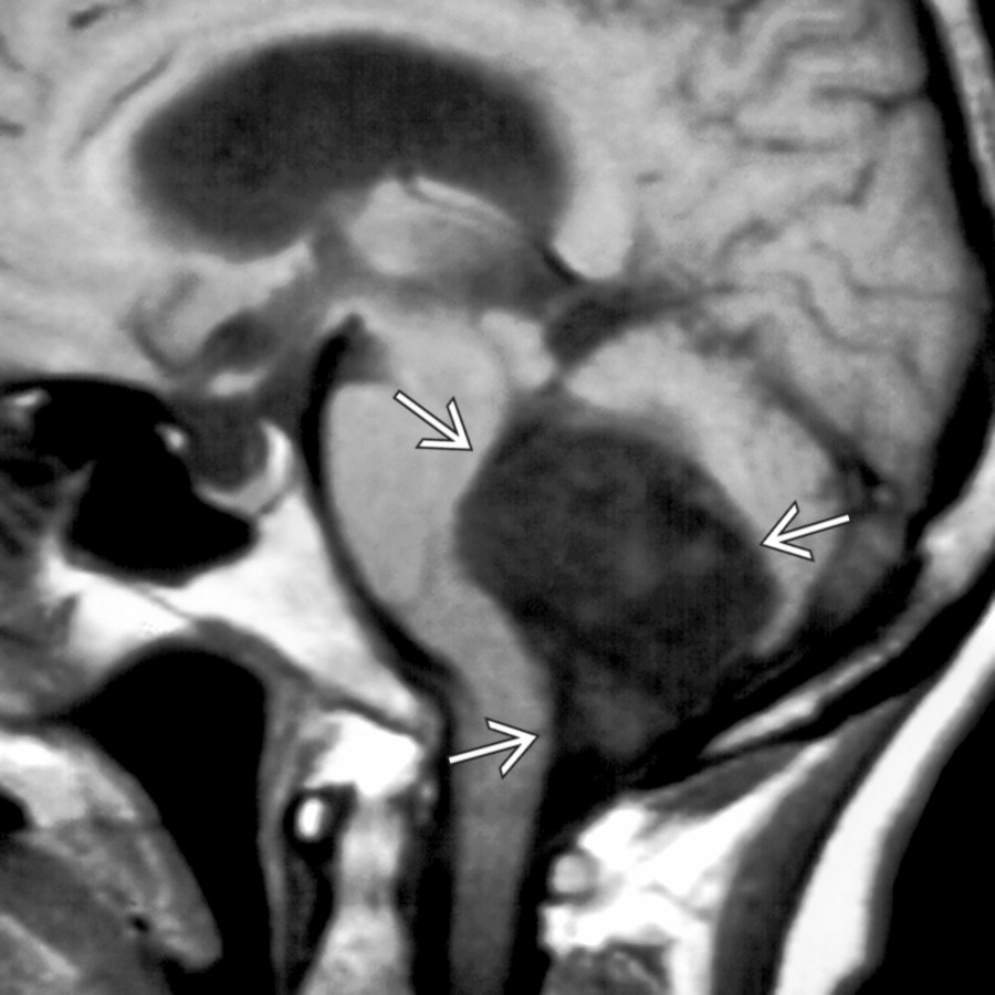
**Epidermoid Cyst**
*Sagittal T1WI MR shows a typical case of a large 4th ventricular epidermoid cyst , which follows CSF intensity but is often slightly brighter and mildly heterogeneous in signal.*

**Dermoid Cyst**
*Axial T2WI MR demonstrates a T2- hyperintense mass encroaching laterally upon the ventral CSF at the foramen magnum. There was evidence of rupture (not shown) causing recurrent meningitis.*

**Hemangioblastoma**
*Sagittal T1WI C+ MR shows an enhancing vermian mass protruding through the foramen magnum . An associated cyst is deforming the 4th ventricle .*

**Neurenteric Cyst**
*Axial T1WI C+ MR shows a neurenteric cyst encroaching upon the prepontine cistern. Although most often these are located anteriorly, they may arise posteriorly at the cisterna magnum.*
### Additional Images

**Chiari 2**
*Sagittal T1WI MR shows a small posterior fossa with extension of the cerebellar peg through the foramen magnum to the level of C3. The cervicomedullary junction and 4th ventricle are low-lying, and there is tectal beaking .*

**Dandy-Walker Continuum**
*Sagittal T1WI MR demonstrates a markedly enlarged posterior fossa with cystic dilatation of the 4th ventricle , upwardly rotated hypoplastic vermis , and low-lying torcular Herophili .*
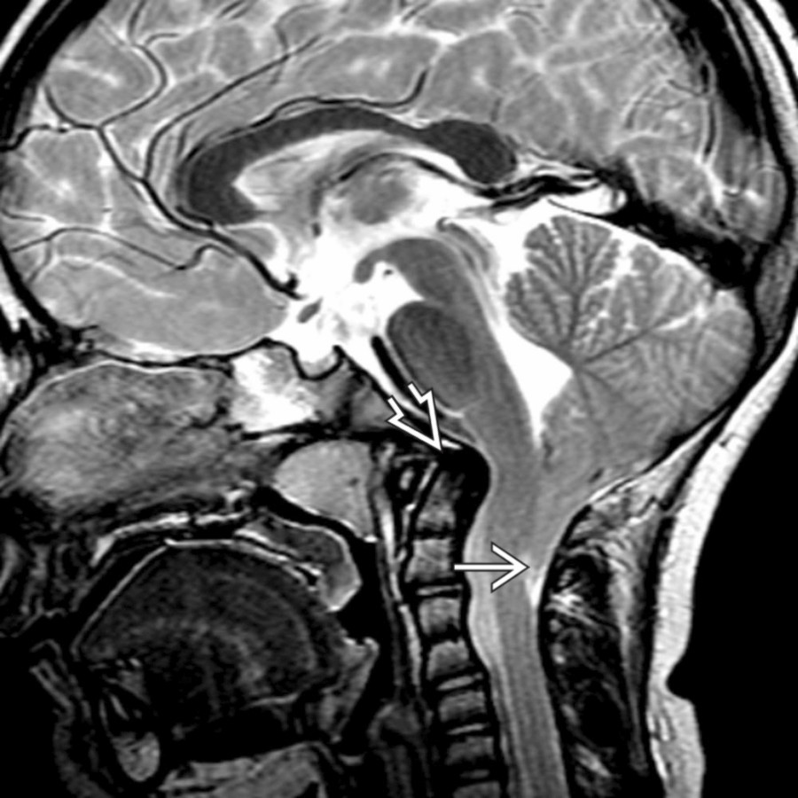
**Chiari 1**
*Sagittal T2 MR in a patient with occipital headaches shows a significantly pointed configuration of a low-lying cerebellar tonsil to the level of the midposterior ring of C2. Notice also the dorsally tilted dens and effacement of CSF at the foramen magnum.*

**Chiari 1**
*Sagittal T2 MR in a patient with occipital headaches shows a significantly pointed configuration of a low-lying cerebellar tonsil to the level of the midposterior ring of C2. Notice also the dorsally tilted dens and effacement of CSF at the foramen magnum.*
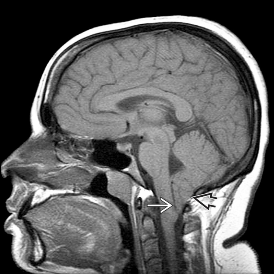
**Chiari 1**
*Sagittal T1WI MR shows inferior extension of the peg-like cerebellar tonsils well below the level of the foramen magnum , consistent with Chiari 1.*

**Chiari 1**
*Sagittal T1WI MR shows inferior extension of the peg-like cerebellar tonsils well below the level of the foramen magnum , consistent with Chiari 1.*

**Herniation Syndromes, Intracranial**
*Axial T2WI MR at the level of the foramen magnum demonstrates downward tonsillar herniation secondary to mass affect from brain death.*
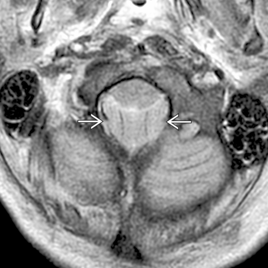
**Herniation Syndromes, Intracranial**
*Axial T2WI MR at the level of the foramen magnum demonstrates downward tonsillar herniation secondary to mass affect from brain death.*

**Herniation Syndromes, Intracranial**
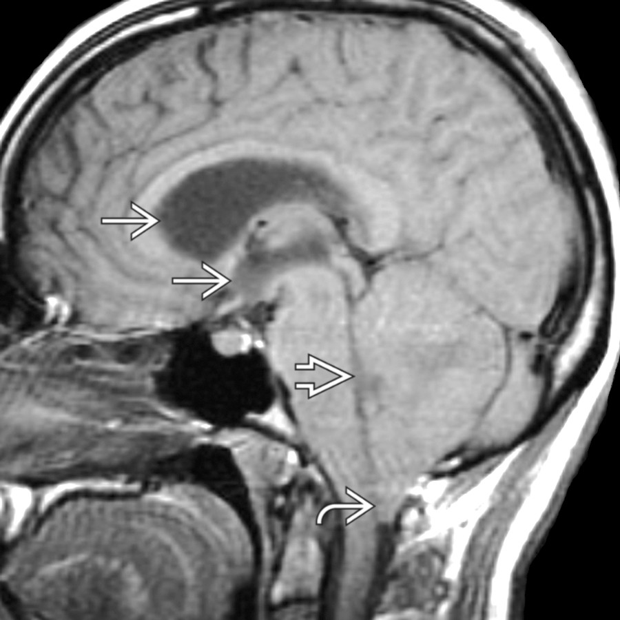
*Sagittal T1WI MR shows cerebellar tonsillar herniation from a large left posterior fossa mass. Note compression of 4th ventricle . Supratentorial ventricles are enlarged .*
**Herniation Syndromes, Intracranial**
*Sagittal T1WI MR shows cerebellar tonsillar herniation from a large left posterior fossa mass. Note compression of 4th ventricle . Supratentorial ventricles are enlarged .*
**Chiari 1**
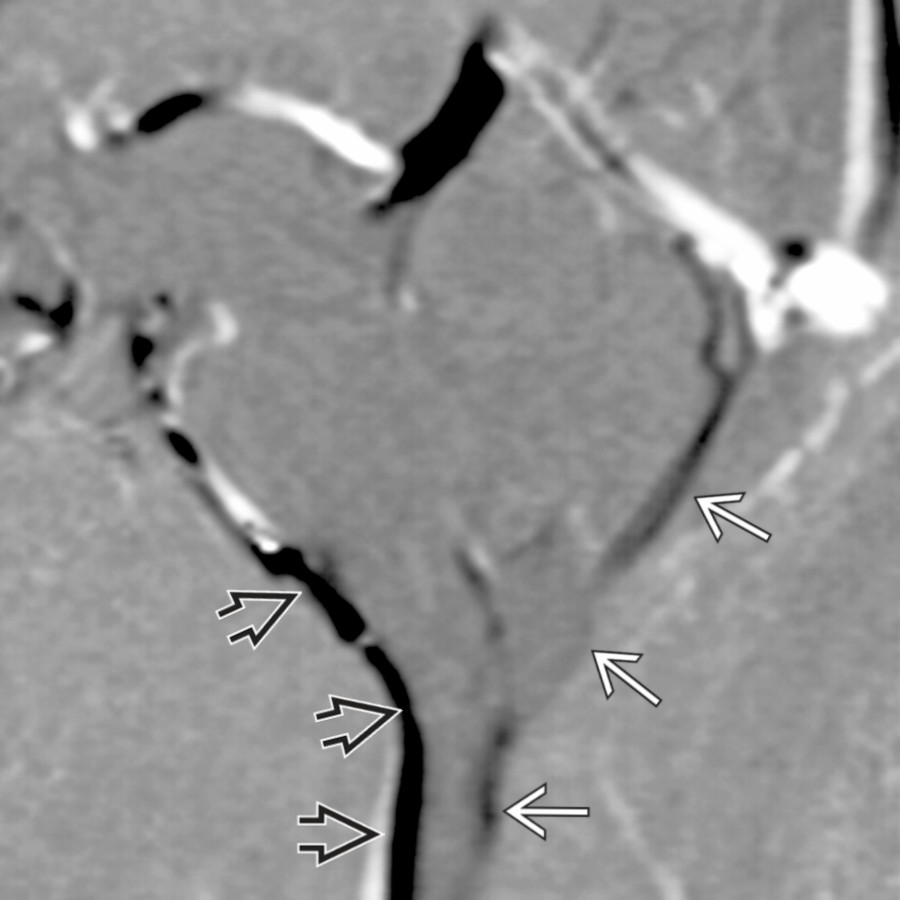
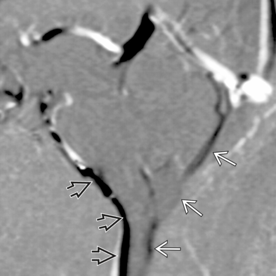
*Sagittal CINE phase-contrast CSF flow - diastolic shows diminished posterior CSF flow compared to anterior CSF flow at the site of tonsillar impaction.*
**Chiari 1**
*Sagittal CINE phase-contrast CSF flow - diastolic shows diminished posterior CSF flow compared to anterior CSF flow at the site of tonsillar impaction.*
**Chiari 1**
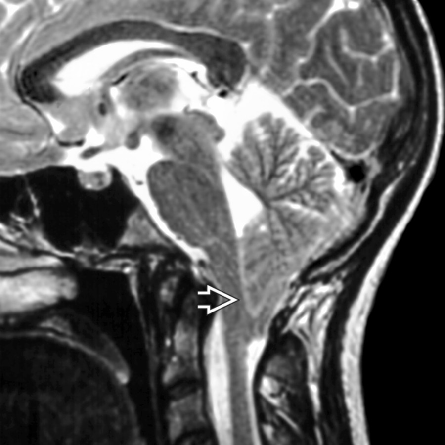
*Sagittal T2WI MR shows a classic case of Chiari 1 with pointed cerebellar tonsils protruding through the foramen magnum and effacing the cisterna magna.*

**Chiari 1**
*Sagittal T2WI MR shows a classic case of Chiari 1 with pointed cerebellar tonsils protruding through the foramen magnum and effacing the cisterna magna.*

**Chiari 2**
*Sagittal T2WI MR shows a small posterior fossa with caudal descent of cerebellar tonsillar tissue , elongation of the 4th ventricle , and cervical cord syrinx .*
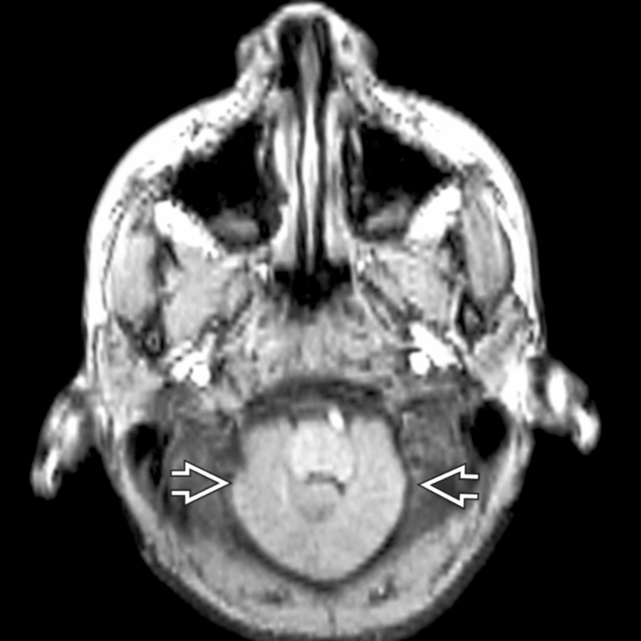
**Chiari 2**
*Axial FSPGR image demonstrates caudally displaced cerebellar tissue wrapping around the brainstem. This is sometimes referred to as a "creeping cerebellum" due to a small posterior fossa.*

**Chiari 2**
*Sagittal T1WI MR shows caudal descent of cerebellar vermian tissue and elongated 4th ventricle as well as callosal dysgenesis and a small posterior fossa.*

**Chiari 2**
*Sagittal T1 C+ MR shows a small posterior fossa with extension of the vermis through the foramen magnum to the level of C1/2 . The 4th ventricle is small , and there is tectal beaking .*

**Dandy-Walker Continuum**
*Axial T2WI MR shows a normal 4th ventricle and vermis; however, there is a prominent retrocerebellar CSF space , which does not cause compression but does remodel endosteum, leading to mega cisterna magna.*
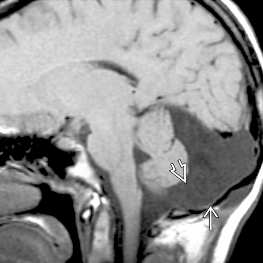
**Arachnoid Cyst**
*Sagittal T1WI MR demonstrates a CSF-intensity arachnoid cyst remodeling the endosteum and filling the cisterna magna .*

**Ependymoma**
*Sagittal T2WI MR shows heterogeneous features of an ependymoma. Focal hyperintense regions correspond to focal necrosis and hypointense regions correlate to calcification .*
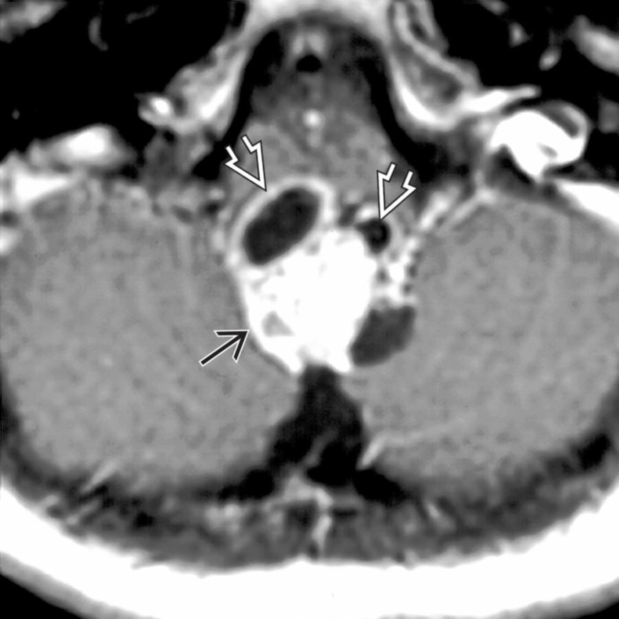
**Ependymoma**
*Axial T1 C+ MR demonstrates a heterogeneously enhancing ependymoma containing cystic elements .*

**Ependymoma**
*Sagittal T1 C+ MR shows an enhancing mass expanding the 4th ventricle . The mass is also distending the obex and protruding through the foramen of Magendie into the cisterna magna .*

**Meningioma**
*Axial T2WI MR demonstrates a posterior fossa meningioma with significant mass effect upon the cerebellum with interposed trapped CSF clefts and vessels .*

**Meningioma**
*Axial T1 C+ MR of a meningioma shows an avidly enhancing, dural-based mass splaying the cerebellar hemispheres, flattening the 4th ventricle , with dural tail .*
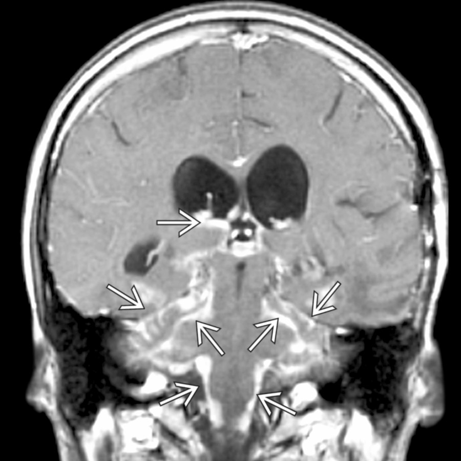
**Metastasis**
*Coronal T1 C+ MR demonstrates the diffuse enhancement of meningeal carcinomatosis from metastatic xanthoastrocytoma spread throughout the posterior fossa, folia, and also more cephalad.*

**Intracranial Hypotension**
*Sagittal T1WI MR shows tonsillar descent and dural thickening secondary to intracranial hypotension.*
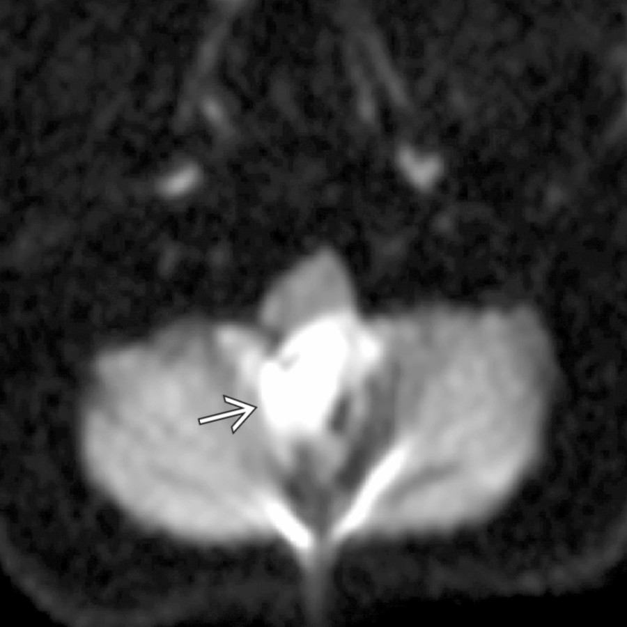
**Epidermoid Cyst**
*Axial DWI MR shows a hyperintense mass in the midline posterior fossa.*
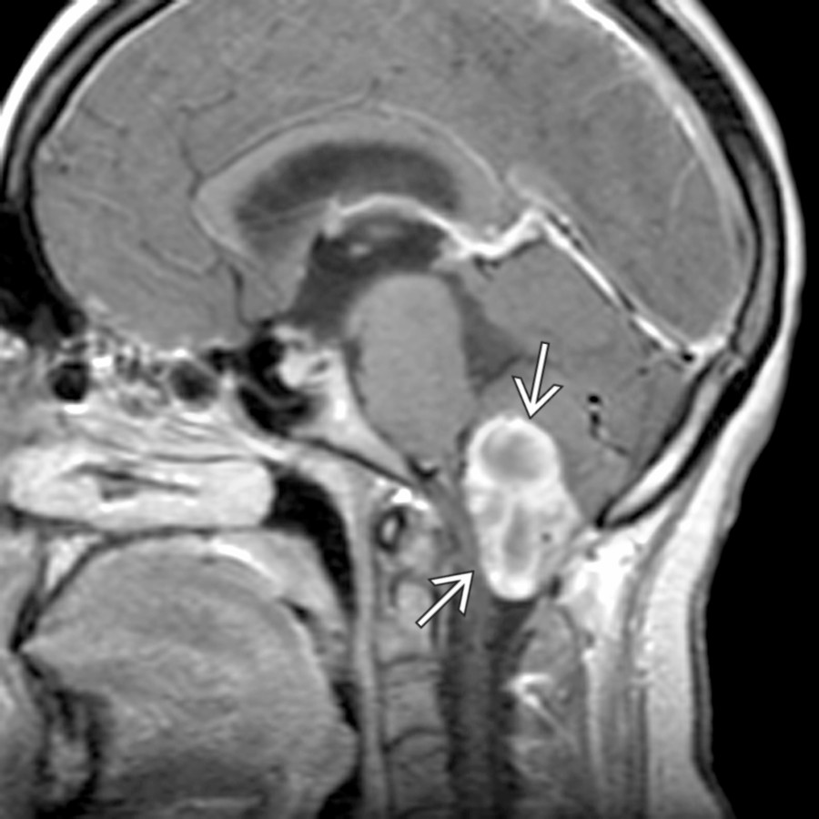
**Hemangioblastoma**
*Sagittal T1 C+ MR demonstrates a mostly solid hemangioblastoma involving the cerebellar tonsils and effacing the cisterna magna.*
with effacement of the cisterna magna.](images/app.statdx.com_image_thumbnail_43f42495-6e64-47ac-8abd-7ca9ca2ff540_annotated_true_size_900_quality_90_a13877a5.jpg)
**Intracranial Hypotension**
*Sagittal T1WI C+ MR shows obliteration of the suprasellar cistern , sagging/fat midbrain with closed angle between the peduncles and the pons , dural enhancement , and tonsillar descent with effacement of the cisterna magna.*

**Subependymoma**
*Sagittal T1WI C+ MR in 40-year-old man shows an enhancing mass at the bottom of the 4th ventricle filling the cisterna magna.*

**Epidermoid Cyst**
*Sagittal T1WI MR shows a typical case of a large 4th ventricular epidermoid cyst , which follows CSF intensity but is often slightly brighter and mildly heterogeneous in signal.*

**Dermoid Cyst**
*Axial T2WI MR demonstrates a T2- hyperintense mass encroaching laterally upon the ventral CSF at the foramen magnum. There was evidence of rupture (not shown) causing recurrent meningitis.*

**Hemangioblastoma**
*Sagittal T1WI C+ MR shows an enhancing vermian mass protruding through the foramen magnum . An associated cyst is deforming the 4th ventricle .*

**Neurenteric Cyst**
*Axial T1WI C+ MR shows a neurenteric cyst encroaching upon the prepontine cistern. Although most often these are located anteriorly, they may arise posteriorly at the cisterna magnum.*
### Additional Images

**Chiari 2**
*Sagittal T1WI MR shows a small posterior fossa with extension of the cerebellar peg through the foramen magnum to the level of C3. The cervicomedullary junction and 4th ventricle are low-lying, and there is tectal beaking .*

**Dandy-Walker Continuum**
*Sagittal T1WI MR demonstrates a markedly enlarged posterior fossa with cystic dilatation of the 4th ventricle , upwardly rotated hypoplastic vermis , and low-lying torcular Herophili .*

**Chiari 1**
*Sagittal T2 MR in a patient with occipital headaches shows a significantly pointed configuration of a low-lying cerebellar tonsil to the level of the midposterior ring of C2. Notice also the dorsally tilted dens and effacement of CSF at the foramen magnum.*

**Chiari 1**
*Sagittal T2 MR in a patient with occipital headaches shows a significantly pointed configuration of a low-lying cerebellar tonsil to the level of the midposterior ring of C2. Notice also the dorsally tilted dens and effacement of CSF at the foramen magnum.*

**Chiari 1**
*Sagittal T1WI MR shows inferior extension of the peg-like cerebellar tonsils well below the level of the foramen magnum , consistent with Chiari 1.*

**Chiari 1**
*Sagittal T1WI MR shows inferior extension of the peg-like cerebellar tonsils well below the level of the foramen magnum , consistent with Chiari 1.*

**Herniation Syndromes, Intracranial**
*Axial T2WI MR at the level of the foramen magnum demonstrates downward tonsillar herniation secondary to mass affect from brain death.*

**Herniation Syndromes, Intracranial**
*Axial T2WI MR at the level of the foramen magnum demonstrates downward tonsillar herniation secondary to mass affect from brain death.*

**Herniation Syndromes, Intracranial**
*Sagittal T1WI MR shows cerebellar tonsillar herniation from a large left posterior fossa mass. Note compression of 4th ventricle . Supratentorial ventricles are enlarged .*

**Herniation Syndromes, Intracranial**
*Sagittal T1WI MR shows cerebellar tonsillar herniation from a large left posterior fossa mass. Note compression of 4th ventricle . Supratentorial ventricles are enlarged .*

**Chiari 1**
*Sagittal CINE phase-contrast CSF flow - diastolic shows diminished posterior CSF flow compared to anterior CSF flow at the site of tonsillar impaction.*

**Chiari 1**
*Sagittal CINE phase-contrast CSF flow - diastolic shows diminished posterior CSF flow compared to anterior CSF flow at the site of tonsillar impaction.*

**Chiari 1**
*Sagittal T2WI MR shows a classic case of Chiari 1 with pointed cerebellar tonsils protruding through the foramen magnum and effacing the cisterna magna.*

**Chiari 1**
*Sagittal T2WI MR shows a classic case of Chiari 1 with pointed cerebellar tonsils protruding through the foramen magnum and effacing the cisterna magna.*

**Chiari 2**
*Sagittal T2WI MR shows a small posterior fossa with caudal descent of cerebellar tonsillar tissue , elongation of the 4th ventricle , and cervical cord syrinx .*

**Chiari 2**
*Axial FSPGR image demonstrates caudally displaced cerebellar tissue wrapping around the brainstem. This is sometimes referred to as a "creeping cerebellum" due to a small posterior fossa.*

**Chiari 2**
*Sagittal T1WI MR shows caudal descent of cerebellar vermian tissue and elongated 4th ventricle as well as callosal dysgenesis and a small posterior fossa.*

**Chiari 2**
*Sagittal T1 C+ MR shows a small posterior fossa with extension of the vermis through the foramen magnum to the level of C1/2 . The 4th ventricle is small , and there is tectal beaking .*

**Dandy-Walker Continuum**
*Axial T2WI MR shows a normal 4th ventricle and vermis; however, there is a prominent retrocerebellar CSF space , which does not cause compression but does remodel endosteum, leading to mega cisterna magna.*

**Arachnoid Cyst**
*Sagittal T1WI MR demonstrates a CSF-intensity arachnoid cyst remodeling the endosteum and filling the cisterna magna .*

**Ependymoma**
*Sagittal T2WI MR shows heterogeneous features of an ependymoma. Focal hyperintense regions correspond to focal necrosis and hypointense regions correlate to calcification .*

**Ependymoma**
*Axial T1 C+ MR demonstrates a heterogeneously enhancing ependymoma containing cystic elements .*

**Ependymoma**
*Sagittal T1 C+ MR shows an enhancing mass expanding the 4th ventricle . The mass is also distending the obex and protruding through the foramen of Magendie into the cisterna magna .*

**Meningioma**
*Axial T2WI MR demonstrates a posterior fossa meningioma with significant mass effect upon the cerebellum with interposed trapped CSF clefts and vessels .*

**Meningioma**
*Axial T1 C+ MR of a meningioma shows an avidly enhancing, dural-based mass splaying the cerebellar hemispheres, flattening the 4th ventricle , with dural tail .*

**Metastasis**
*Coronal T1 C+ MR demonstrates the diffuse enhancement of meningeal carcinomatosis from metastatic xanthoastrocytoma spread throughout the posterior fossa, folia, and also more cephalad.*

**Intracranial Hypotension**
*Sagittal T1WI MR shows tonsillar descent and dural thickening secondary to intracranial hypotension.*

**Epidermoid Cyst**
*Axial DWI MR shows a hyperintense mass in the midline posterior fossa.*

**Hemangioblastoma**
*Sagittal T1 C+ MR demonstrates a mostly solid hemangioblastoma involving the cerebellar tonsils and effacing the cisterna magna.*