---
title: "CPA Mass, Adult"
docid: "f3cd22f6-53b9-4392-be23-512d221d2e02"
authors:
- key: "07a2c087-6202-49e7-870b-7aa162d18f06"
value: "Bronwyn E. Hamilton, MD"
breadcrumbs:
-
name: "Head and Neck"
slug: "head-and-neck"
treeNodeId: "ed24ed8c-5d57-4629-879b-447b82d2973d"
-
name: "Differential Diagnosis"
slug: "differential-diagnosis"
treeNodeId: "40d68862-8975-4dde-ac2b-ebc43ab0fb5c"
-
name: "CPA-IAC and Posterior Fossa"
slug: "cpa-iac-and-posterior-fossa"
treeNodeId: "c590eedb-4a3b-4158-a04f-ad880564c992"
-
name: "Anatomically Based Differentials"
slug: "anatomically-based-differentials"
treeNodeId: "debfb06c-8656-4f5d-92c1-eaa468185d78"
-
name: "CPA Mass, Adult"
slug: "cpa-mass-adult"
treeNodeId: null
category: "Head and Neck"
documentVersionId: "3389aa3f-4eea-4b0c-aab3-7b7265e22a43"
imageCount: 25
lastUpdated: "01/18/24"
pageDescription: "CPA Mass, Adult"
pageKeywords: "Head and Neck, Differential Diagnosis, CPA-IAC and Posterior Fossa, Anatomically Based Differentials, CPA Mass, Adult"
pageTitle: "CPA Mass, Adult | STATdx"
enhancedTitle: "CPA Mass, Adult"
type: "DDX"
references: true
breadcrumbs:
- "Head and Neck"
- "Differential Diagnosis"
- "CPA-IAC and Posterior Fossa"
- "Anatomically Based Differentials"
- "CPA Mass, Adult"
---
# ESSENTIAL INFORMATION
- ## Key Differential Diagnosis Issues
- Idealized imaging protocol in evaluating CPA mass lesions
- T1 C+ fat-saturated MR is gold standard
- Fat saturation differentiates lipoma from vestibular schwannoma
- Add DWI for possible epidermoid cyst
- Add GRE for aneurysm wall clot & calcification; also useful for tumor calcifications
- T2 thin-section, high-resolution MR gives more surgical data when vestibular schwannoma is diagnosed
- CISS or FIESTA most commonly used
- Helps define amount of CSF cap in lateral IAC
- Assesses relationship of cochlear nerve canal to lesion
- If small schwannoma, may define nerve of origin (superior vs. inferior vestibular schwannoma)
- Knowledge of relative incidence of lesions key in CPA-IAC lesion assessment
- Vestibular schwannoma: ~ **90%** of all CPA-IAC masses
- Meningioma, epidermoid cyst, aneurysm, & arachnoid cyst together represent ~ **8%** of all CPA-IAC masses
- All other diagnoses in differential list: ~ **2%**
- Other factors relevant to imaging CPA masses
- 3D facial nerve tractography for CPA masses may aid surgical planning to reduce risk of facial nerve injury
- CPA tumors in women independent risk factor for intracranial aneurysms
- ## Helpful Clues for Common Diagnoses
- **Vestibular Schwannoma**
- Morphology
- Ovoid intracanalicular mass (IAC)
- Ice cream on cone shape (CPA-IAC)
- T1 C+ MR
- Enhancing well-defined tumor ± intramural cysts
- High-resolution thin 0.6-mm 3D T2 MR alternative screening modality for schwannomas without contrast
- SWI
- Intratumoral microhemorrhages favor schwannoma, rare in meningioma
- ## Helpful Clues for Less Common Diagnoses
- **Meningioma in CPA-IAC**
- Morphology
- Mushroom-shaped dural-based mass capping IAC asymmetrically
- T1 C+ MR
- Enhancing mass, ± dural tails, ± CSF-vascular cleft if CPA component is larger
- 25% of CPA meningiomas have direct extension or dural tail projecting into IAC
- **Epidermoid Cyst in CPA-IAC**
- Morphology
- Insinuating ± scalloping brainstem margin
- MR imaging
- T1 C+ MR: Nonenhancing
- If known or suspected epidermoid develops enhancing margins, consider rare malignant transformation (squamous cell carcinoma); PET avidity can be helpful to confirm suspicion
- DWI: Restricted diffusion (high signal) makes diagnosis
- T2 MR: Follows fluid signal intensity
- FLAIR: Lack of complete fluid suppression
- **Aneurysm****in****CPA-IAC**
- Morphology
- Ovoid or fusiform; rarely IAC
- MR imaging
- T1 & T1 C+ MR: Complex signal mass from wall calcification, clot, & flow
- MRA (CTA, angiography) sorts out diagnosis
- **Arachnoid Cyst****in****CPA**
- Morphology
- Fills cistern with rounded margins
- MR imaging
- T1 C+ MR: No enhancement
- FLAIR: Lesion attenuates
- DWI: No restricted diffusion
- **Metastases in CPA-IAC**
- Morphology
- Irregular invasive margins
- MR imaging
- T1 C+ MR: Single or multiple enhancing masses in CPA
- 4 sites primarily involved: Flocculus, choroid plexus, arachnoid-dura, or pia
- ## Helpful Clues for Rare Diagnoses
- **Neurofibromatosis Type****2**
- Morphology
- Bilateral ovoid IAC or ice cream on cone-shaped CPA-IAC masses
- MR imaging
- T1 C+ MR: Bilateral enhancing CPA-IAC masses
- ± additional schwannomas & meningiomas
- **Sarcoidosis****in****CPA-IAC**
- Laboratory
- CSF lymphocytosis
- ↑ blood angiotensin converting enzyme
- Morphology
- En plaque or nodular dural lesion(s)
- MR imaging
- T1 C+ MR: Enhancing multifocal dural-based lesions
- **Choroid Plexus Papilloma****in****CPA**
- Morphology
- Dumbbell shape with 4th ventricle & CPA cistern components
- Pear-shaped if begins in foramen of Luschka
- MR imaging
- T1 C+ MR: Avidly enhancing mass in 4th ventricle projecting through foramen of Luschka into CPA
- **Lipoma****in****CPA-IAC**
- Morphology
- Ovoid if IAC only
- CPA lesion may be broad-based against lateral pons
- Nonenhanced CT
- Fat-density lesion of CPA ± IAC ± inner ear
- MR imaging
- T1 MR: High-signal lesion, suppresses with fat saturation
- Caveat: If T1 C+ without fat saturation, lipoma may be mistaken for vestibular schwannoma
- **Ependymoma in CPA**
- Morphology
- Irregular soft tumor squeezes out through 4th ventricle foramen of Luschka into CPA
- Tumor margins amorphous
- Bone CT
- Calcifications in 50% of cases
- MR imaging
- T1 C+ MR: Heterogeneous enhancement of solid tumor components
- Marginal enhancement of tumor cyst wall
- **Hypertrophic Pachymeningitis**
- Varied causes require tissue diagnosis
- IgG4-related disease (↑ plasma or tissue IgG4)
- Granulomatosis with polyangiitis: Antineutrophil cytoplasmic antibody (ANCA) (+) associated vasculitis
- Idiopathic (a.k.a. intracranial idiopathic inflammatory pseudotumor)
- Morphology
- En plaque
- MR imaging
- T1 C+ MR: Thickened enhancing dura
- Caveat: May mimic meningioma, sarcoidosis, or metastatic disease
- **F****acial Nerve****Schwannoma in CPA-IAC**
- Morphology
- CPA-IAC mass with labyrinthine tail
- Bone CT
- Labyrinthine segment of CNVII may be enlarged
- MR imaging
- T1 C+ MR: Enhancing tubular mass in CPA-IAC & labyrinthine segment of CNVII
- Caveat: If labyrinthine segment of CNVII not involved, cannot be differentiated from vestibular schwannoma
- **Jugular Foramen Schwannoma**
- Morphology
- Lobular mass projects superomedially from jugular foramen toward lateral brainstem
- MR imaging
- T1 C+ MR: Enhancing mass arising from jugular foramen
- **IAC Venous Malformation ("Hemangioma")**
- Morphology
- Poorly marginated IAC mass with punctate calcifications
- Bone CT
- Punctate calcifications in IAC mass
- MR imaging
- T1 C+ MR: Enhancing IAC mass with focal low-signal foci (calcifications)
- **Neurenteric Cyst**
- Morphology
- Rounded ovoid mass in prepontine cistern
- MR imaging
- T1: Intermediate to high signal T1 prepontine mass
- Nonenhancing
- Caveat: ↑ T1 signal differentiates from epidermoid cyst
## References
# Selected References
1. [Shimanuki MN et al: Imaging of temporal bone mass lesions: a pictorial review. Diagnostics (Basel). 13(16), 2023](http://www.ncbi.nlm.nih.gov/pubmed/?term=37627924%5Bpmid%5D)
1. [Shimojima Y et al: Hypertrophic pachymeningitis in ANCA-associated vasculitis: clinical and immunopathological features and insights. Autoimmun Rev. 22(6):103338, 2023](http://www.ncbi.nlm.nih.gov/pubmed/?term=37062439%5Bpmid%5D)
1. [Ota Y et al: Advanced MRI to differentiate schwannomas and metastases in the cerebellopontine angle/internal auditory canal. J Neuroimaging. 32(6):1177-84, 2022](http://www.ncbi.nlm.nih.gov/pubmed/?term=35879866%5Bpmid%5D)
1. [Ozaki K et al: Arachnoid cyst alone causes hemifacial spasm: illustrative case. J Neurosurg Case Lessons. 3(15), 2022](http://www.ncbi.nlm.nih.gov/pubmed/?term=36303502%5Bpmid%5D)
1. [Sakamoto H et al: Radio-pathological characteristics of malignant transformation of an epidermoid cyst in the cerebellopontine angle: a case report. Surg Neurol Int. 13:135, 2022](http://www.ncbi.nlm.nih.gov/pubmed/?term=35509542%5Bpmid%5D)
1. [Banerjee P et al: Role of neuroimaging in cases of primary and secondary hemifacial spasm. Indian J Ophthalmol. 69(2):253-6, 2021](http://www.ncbi.nlm.nih.gov/pubmed/?term=33463567%5Bpmid%5D)
1. [Connor SEJ: Imaging of the vestibular schwannoma: diagnosis, monitoring, and treatment planning. Neuroimaging Clin N Am. 31(4):451-71, 2021](http://www.ncbi.nlm.nih.gov/pubmed/?term=34689927%5Bpmid%5D)
1. [Saigal G et al: Utility of Microhemorrhage as a diagnostic tool in distinguishing vestibular schwannomas from other cerebellopontine angle (CPA) Tumors. Indian J Otolaryngol Head Neck Surg. 73(3):321-6, 2021](http://www.ncbi.nlm.nih.gov/pubmed/?term=34471620%5Bpmid%5D)
1. [Totten DJ et al: Cerebellopontine angle and internal auditory canal lipomas: case series and systematic review. Laryngoscope. 131(9):2081-7, 2021](http://www.ncbi.nlm.nih.gov/pubmed/?term=33567134%5Bpmid%5D)
1. [Pamela Ferreira Neto B et al: Noncystic cerebellopontine angle hemangioblastoma: A case of an atypical location. Int J Surg Case Rep. 74:234-7, 2020](http://www.ncbi.nlm.nih.gov/pubmed/?term=32892127%5Bpmid%5D)
1. [Melenotte C et al: Clinical presentation, treatment and outcome of IgG4-related pachymeningitis: from a national case registry and literature review. Semin Arthritis Rheum. 49(3):430-7, 2019](http://www.ncbi.nlm.nih.gov/pubmed/?term=31155444%5Bpmid%5D)
1. [Zheng SF et al: Cerebellopontine angle tumors are associated with a greater incidence of unruptured intracranial aneurysms. World Neurosurg. 122:e561-8, 2019](http://www.ncbi.nlm.nih.gov/pubmed/?term=31108072%5Bpmid%5D)
1. [Dunn IF et al: Congress of neurological surgeons systematic review and evidence-based guidelines on the role of imaging in the diagnosis and management of patients with vestibular schwannomas. Neurosurgery. 82(2):E32-4, 2018](http://www.ncbi.nlm.nih.gov/pubmed/?term=29309686%5Bpmid%5D)
1. [Prabhu V et al: Preserved cochlear CISS signal is a predictor for hearing preservation in patients treated for vestibular schwannoma with stereotactic radiosurgery. Otol Neurotol. 39(5):628-31, 2018](http://www.ncbi.nlm.nih.gov/pubmed/?term=29561382%5Bpmid%5D)
1. [Mishra A et al: Susceptibility weighted imaging - a problem-solving tool in differentiation of cerebellopontine angle schwannomas and meningiomas. Neuroradiol J. 30(3):253-8, 2017](http://www.ncbi.nlm.nih.gov/pubmed/?term=28627983%5Bpmid%5D)
1. [Schulze M et al: Improvement in imaging common temporal bone pathologies at 3 T MRI: small structures benefit from a small field of view. Clin Radiol. 72(3):267.e1-12, 2017](http://www.ncbi.nlm.nih.gov/pubmed/?term=28034444%5Bpmid%5D)
1. [Rueckriegel SM et al: Probabilistic fiber-tracking reveals degeneration of the contralateral auditory pathway in patients with vestibular schwannoma. AJNR Am J Neuroradiol. 37(9):1610-6, 2016](http://www.ncbi.nlm.nih.gov/pubmed/?term=27256855%5Bpmid%5D)
1. [Watanabe N et al: Imaging alterations due to squamous metaplasia in intracranial neurenteric cysts: A report of two cases. Neuroradiol J. 29(3):187-92, 2016](http://www.ncbi.nlm.nih.gov/pubmed/?term=27009777%5Bpmid%5D)
1. [Mukherjee P et al: Intracranial lipomas affecting the cerebellopontine angle and internal auditory canal: a case series. Otol Neurotol. 32(4):670-5, 2011](http://www.ncbi.nlm.nih.gov/pubmed/?term=21358448%5Bpmid%5D)
1. [Warren FM et al: Imaging characteristics of metastatic lesions to the cerebellopontine angle. Otol Neurotol. 29(6):835-8, 2008](http://www.ncbi.nlm.nih.gov/pubmed/?term=18636029%5Bpmid%5D)
1. [Barrera JE et al: Cavernous hemangioma of the internal auditory canal: a case report and review of the literature. Am J Otolaryngol. 25(3):199-203, 2004](http://www.ncbi.nlm.nih.gov/pubmed/?term=15124171%5Bpmid%5D)
1. [Nakamura M et al: Meningiomas of the internal auditory canal. Neurosurgery. 55(1):119-27; discussion 127-8, 2004](http://www.ncbi.nlm.nih.gov/pubmed/?term=15214980%5Bpmid%5D)
1. [Swartz JD: Lesions of the cerebellopontine angle and internal auditory canal: diagnosis and differential diagnosis. Semin Ultrasound CT MR. 25(4):332-52, 2004](http://www.ncbi.nlm.nih.gov/pubmed/?term=15497614%5Bpmid%5D)
1. [Daniels RL et al: Causes of unilateral sensorineural hearing loss screened by high-resolution fast spin echo magnetic resonance imaging: review of 1,070 consecutive cases. Am J Otol. 21(2):173-80, 2000](http://www.ncbi.nlm.nih.gov/pubmed/?term=10733180%5Bpmid%5D)
1. [Kohan D et al: Uncommon lesions presenting as tumors of the internal auditory canal and cerebellopontine angle. Am J Otol. 18(3):386-92, 1997](http://www.ncbi.nlm.nih.gov/pubmed/?term=9149836%5Bpmid%5D)
1. [Smirniotopoulos JG et al: Cerebellopontine angle masses: radiologic-pathologic correlation. Radiographics. 13(5):1131-47, 1993](http://www.ncbi.nlm.nih.gov/pubmed/?term=8210595%5Bpmid%5D)
## Images
### Selected Images
 . A small amount of tumor extension is visible extending into the IAC
  .](images/app.statdx.com_image_thumbnail_8b05ff12-1b6e-4f10-b2ef-b957b56a3fb7_annotated_true_size_900_quality_90_4ed330ffaef11f383ae38df21c6c60eaec019564.jpg)
**Vestibular Schwannoma**
*Axial T1 C+ MR demonstrates a mixed solid and cystic enhancing mass in the right CPA cistern . A small amount of tumor extension is visible extending into the IAC .*
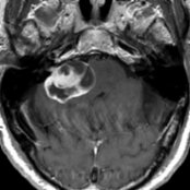
**Vestibular Schwannoma**
*Axial T1 C+ MR demonstrates a mixed solid and cystic enhancing mass in the right CPA cistern . A small amount of tumor extension is visible extending into the IAC .*

**Vestibular Schwannoma**
*Axial T1 C+ MR demonstrates a mixed solid and cystic enhancing mass in the right CPA cistern . A small amount of tumor extension is visible extending into the IAC .*

**Meningioma in CPA-IAC**
*Axial T1 C+ MR demonstrates a homogeneously enhancing meningioma in the right CPA . There was no substantial IAC component, and a dural tail was present on additional images (not shown).*

**Epidermoid Cyst in CPA-IAC**
*Axial T2 MR shows a heterogeneous right CPA mass with insinuating contours around the brainstem and cerebellum , typical of an epidermoid cyst. Corresponding DWI showed bright signal (restricted diffusion), and FLAIR showed lack of fluid suppression (not shown).*

**Vestibular Schwannoma**
*Axial T1 C+ MR demonstrates a mixed solid and cystic enhancing mass in the right CPA cistern . A small amount of tumor extension is visible extending into the IAC .*

**Vestibular Schwannoma**
*Axial T1 C+ MR demonstrates a mixed solid and cystic enhancing mass in the right CPA cistern . A small amount of tumor extension is visible extending into the IAC .*

**Vestibular Schwannoma**
*Axial T1 C+ MR demonstrates a mixed solid and cystic enhancing mass in the right CPA cistern . A small amount of tumor extension is visible extending into the IAC .*

**Meningioma in CPA-IAC**
*Axial T1 C+ MR demonstrates a homogeneously enhancing meningioma in the right CPA . There was no substantial IAC component, and a dural tail was present on additional images (not shown).*

**Epidermoid Cyst in CPA-IAC**
*Axial T2 MR shows a heterogeneous right CPA mass with insinuating contours around the brainstem and cerebellum , typical of an epidermoid cyst. Corresponding DWI showed bright signal (restricted diffusion), and FLAIR showed lack of fluid suppression (not shown).*
 .](677e1c8f-7355-4a17-b864-e21f51d64ae4)
**Aneurysm in CPA-IAC**
*Axial T1 C+ MR demonstrates a large enhancing distal vertebral artery aneurysm projecting up into the CPA cistern and compressing the area where CNVII and CNVIII exit the brainstem .*

**Arachnoid Cyst in CPA**
*Axial T2 FS MR shows a high-signal cystic mass in the low CPA cistern. Note the anterior displacement of the proximal vestibulocochlear nerve by the arachnoid cyst . The high signal results from the absence of CSF flow-related artifact.*

**Metastases in CPA-IAC**
*Axial T1 C+ FS MR reveals an inhomogeneously enhancing metastatic focus arising from the dura along the prepontine cistern. This metastasis reaches the anterior margin of the porus acusticus .*

**Neurofibromatosis Type 2**
*Axial T1 C+ MR shows bilateral enhancing CPA-IAC schwannomas . The left-sided schwannoma involves the intratemporal anterior genu of facial nerve , indicating it is most likely a facial nerve schwannoma.*

**Sarcoidosis in CPA-IAC**
*Axial T1 C+ MR shows a heaped-up, dural-based sarcoid deposit in the right CPA cistern that enters the IAC . The Meckel cave is also affected. This lesion mimics meningioma.*

**Choroid Plexus Papilloma in CPA**
*Axial T1 C+ MR reveals a pear-shaped, inhomogeneously enhancing papilloma projecting from the lateral recess of the 4th ventricle through the foramen of Luschka into the low CPA cistern .*

**Lipoma in CPA-IAC**
*Axial NECT performed for trauma shows large, bilateral, fat-attenuation masses within both CPA cisterns , compatible with lipomas. The patient has a longstanding history of deafness. Surgery is not indicated, since hearing does not improve with resection.*

**Ependymoma in CPA**
*Axial T1 C+ MR demonstrates an aggressive mixed cystic-solid enhancing ependymoma of the right CPA cistern , 4th ventricle , and cerebellar hemisphere .*

**Hypertrophic Pachymeningitis**
*Axial T1 C+ MR demonstrates an extensive area of enhancing dural thickening along the right low CPA cistern. Pachymeningitis also involves the subjacent jugular foramen .*

**Facial Nerve Schwannoma in CPA-IAC**
*Axial T1 C+ MR shows a variant facial nerve schwannoma with a solid enhancing CPA-IAC component extending into the geniculate ganglion . Note the associated arachnoid cyst .*

**Jugular Foramen Schwannoma**
*Axial T1 C+ FS MR shows a bilobed intensely enhancing mass in the right CPA cistern distorting adjacent brain. A large enhancing component within the enlarged right jugular foramen indicated the site of origin. No extension into the adjacent IAC (not shown) was seen.*
### Additional Images

**Vestibular Schwannoma**
*Axial T1 C+ MR shows a heterogeneous enhancing CPA mass filling and expanding the right IAC . Note characteristic internal cystic foci .*

**Vestibular Schwannoma**
*Axial T1 C+ MR shows an typical, heterogeneously enhancing right CPA schwannoma with a colocated arachnoid cyst .*

**Epidermoid Cyst in CPA-IAC**
*Axial T2 MR shows a hyperintense mass in the right CPA with "insinuating" margins , typical of epidermoid cysts. No enhancement was noted on postcontrast imaging and DWI (not shown) showed lesion restriction.*

**IAC Venous Malformation ("Hemangioma")**
*Axial T1 C+ MR shows an enhancing IAC mass with multiple punctate low-signal foci . CT showed that calcifications were present, supporting the diagnosis of IAC hemangioma.*

**Neurenteric Cyst**
*Axial FLAIR MR demonstrates an area of high signal in the low CPA cistern that was found to be a neurenteric cyst at surgery.*

**Vestibular Schwannoma**
*Axial T1 C+ MR reveals an enhancing mass filling the CPA and IAC . Note that the cochlear nerve canal is involved , making resection with hearing preservation difficult.*

**Meningioma in CPA-IAC**
*Axial T1 C+ FS MR reveals an enhancing dural-based mass centered over the IAC but with minimal IAC involvement . The shape and the associated dural tail make meningioma the diagnosis.*

**Epidermoid Cyst in CPA-IAC**
*Axial T1WI MR shows a low-signal mass in the right CPA cistern that insinuates and enlarges the foramen of Luschka and scallops the ventral cerebellar hemisphere .*

**Lipoma in CPA-IAC**
*Axial T1WI MR shows a variant 3-part lipoma affecting the CPA cistern , the high anterior jugular foramen , and the vestibule of the inner ear . Surgical resection is not performed for such lesions.*

**Jugular Foramen Schwannoma**
*Coronal T1 C+ FS MR reveals a schwannoma projecting cephalad from the jugular foramen into the CPA cistern. Note that the normal IAC is at the level of the upper margin of the tumor.*

**Epidermoid Cyst in CPA-IAC**
*Axial T1 C+ MR shows a partially cystic prepontine and left CPA mass with marginal nodular areas of enhancement due to epidermoid cyst, which in this case was complicated by rare malignant transformation into squamous cell carcinoma. Areas of diffusion restriction were noted in the cystic component; however, soft tissue enhancement along the margins are unexpected for epidermoid, and raise concern for malignancy.*
.](677e1c8f-7355-4a17-b864-e21f51d64ae4)
**Aneurysm in CPA-IAC**
*Axial T1 C+ MR demonstrates a large enhancing distal vertebral artery aneurysm projecting up into the CPA cistern and compressing the area where CNVII and CNVIII exit the brainstem .*

**Arachnoid Cyst in CPA**
*Axial T2 FS MR shows a high-signal cystic mass in the low CPA cistern. Note the anterior displacement of the proximal vestibulocochlear nerve by the arachnoid cyst . The high signal results from the absence of CSF flow-related artifact.*

**Metastases in CPA-IAC**
*Axial T1 C+ FS MR reveals an inhomogeneously enhancing metastatic focus arising from the dura along the prepontine cistern. This metastasis reaches the anterior margin of the porus acusticus .*

**Neurofibromatosis Type 2**
*Axial T1 C+ MR shows bilateral enhancing CPA-IAC schwannomas . The left-sided schwannoma involves the intratemporal anterior genu of facial nerve , indicating it is most likely a facial nerve schwannoma.*

**Sarcoidosis in CPA-IAC**
*Axial T1 C+ MR shows a heaped-up, dural-based sarcoid deposit in the right CPA cistern that enters the IAC . The Meckel cave is also affected. This lesion mimics meningioma.*

**Choroid Plexus Papilloma in CPA**
*Axial T1 C+ MR reveals a pear-shaped, inhomogeneously enhancing papilloma projecting from the lateral recess of the 4th ventricle through the foramen of Luschka into the low CPA cistern .*

**Lipoma in CPA-IAC**
*Axial NECT performed for trauma shows large, bilateral, fat-attenuation masses within both CPA cisterns , compatible with lipomas. The patient has a longstanding history of deafness. Surgery is not indicated, since hearing does not improve with resection.*

**Ependymoma in CPA**
*Axial T1 C+ MR demonstrates an aggressive mixed cystic-solid enhancing ependymoma of the right CPA cistern , 4th ventricle , and cerebellar hemisphere .*

**Hypertrophic Pachymeningitis**
*Axial T1 C+ MR demonstrates an extensive area of enhancing dural thickening along the right low CPA cistern. Pachymeningitis also involves the subjacent jugular foramen .*

**Facial Nerve Schwannoma in CPA-IAC**
*Axial T1 C+ MR shows a variant facial nerve schwannoma with a solid enhancing CPA-IAC component extending into the geniculate ganglion . Note the associated arachnoid cyst .*

**Jugular Foramen Schwannoma**
*Axial T1 C+ FS MR shows a bilobed intensely enhancing mass in the right CPA cistern distorting adjacent brain. A large enhancing component within the enlarged right jugular foramen indicated the site of origin. No extension into the adjacent IAC (not shown) was seen.*
### Additional Images

**Vestibular Schwannoma**
*Axial T1 C+ MR shows a heterogeneous enhancing CPA mass filling and expanding the right IAC . Note characteristic internal cystic foci .*

**Vestibular Schwannoma**
*Axial T1 C+ MR shows an typical, heterogeneously enhancing right CPA schwannoma with a colocated arachnoid cyst .*

**Epidermoid Cyst in CPA-IAC**
*Axial T2 MR shows a hyperintense mass in the right CPA with "insinuating" margins , typical of epidermoid cysts. No enhancement was noted on postcontrast imaging and DWI (not shown) showed lesion restriction.*

**IAC Venous Malformation ("Hemangioma")**
*Axial T1 C+ MR shows an enhancing IAC mass with multiple punctate low-signal foci . CT showed that calcifications were present, supporting the diagnosis of IAC hemangioma.*

**Neurenteric Cyst**
*Axial FLAIR MR demonstrates an area of high signal in the low CPA cistern that was found to be a neurenteric cyst at surgery.*

**Vestibular Schwannoma**
*Axial T1 C+ MR reveals an enhancing mass filling the CPA and IAC . Note that the cochlear nerve canal is involved , making resection with hearing preservation difficult.*

**Meningioma in CPA-IAC**
*Axial T1 C+ FS MR reveals an enhancing dural-based mass centered over the IAC but with minimal IAC involvement . The shape and the associated dural tail make meningioma the diagnosis.*

**Epidermoid Cyst in CPA-IAC**
*Axial T1WI MR shows a low-signal mass in the right CPA cistern that insinuates and enlarges the foramen of Luschka and scallops the ventral cerebellar hemisphere .*

**Lipoma in CPA-IAC**
*Axial T1WI MR shows a variant 3-part lipoma affecting the CPA cistern , the high anterior jugular foramen , and the vestibule of the inner ear . Surgical resection is not performed for such lesions.*

**Jugular Foramen Schwannoma**
*Coronal T1 C+ FS MR reveals a schwannoma projecting cephalad from the jugular foramen into the CPA cistern. Note that the normal IAC is at the level of the upper margin of the tumor.*

**Epidermoid Cyst in CPA-IAC**
*Axial T1 C+ MR shows a partially cystic prepontine and left CPA mass with marginal nodular areas of enhancement due to epidermoid cyst, which in this case was complicated by rare malignant transformation into squamous cell carcinoma. Areas of diffusion restriction were noted in the cystic component; however, soft tissue enhancement along the margins are unexpected for epidermoid, and raise concern for malignancy.*