---
title: "Craniocervical Junction Acute Injury"
docid: "89ec23bf-2ed4-4694-98a7-7064d22cbf16"
authors:
- key: "bee1f359-33fb-4cba-9e6b-ed1ca1842439"
value: "Jeffrey S. Ross, MD"
- key: "86b8c311-8667-4afd-9b2b-0c2036a02b8a"
value: "Julia R. Crim, MD"
breadcrumbs:
-
name: "Spine"
slug: "spine"
treeNodeId: "b337a156-914a-4696-a77c-af206720fab5"
-
name: "Differential Diagnosis"
slug: "differential-diagnosis"
treeNodeId: "ef5fd925-2033-4f3b-aa7c-640fef9aa956"
-
name: "Craniovertebral Junction"
slug: "craniovertebral-junction"
treeNodeId: "1b500928-e185-4268-95e4-d472fc416f2c"
-
name: "Anatomically Based Differentials"
slug: "anatomically-based-differentials"
treeNodeId: "13201c88-825d-423b-a917-f0e7cce4a599"
-
name: "Craniocervical Junction Acute Injury"
slug: "craniocervical-junction-acute-inju-"
treeNodeId: null
category: "Spine"
documentVersionId: "a58ff524-e50c-41ce-8979-65b534939534"
imageCount: 21
lastUpdated: "02/02/23"
pageDescription: "Craniocervical Junction Acute Injury"
pageKeywords: "Spine, Differential Diagnosis, Craniovertebral Junction, Anatomically Based Differentials, Craniocervical Junction Acute Injury"
pageTitle: "Craniocervical Junction Acute Injury | STATdx"
enhancedTitle: "Craniocervical Junction Acute Injury"
type: "DDX"
breadcrumbs:
- "Spine"
- "Differential Diagnosis"
- "Craniovertebral Junction"
- "Anatomically Based Differentials"
- "Craniocervical Junction Acute Injury"
---
# ESSENTIAL INFORMATION
- ## Key Differential Diagnosis Issues
- Coronal, sagittal CT reformations essential for full evaluation of injury
- MR very useful to evaluate for ligament injuries, cord injury, disc herniations
- CT angiogram equally accurate (and faster) than MR angiogram for vertebral dissection
- Time is often of essence in these patients, who tend to have multiple injuries
- ## Helpful Clues for Common Diagnoses
- **Occipital Condyle Fracture**
- CT best method of diagnosis
- Symptoms and outcome determined by associated head injury
- Cranial nerve deficits in 30%
- **Jefferson C1 Fracture**
- If combined displacement of lateral masses > 6.9 mm, unstable
- High likelihood of other fractures: Spine, skull, pelvis, lower extremity
- **Odontoid C2 Fracture**
- Usually low-velocity injury in older adult
- Type II most frequent
- **Chronic Injury Mimics**
- **Os odontoideum**
- Chronic, nonunited odontoid fracture
- Smooth corticated margins
- **Nontraumatic Mimics**
- **Pseudosubluxation C2-C3**
- Children < 10 years old; anterolisthesis may measure up to 4 mm
- ## Helpful Clues for Less Common Diagnoses
- **Atlantoccipital Dislocation**
- High incidence of cord injury
- Formerly usually fatal; now patients often survive to hospital
- **Atlantoaxial Dislocation**
- Normal atlantoaxial joint space < 3.4 mm in adults on CT
- **Spinal Cord Injury Without Radiographic Abnormality (SCIWORA)**
- Occurs primarily in children
- MR: Injuries to cord, ligaments, intervertebral discs, cartilaginous endplates
- 2/3 of severe cervical injuries in children < 8 years are SCIWORA
## Images
### Selected Images
 as well as a C4 articular pillar fracture
  , reflecting lateral flexion injury.](images/app.statdx.com_image_thumbnail_22402513-d562-489c-8431-9e1d973aedc7_annotated_true_size_900_quality_90_19489e91.jpg)
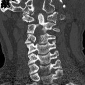
**Occipital Condyle Fracture**
*Coronal NECT shows a nondisplaced occipital condyle fracture as well as a C4 articular pillar fracture , reflecting lateral flexion injury.*
**Occipital Condyle Fracture**
*Coronal NECT shows a nondisplaced occipital condyle fracture as well as a C4 articular pillar fracture , reflecting lateral flexion injury.*
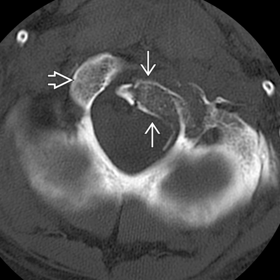
**Occipital Condyle Fracture**
*Axial CT shows a type 3 condyle fracture rotated into the foramen magnum. Note the normal position of the right condyle .*
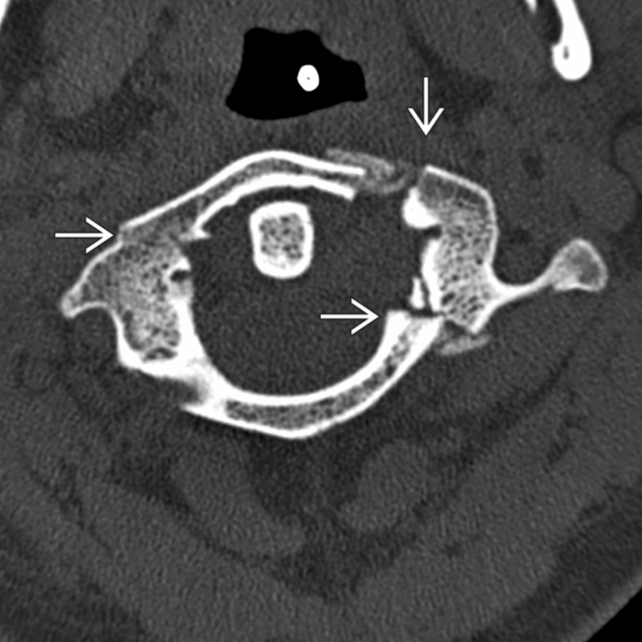
**Jefferson C1 Fracture**
*Axial bone CT shows multiple fractures of the C1 ring . Lateral displacement in this patient indicates rupture of the transverse ligament of dens and resultant instability.*
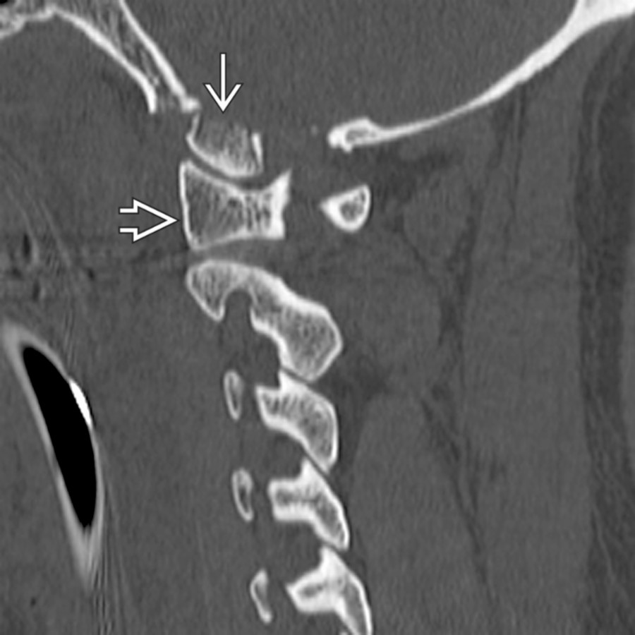
**Occipital Condyle Fracture**
*Parasagittal CT shows a condyle fracture that has maintained its relationship with the lateral mass of C1 despite the posterior displacement.*
**Odontoid C2 Fracture**
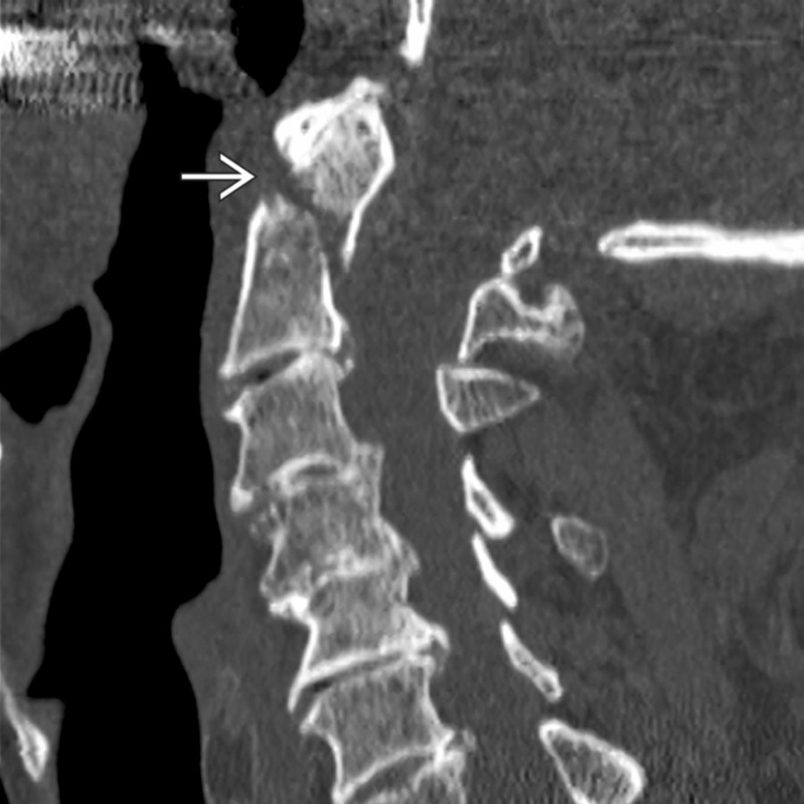
*Sagittal NECT shows a type 2 dens fracture in an osteoporotic patient. Soft tissue swelling is mild. These fractures are commonly subtle on radiographs and best seen on lateral (not odontoid) view. If there is any question, perform CT.*
**Odontoid C2 Fracture**
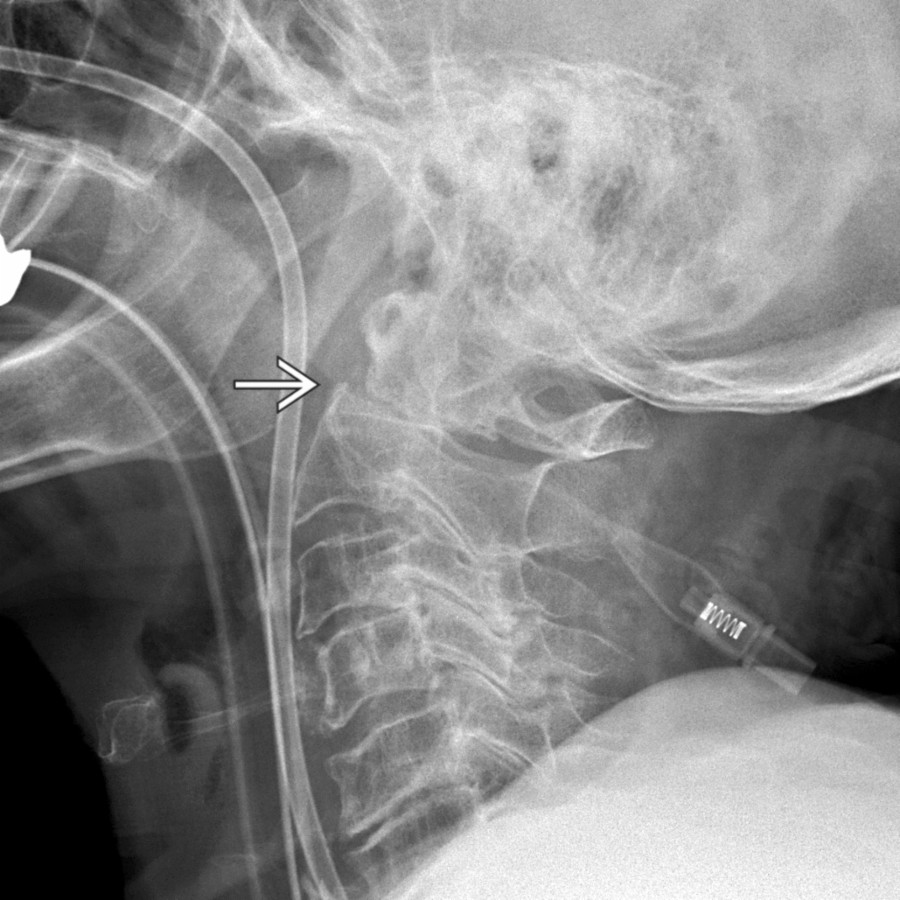
*Plain radiograph shows a fracture through the base of the odontoid , which is displaced posteriorly with slight extension.*
**Burst Fracture, C2**
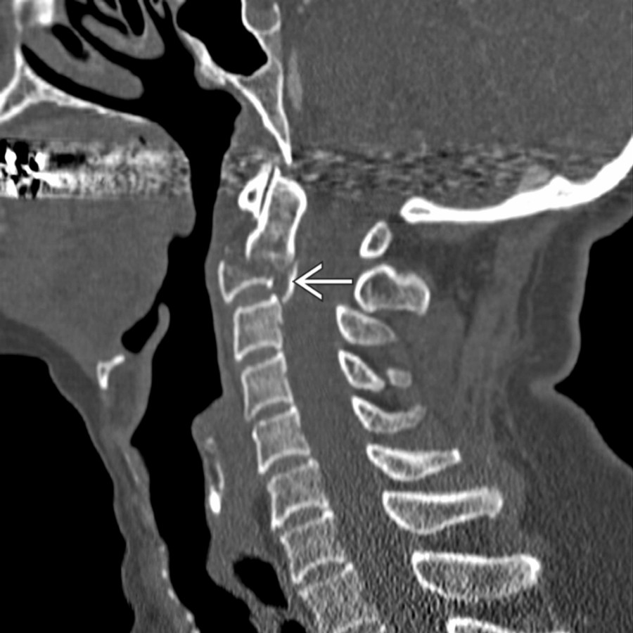
*Sagittal bone CT shows a comminuted C2 body fracture with characteristic retropulsion of the posterior cortex . Additional burst fractures are commonly present elsewhere in the spine.*
**Odontoid C2 Fracture**
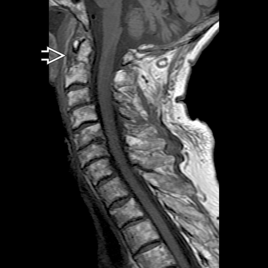
*Sagittal T1WI MR shows a well-defined fracture line . MR is useful for defining soft tissue abnormalities but should not be relied upon for pure bony fracture identification.*
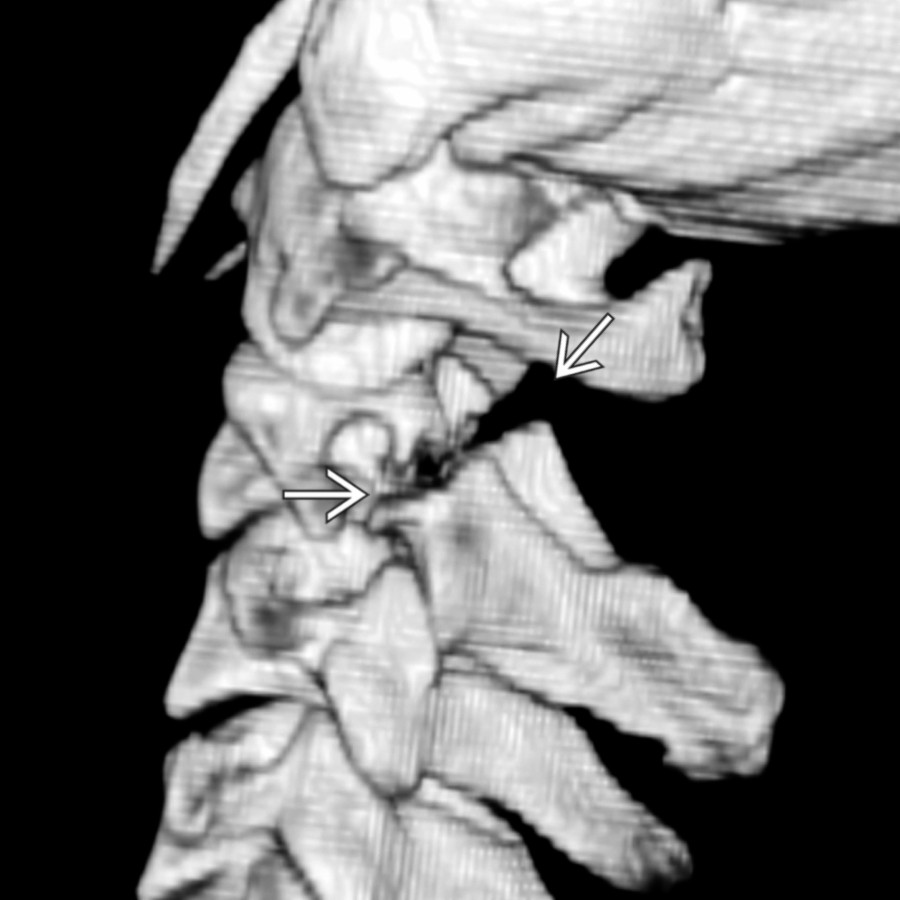
**Hangman's C2 Fracture**
*Sagittal oblique 3D CT shows bilateral C2 pedicle fractures without fracture of the vertebral body. Effendi classification uses the presence of disruption of the C2-C3 disc and facet joints as a measure of the severity of injury.*
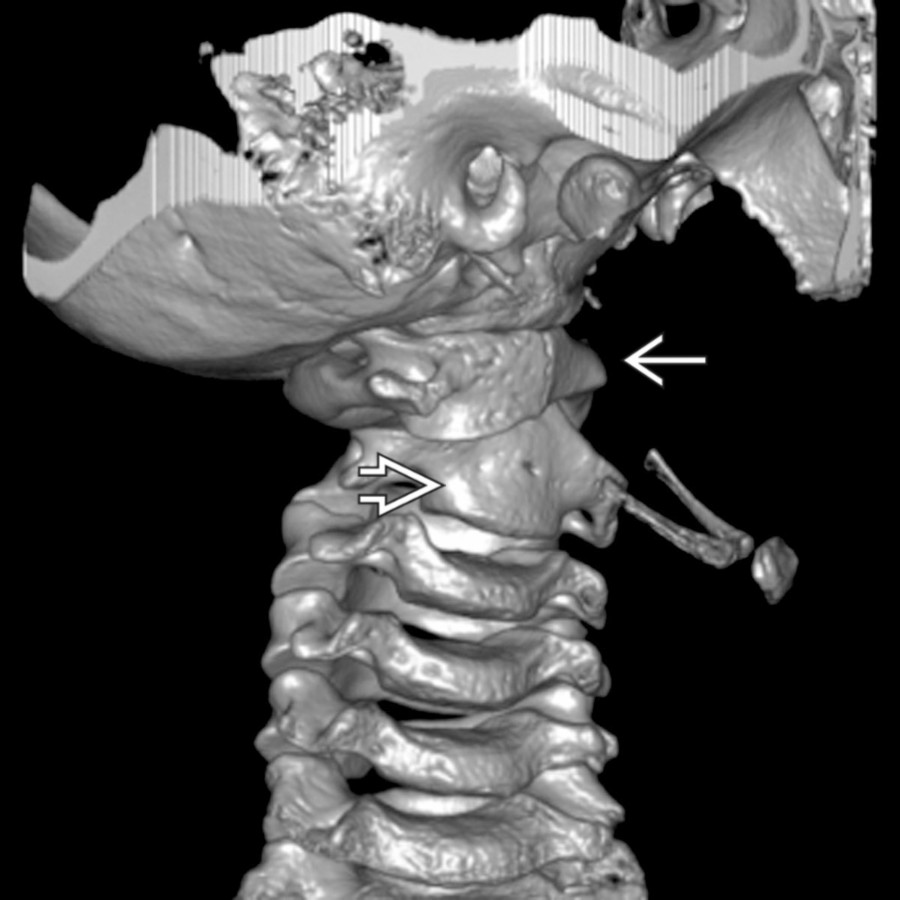
**Atlantoaxial Rotary Subluxation/Fixation**
*3D CT reconstruction visualizes the markedly left rotated C1 with respect to the mildly rotated C2 . Findings are typical of a Fielding-Hawkins type I atlantoaxial rotatory fixation.*
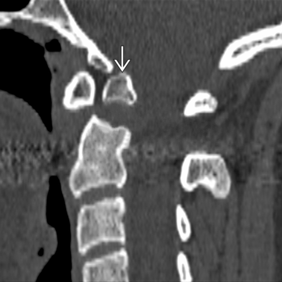
**Os Odontoideum**
*Sagittal bone CT shows a chronic, nonunited dens fracture , so-called os odontoideum. This may be unstable, and flexion-extension views should be performed.*
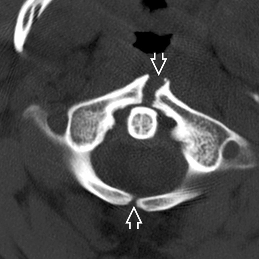
**Craniovertebral Junction Variants**
*Axial bone CT shows anterior and posterior clefts of C1. Smooth, corticated margins are signs distinguishing this from trauma.*
**Craniovertebral Junction Variants**
*Sagittal bone CT shows the anterior arch of C1 fused to the clivus and posterior arch fused to C2 .*
**Atlantooccipital Dislocation**
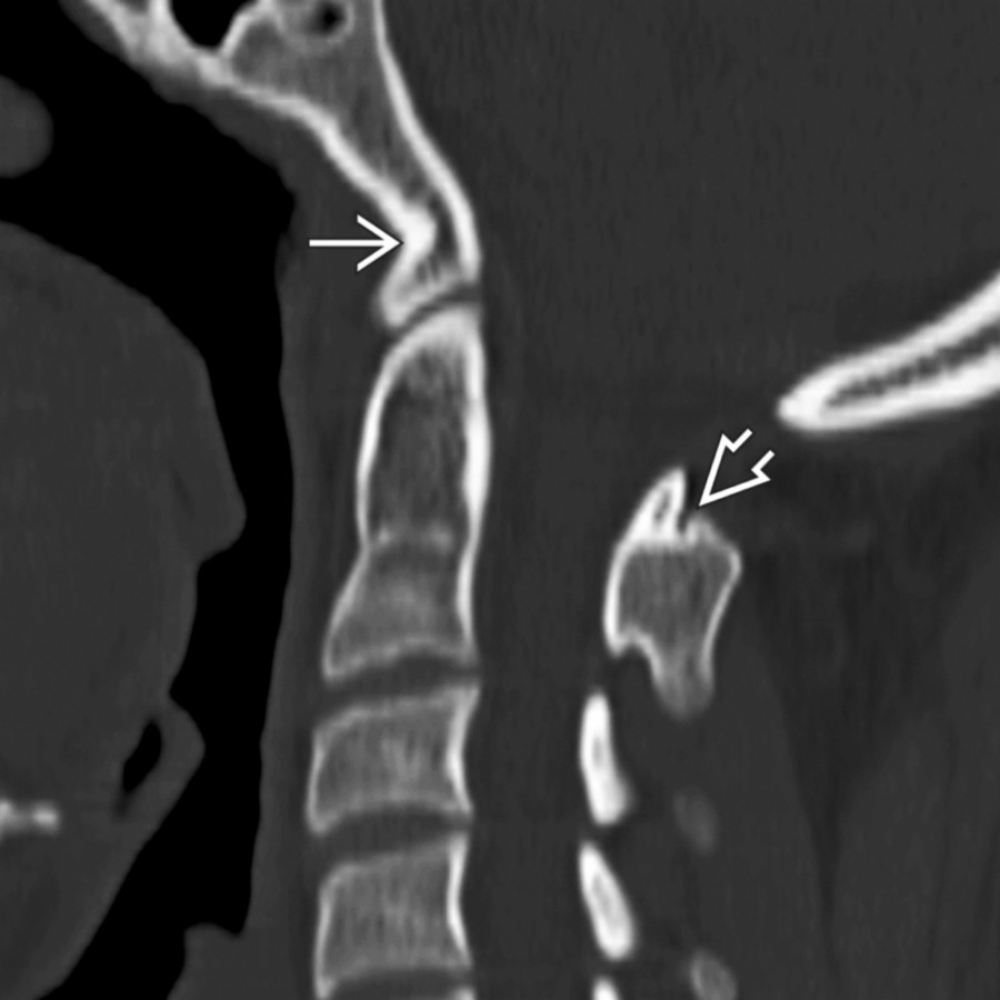
*Parasagittal CT shows posterior displacement of the lateral mass of C1 with respect to the occipital condyle . The normal condyle would be closely applied to the lateral mass of C1.*
**Occipital Condyle Fracture**
*Coronal NECT shows a nondisplaced occipital condyle fracture as well as a C4 articular pillar fracture , reflecting lateral flexion injury.*

**Occipital Condyle Fracture**
*Coronal NECT shows a nondisplaced occipital condyle fracture as well as a C4 articular pillar fracture , reflecting lateral flexion injury.*

**Occipital Condyle Fracture**
*Axial CT shows a type 3 condyle fracture rotated into the foramen magnum. Note the normal position of the right condyle .*

**Jefferson C1 Fracture**
*Axial bone CT shows multiple fractures of the C1 ring . Lateral displacement in this patient indicates rupture of the transverse ligament of dens and resultant instability.*

**Occipital Condyle Fracture**
*Parasagittal CT shows a condyle fracture that has maintained its relationship with the lateral mass of C1 despite the posterior displacement.*

**Odontoid C2 Fracture**
*Sagittal NECT shows a type 2 dens fracture in an osteoporotic patient. Soft tissue swelling is mild. These fractures are commonly subtle on radiographs and best seen on lateral (not odontoid) view. If there is any question, perform CT.*

**Odontoid C2 Fracture**
*Plain radiograph shows a fracture through the base of the odontoid , which is displaced posteriorly with slight extension.*

**Burst Fracture, C2**
*Sagittal bone CT shows a comminuted C2 body fracture with characteristic retropulsion of the posterior cortex . Additional burst fractures are commonly present elsewhere in the spine.*

**Odontoid C2 Fracture**
*Sagittal T1WI MR shows a well-defined fracture line . MR is useful for defining soft tissue abnormalities but should not be relied upon for pure bony fracture identification.*

**Hangman's C2 Fracture**
*Sagittal oblique 3D CT shows bilateral C2 pedicle fractures without fracture of the vertebral body. Effendi classification uses the presence of disruption of the C2-C3 disc and facet joints as a measure of the severity of injury.*

**Atlantoaxial Rotary Subluxation/Fixation**
*3D CT reconstruction visualizes the markedly left rotated C1 with respect to the mildly rotated C2 . Findings are typical of a Fielding-Hawkins type I atlantoaxial rotatory fixation.*

**Os Odontoideum**
*Sagittal bone CT shows a chronic, nonunited dens fracture , so-called os odontoideum. This may be unstable, and flexion-extension views should be performed.*

**Craniovertebral Junction Variants**
*Axial bone CT shows anterior and posterior clefts of C1. Smooth, corticated margins are signs distinguishing this from trauma.*

**Craniovertebral Junction Variants**
*Sagittal bone CT shows the anterior arch of C1 fused to the clivus and posterior arch fused to C2 .*

**Atlantooccipital Dislocation**
*Parasagittal CT shows posterior displacement of the lateral mass of C1 with respect to the occipital condyle . The normal condyle would be closely applied to the lateral mass of C1.*
 (i.e., abnormal Wackenheim clival line).](images/app.statdx.com_image_thumbnail_a1a12cf6-aa3c-46c9-a6a9-4d2a23636c49_annotated_true_size_900_quality_90_26665e78.jpg)
**Atlantooccipital Dislocation**
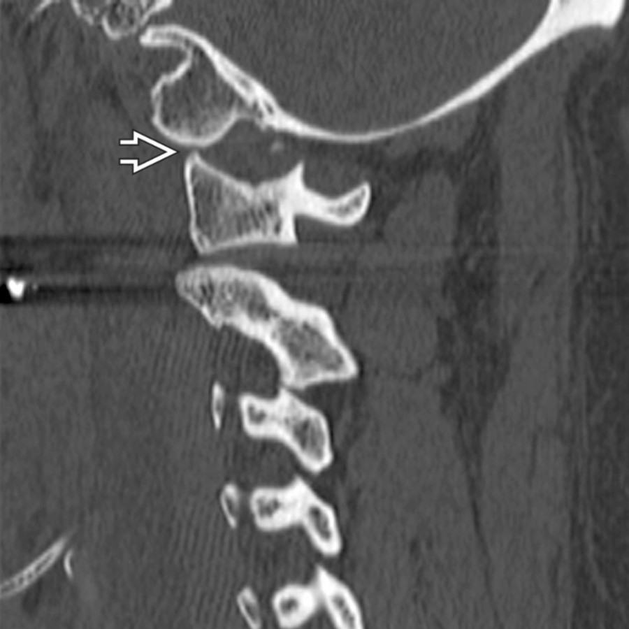
*Sagittal CT shows widening of the basion-dental interval and slight posterior displacement of the odontoid with respect to the foramen magnum (i.e., abnormal Wackenheim clival line).*
**Atlantooccipital Dislocation**
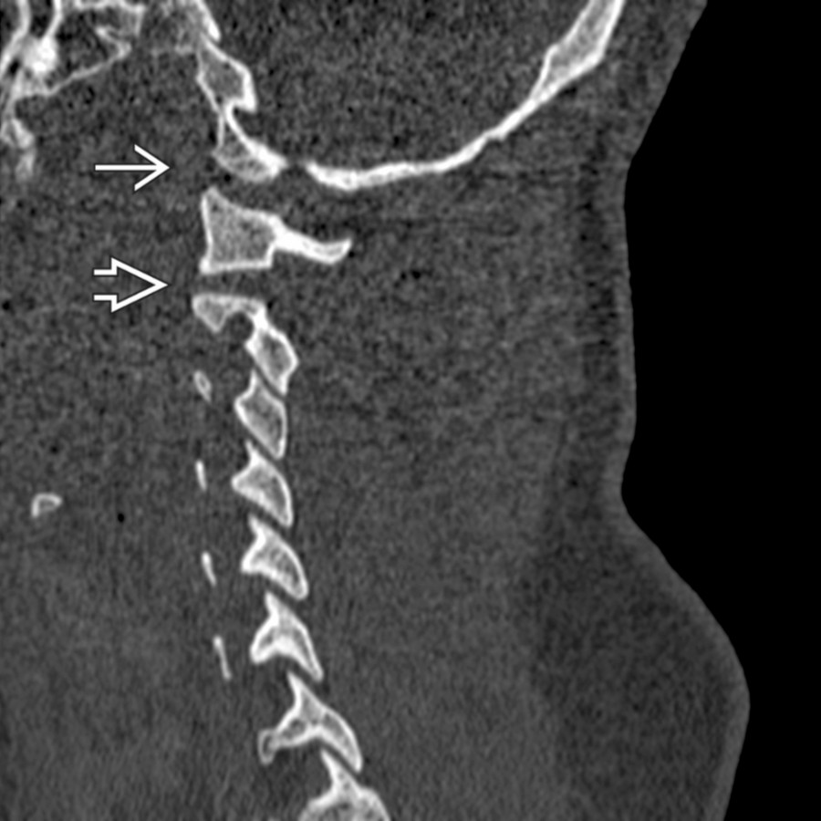
*Parasagittal CT shows slight widening of both C0-C1 and C1-C2 articulations in this patient with both atlantooccipital and atlantoaxial subluxations.*
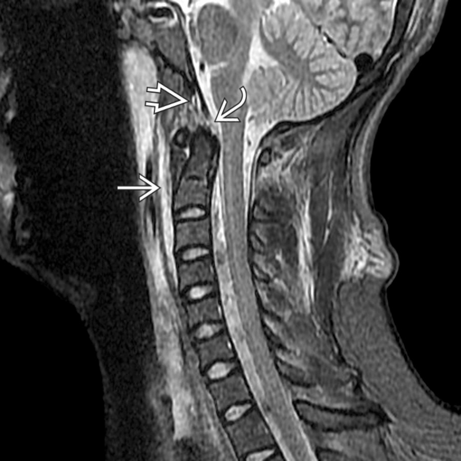
**Atlantooccipital Dislocation**
*Sagittal STIR MR shows diffuse prevertebral high signal from edema and a widened basion-dental interval . Note the disruption of the tectorial membrane .*
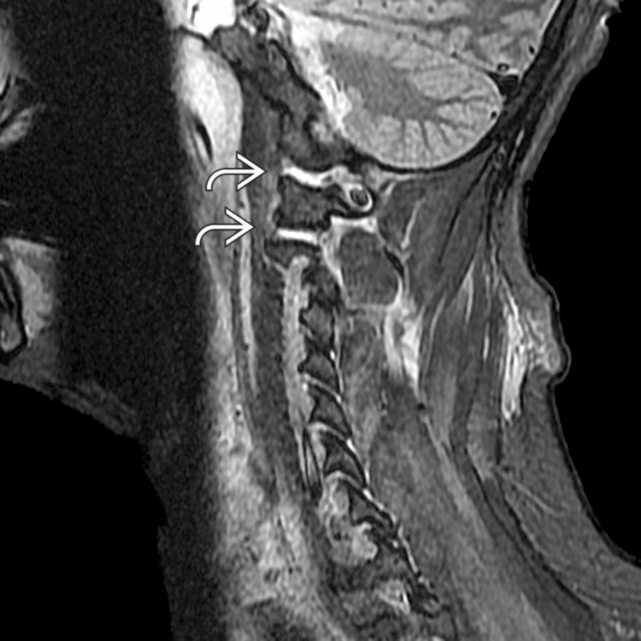
**Atlantoaxial Dislocation**
*Sagittal STIR MR shows abnormal hyperintensity within the C0-C1 and C1-C2 articulations due to simultaneous atlantooccipital and atlantoaxial subluxations. The 2-level abnormality is important to identify since the posterior fusion will be extended more inferiorly.*
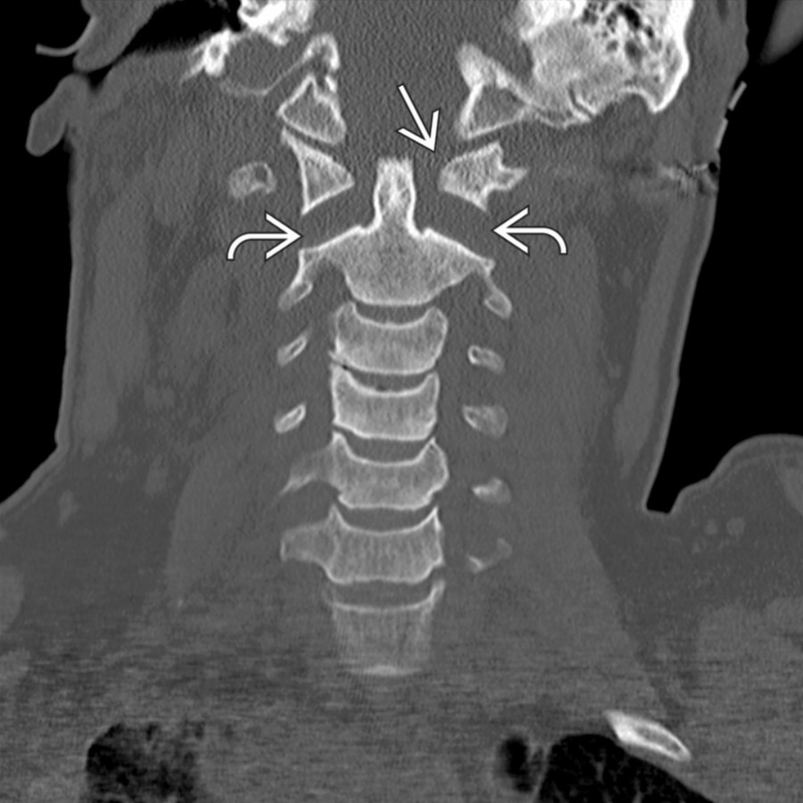
**Atlantoaxial Dislocation**
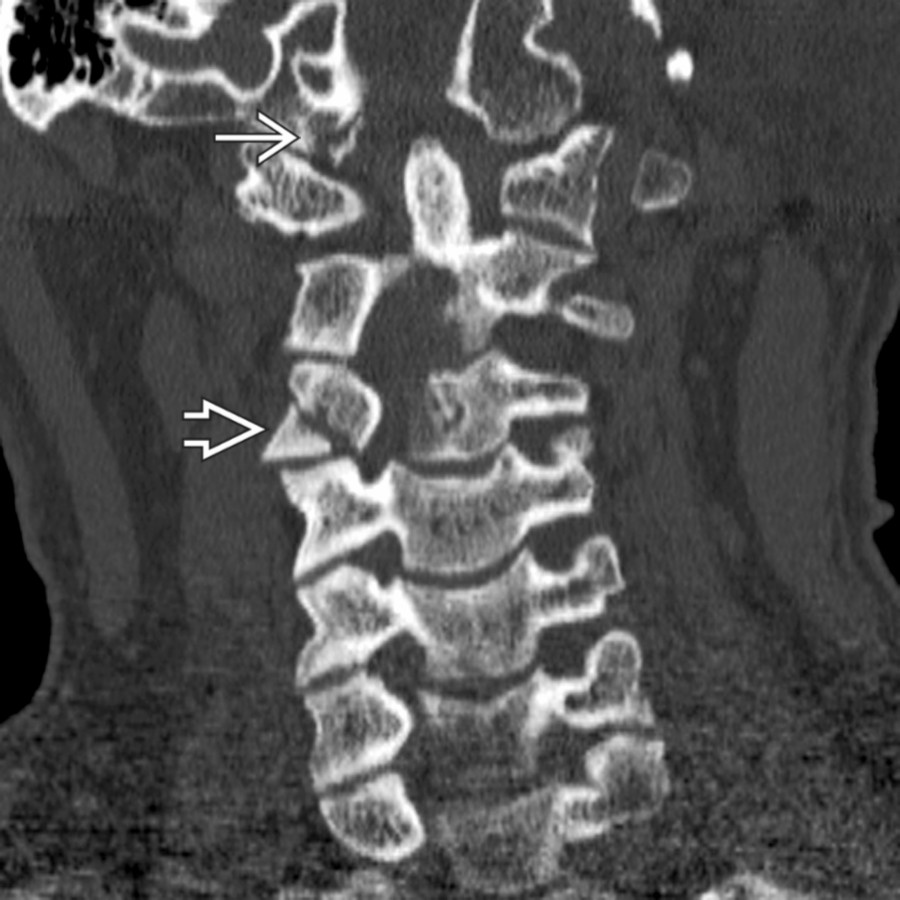
*Coronal CT shows the disrupted spinal articulations at C1-C2 (atlantoaxial dissociation). There is widening of the atlantoodontoid articulation and widening/dislocation of the lateral masses of C1 and C2 .*
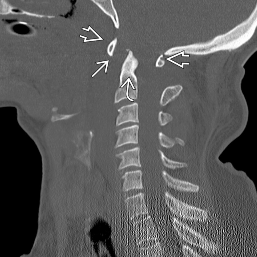
**Atlantoaxial Dislocation**
*Sagittal CT shows gross widening of the atlantoodontoid articulation . The ring of C1 has maintained position with respect to the foramen magnum , but C2 is displaced posteriorly .*
### Additional Images
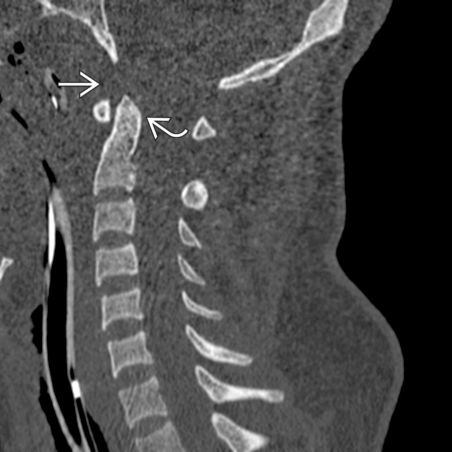
**Atlantooccipital Dislocation**
*Sagittal CT shows widening of the basion-dental interval and slight posterior displacement of the odontoid with respect to the foramen magnum (i.e., abnormal Wackenheim clival line).*

**Atlantooccipital Dislocation**
*Parasagittal CT shows slight widening of both C0-C1 and C1-C2 articulations in this patient with both atlantooccipital and atlantoaxial subluxations.*

**Atlantooccipital Dislocation**
*Sagittal STIR MR shows diffuse prevertebral high signal from edema and a widened basion-dental interval . Note the disruption of the tectorial membrane .*

**Atlantoaxial Dislocation**
*Sagittal STIR MR shows abnormal hyperintensity within the C0-C1 and C1-C2 articulations due to simultaneous atlantooccipital and atlantoaxial subluxations. The 2-level abnormality is important to identify since the posterior fusion will be extended more inferiorly.*

**Atlantoaxial Dislocation**
*Coronal CT shows the disrupted spinal articulations at C1-C2 (atlantoaxial dissociation). There is widening of the atlantoodontoid articulation and widening/dislocation of the lateral masses of C1 and C2 .*

**Atlantoaxial Dislocation**
*Sagittal CT shows gross widening of the atlantoodontoid articulation . The ring of C1 has maintained position with respect to the foramen magnum , but C2 is displaced posteriorly .*
### Additional Images
 widely separated from lateral mass of C1 , indicating longitudinal atlantooccipital dislocation. Dislocation may also be anterior or posterior in direction.](images/app.statdx.com_image_thumbnail_63bcd917-55ed-466c-b898-55064cf3cd8a_annotated_true_size_900_quality_90_234d1fdf.jpg)
**Atlantooccipital Dislocation**
*Sagittal bone CT shows occipital condyle widely separated from lateral mass of C1 , indicating longitudinal atlantooccipital dislocation. Dislocation may also be anterior or posterior in direction.*
widely separated from lateral mass of C1 , indicating longitudinal atlantooccipital dislocation. Dislocation may also be anterior or posterior in direction.](images/app.statdx.com_image_thumbnail_63bcd917-55ed-466c-b898-55064cf3cd8a_annotated_true_size_900_quality_90_234d1fdf.jpg)
**Atlantooccipital Dislocation**
*Sagittal bone CT shows occipital condyle widely separated from lateral mass of C1 , indicating longitudinal atlantooccipital dislocation. Dislocation may also be anterior or posterior in direction.*