---
title: "Craniopharyngioma"
docid: "00e66680-6731-4287-b5a1-3f0b3f09053b"
authors:
- key: "8d5254e9-8dda-478b-8f08-bdee97a32c79"
value: "Karen L. Salzman, MD, FACR"
breadcrumbs:
-
name: "Brain"
slug: "brain"
treeNodeId: "6d8829f1-14d7-45af-8675-255189aa526a"
-
name: "Diagnosis"
slug: "diagnosis"
treeNodeId: "51c00394-446e-4a38-94af-d3b1d14d34e8"
-
name: "Anatomy-Based Diagnoses"
slug: "anatomy-based-diagnoses"
treeNodeId: "529d3e33-f508-498c-bc70-cf962e81e629"
-
name: "Sella and Pituitary"
slug: "sella-and-pituitary"
treeNodeId: "9afaeeb6-661c-49be-b55f-5bdc1c98a53e"
-
name: "Neoplasms"
slug: "neoplasms"
treeNodeId: "87c53ba9-d217-4dc9-be90-15cfe4d7766c"
-
name: "Craniopharyngioma"
slug: "craniopharyngioma"
treeNodeId: null
category: "Brain"
documentVersionId: "fba662fa-55e8-45a2-8081-e59d71a73b51"
imageCount: 19
lastUpdated: "08/10/20"
pageDescription: "Craniopharyngioma"
pageKeywords: "Brain, Diagnosis, Anatomy-Based Diagnoses, Sella and Pituitary, Neoplasms, Craniopharyngioma"
pageTitle: "Craniopharyngioma | STATdx"
enhancedTitle: "Craniopharyngioma"
type: "DX"
references: true
breadcrumbs:
- "Brain"
- "Diagnosis"
- "Anatomy-Based Diagnoses"
- "Sella and Pituitary"
- "Neoplasms"
- "Craniopharyngioma"
---
# KEY FACTS
- ## Terminology
- Benign, partially cystic sellar region tumor derived from remnants of craniopharyngeal duct/Rathke pouch epithelium
- 2 types
- Adamantinomatous (cystic mass in childhood)
- Papillary (solid mass in older adults)
- ## Imaging
- General features
- Multilobulated, often large (> 5 cm)
- Occasionally giant, multicompartmental
- CT: Cystic (90%), Ca⁺⁺ (90%), enhancing (90%)
- MR: Signal varies with cyst contents
- Cysts variably hyperintense on T1WI and T2WI
- Solid portions enhance heterogeneously; cyst walls enhance strongly
- Cyst contents show broad lipid peak (0.9-1.5 ppm) on MR spectroscopy
- ## Pathology
- Most common pediatric intracranial tumor of nonglial origin
- WHO grade 1
- ## Clinical Issues
- Bimodal age distribution
- Peak 5-15 years; adults 45-60 years (commonly papillary)
- Pediatric patient with morning headache, visual defect, short stature
- Endocrine disturbances include growth hormone (GH) deficiency, luteinizing hormone (LH)/follicle-stimulating hormone (FSH) deficiency
- Others = hypothyroidism > adrenal failure > diabetes insipidus
- Surgical resection is primary therapy
- Surgery, radiation therapy, or cyst aspiration for recurrent tumors
# TERMINOLOGY
- ## Abbreviations
- Craniopharyngioma (CP)
- ## Synonyms
- Craniopharyngeal duct tumor, Rathke pouch tumor, adamantinoma
- ## Definitions
- Benign, partially cystic sellar region tumor derived from Rathke pouch epithelium
- 2 histologies: Adamantinomatous and papillary
# IMAGING
- ## General Features
- ### Best diagnostic clue
- CT: Partially Ca⁺⁺ mixed solid/cystic suprasellar mass in child
- MR: Complex signal intensity suprasellar mass
- ### Location
- Surgical division of CPs into 3 groups
- Sellar
- Prechiasmatic
- Retrochiasmatic
- Imaging locations of CPs (adamantinomatous type)
- Suprasellar (75%)
- Suprasellar + intrasellar component (21%)
- Entirely intrasellar (4%)
- Often extends into multiple cranial fossae: Anterior (30%), middle (23%), posterior, &/or retroclival (20%)
- Rare ectopic locations
- Optic chiasm, 3rd ventricle
- Other: Nasopharynx, paranasal sinuses, pineal gland, sphenoid (clivus), cerebellopontine angle
- ### Size
- Variable; often large at presentation (> 5 cm)
- Occasionally giant, multicompartmental
- ### Morphology
- Multilobulated, multicystic
- ## CT Findings
- ### NECT
- Adamantinomatous type (90% rule)
- 90% mixed solid (isodense), cystic (hypodense)
- 90% calcify
- 90% enhance (solid = nodule; rim = capsule)
- Papillary type: Often solid, isodense, rarely calcifies
- ## MR Findings
- ### T1WI
- Signal varies with cyst contents
- Short T1 due to high protein content
- Classic (adamantinomatous type)
- Hyperintense cyst + heterogeneous nodule
- Less common (papillary type)
- Isointense solid component
- ### T2WI
- Cysts are variably hyperintense
- Solid component = heterogeneous (iso-/hyperintense, Ca⁺⁺ portions hypointense)
- Hyperintense signal in brain parenchyma adjacent to tumor may indicate
- Gliosis, tumor invasion, irritation from leaking cyst fluid
- Edema from compression of optic chiasm/tracts
- Hypointense T2* = Ca⁺⁺
- ### FLAIR
- Cyst contents typically hyperintense
- ### DWI
- Variable depending upon character of cyst fluid
- ### T1WI C+
- Solid portions enhance heterogeneously; cyst walls enhance strongly
- ### MRA
- Vascular displacement &/or encasement
- ### MRS
- Cyst contents show broad lipid spectrum (0.9-1.5 ppm)
- ## Imaging Recommendations
- ### Best imaging tool
- MR with thin sagittal, coronal sequences
- ### Protocol advice
- Pre-/postcontrast T1WI, T2, FLAIR, GRE, DWI, MRS
# DIFFERENTIAL DIAGNOSIS
- [Rathke Cleft Cyst](/document/rathke-cleft-cyst/8f1561f7-92a7-485c-a0ae-2e2d5c8c1628)
- Noncalcified, less heterogeneous
- Look for intracystic nodule on T2
- Does not enhance
- Claw sign (enhancing pituitary draped around cyst)
- Small Rathke cleft cyst (RCC) may be indistinguishable from rare intrasellar CP
- RCCs express CK8 and CK20 (CPs generally do not)
- ## Suprasellar Arachnoid Cyst
- No Ca⁺⁺, enhancement
- ## Hypothalamic/Chiasmatic Astrocytoma
- Solid or with small cystic/necrotic components
- Ca⁺⁺ is rare; robust enhancement is common
- ## Pituitary Adenoma
- Rare in prepubescent children
- Isointense with brain
- Enhances strongly
- Can mimic CP when cystic and hemorrhagic
- ## Epidermoid/Dermoid Tumors
- Minimal or no enhancement
- ## Thrombosed Aneurysm
- Contains blood products; use SWI
- Look for residual patent lumen, phase artifact
- [Germinoma or Mixed Germ Cell Tumor With Cystic Component(s)](/document/germinoma/078b68a2-67de-457e-818a-63655cec95aa)
- Cerebrospinal fluid spread is common, Ca⁺⁺ is rare
# PATHOLOGY
- ## General Features
- ### Etiology
- 2 proposed theories
- CPs arise from remnants of craniopharyngeal duct and Rathke pouch epithelium
- CPs arise from squamous epithelial cells in pars tuberalis of adenohypophysis
- ### Genetics
- No known genetic susceptibility (rare reports of siblings, parent-child)
- Small subset of CPs are monoclonal tumors that arise from oncogenes at specific loci
- Adamantinomatous: *CTNNB1* mutations and aberrant nuclear expression of β-catenin in up to 95% of cases
- Papillary:*BRAF* V600E mutations in 81-95% of cases
- ## Staging, Grading, & Classification
- WHO grade 1
- MIB-1 labeling index > 7% predicts recurrence
- ## Gross Pathologic & Surgical Features
- Solid tumor with variable cysts
- Adamantinomatous cysts often contain thick "crankcase oil" fluid
- Epithelial fronds penetrate adjacent hypothalamus/chiasm
- ## Microscopic Features
- Adamantinomatous (mostly pediatric)
- Multistratified squamous epithelium with nuclear palisading
- Nodules of "wet" keratin
- Dystrophic Ca⁺⁺
- Papillary (mostly adults)
- Sheets of squamous epithelium form pseudopapillae
- Villous fibrovascular stroma
- Malignant transformation, distant metastases rare
- May occur with varied histologies, resulting in poor prognosis
# CLINICAL ISSUES
- ## Presentation
- ### Most common signs/symptoms
- Symptoms vary with location, size of tumor, age of patient
- Visual disturbances (60-85%)
- Bitemporal hemianopsia
- ### Other signs/symptoms
- Endocrine disturbances (52-87%)
- Growth hormone (GH) deficiency (75%) > luteinizing hormone (LH)/follicle-stimulating hormone (FSH) deficiency > hypothyroidism > adrenal failure > diabetes insipidus
- Headaches
- Cognitive impairment (~ 50%)
- ### Clinical profile
- Pediatric patient with morning headache, visual defect, short stature
- ## Demographics
- ### Age
- Bimodal distribution (peak 5-15 years, with smaller peak 45-60 years)
- Papillary CP: 40-55 years
- ### Sex
- M = F
- ### Ethnicity
- More common in Japanese children
- ### Epidemiology
- Most common pediatric intracranial tumor of nonglial origin
- Comprise 1.2-4.6% of all intracranial tumors across all ages
- 6-11% of all pediatric intracranial tumors
- Incidence = 0.5-2.5 new cases per 1 million per year
- ~ 54% of all pediatric sellar/chiasmatic region tumors are CPs
- ## Natural History & Prognosis
- Typically slow-growing benign neoplasm
- Prognosis based upon size, extent of tumor at presentation
- < 5 cm, recurrence rate: 20%
- > 5 cm, recurrence rate: 83%
- Overall 10-year survival: 64-96%
- ## Treatment
- Methods of primary treatment
- Radical surgery = gross total resection
- Complications = hypothalamic injury, endocrine symptoms, vasa vasorum injury, and pseudoaneurysm
- Surgery may occur via craniotomy, transnasal, transorbital, or endoscopic routes
- Less invasive surgery = subtotal resection + radiation therapy
- Biopsy, cyst drainage, and radiation therapy
- Treatment for residual or recurrent tumor
- Surgery, radiation therapy, or cyst aspiration
- Cyst instillation with intracavitary radioisotopes, bleomycin, or other sclerosing agents
# DIAGNOSTIC CHECKLIST
- ## Consider
- Preoperative ophthalmologic and endocrine evaluations
- ## Image Interpretation Pearls
- Use NECT to detect Ca⁺⁺ if MR diagnosis is in question
- Adamantinomatous CP = 90% rule (90% cystic, calcified, enhancing)
- Papillary CP is typically solid and primarily adult neoplasm
c497473c-5835-4221-bfa1-0d2be04bee73
## References
# Selected References
1. [Azuma M et al: Usefulness of contrast-enhanced 3D-FLAIR MR imaging for differentiating Rathke cleft cyst from cystic craniopharyngioma. AJNR Am J Neuroradiol. 41(1):106-10, 2020](http://www.ncbi.nlm.nih.gov/pubmed/?term=31857323%5Bpmid%5D)
1. [Fouda MA et al: Sixty years single institutional experience with pediatric craniopharyngioma: between the past and the future. Childs Nerv Syst. 36(2):291-6, 2020](http://www.ncbi.nlm.nih.gov/pubmed/?term=31292757%5Bpmid%5D)
1. [Goldman S et al: Phase II study of peginterferon alpha-2b for patients with unresectable or recurrent craniopharyngiomas: a Pediatric Brain Tumor Consortium report. Neuro Oncol. ePub, 2020](http://www.ncbi.nlm.nih.gov/pubmed/?term=32393959%5Bpmid%5D)
1. [Prince E et al: Transcriptional analyses of adult and pediatric adamantinomatous craniopharyngioma reveals similar expression signatures regarding potential therapeutic targets. Acta Neuropathol Commun. 8(1):68, 2020](http://www.ncbi.nlm.nih.gov/pubmed/?term=32404202%5Bpmid%5D)
1. [Sadashivam S et al: Adult craniopharyngioma: the role of extent of resection in tumor recurrence and long-term functional outcome. Clin Neurol Neurosurg. 192:105711, 2020](http://www.ncbi.nlm.nih.gov/pubmed/?term=32036264%5Bpmid%5D)
1. [Soldozy S et al: Endoscopic endonasal surgery outcomes for pediatric craniopharyngioma: a systematic review. Neurosurg Focus. 48(1):E6, 2020](http://www.ncbi.nlm.nih.gov/pubmed/?term=31896083%5Bpmid%5D)
1. [Drapeau A et al: Pediatric craniopharyngioma. Childs Nerv Syst. 35(11):2133-45, 2019](http://www.ncbi.nlm.nih.gov/pubmed/?term=31385085%5Bpmid%5D)
1. [Madsen PJ et al: Endoscopic endonasal resection versus open surgery for pediatric craniopharyngioma: comparison of outcomes and complications. J Neurosurg Pediatr. 1-10, 2019](http://www.ncbi.nlm.nih.gov/pubmed/?term=31174192%5Bpmid%5D)
1. [Marcus HJ et al: Craniopharyngioma in children: trends from a third consecutive single-center cohort study. J Neurosurg Pediatr. 1-9, 2019](http://www.ncbi.nlm.nih.gov/pubmed/?term=31860822%5Bpmid%5D)
1. [Whelan R et al: Interrater reliability of a method to assess hypothalamic involvement in pediatric adamantinomatous craniopharyngioma. J Neurosurg Pediatr. 1-6, 2019](http://www.ncbi.nlm.nih.gov/pubmed/?term=31604324%5Bpmid%5D)
1. Buslei et al: Craniopharyngioma. In Louis DN et al: WHO Classification of Tumors of the Central Nervous System. IARC. 324-8, 2016
1. [Greenfield BJ et al: Long-term disease control and toxicity outcomes following surgery and intensity modulated radiation therapy (IMRT) in pediatric craniopharyngioma. Radiother Oncol. 114(2):224-9, 2015](http://www.ncbi.nlm.nih.gov/pubmed/?term=25542650%5Bpmid%5D)
1. [Kim JH et al: BRAF V600E mutation is a useful marker for differentiating Rathke's cleft cyst with squamous metaplasia from papillary craniopharyngioma. J Neurooncol. 123(1):189-91, 2015](http://www.ncbi.nlm.nih.gov/pubmed/?term=25820214%5Bpmid%5D)
1. [Lee HJ et al: Pretreatment diagnosis of suprasellar papillary craniopharyngioma and germ cell tumors of adult patients. AJNR Am J Neuroradiol. 36(3):508-17, 2015](http://www.ncbi.nlm.nih.gov/pubmed/?term=25339645%5Bpmid%5D)
1. [Sterkenburg AS et al: Survival, hypothalamic obesity, and neuropsychological/psychosocial status after childhood-onset craniopharyngioma: newly reported long-term outcomes. Neuro Oncol. 17(7):1029-38, 2015](http://www.ncbi.nlm.nih.gov/pubmed/?term=25838139%5Bpmid%5D)
1. [Lee CC et al: Gamma Knife surgery for craniopharyngioma: report on a 20-year experience. J Neurosurg. 121 Suppl:167-78, 2014](http://www.ncbi.nlm.nih.gov/pubmed/?term=25434950%5Bpmid%5D)
1. [Clark AJ et al: A systematic review of the results of surgery and radiotherapy on tumor control for pediatric craniopharyngioma. Childs Nerv Syst. 29(2):231-8, 2013](http://www.ncbi.nlm.nih.gov/pubmed/?term=23089933%5Bpmid%5D)
1. [Müller HL: Childhood craniopharyngioma. Pituitary. 16(1):56-67, 2013](http://www.ncbi.nlm.nih.gov/pubmed/?term=22678820%5Bpmid%5D)
1. [Chentli F et al: Congenital craniopharyngioma: a case report and literature review. J Pediatr Endocrinol Metab. 25(11-12):1181-3, 2012](http://www.ncbi.nlm.nih.gov/pubmed/?term=23329768%5Bpmid%5D)
1. [Clark AJ et al: Treatment-related morbidity and the management of pediatric craniopharyngioma: a systematic review. J Neurosurg Pediatr. 10(4):293-301, 2012](http://www.ncbi.nlm.nih.gov/pubmed/?term=22920295%5Bpmid%5D)
1. [İnci MF et al: A rare presentation of craniopharyngioma: delayed puberty. BMJ Case Rep. 2012, 2012](http://www.ncbi.nlm.nih.gov/pubmed/?term=23195827%5Bpmid%5D)
1. [Shi Z et al: Transient enlargement of craniopharyngioma after radiation therapy: pattern of magnetic resonance imaging response following radiation. J Neurooncol. 109(2):349-55, 2012](http://www.ncbi.nlm.nih.gov/pubmed/?term=22692563%5Bpmid%5D)
1. [Boongird A et al: Malignant craniopharyngioma; case report and review of the literature. Neuropathology. 29(5):591-6, 2009](http://www.ncbi.nlm.nih.gov/pubmed/?term=19077042%5Bpmid%5D)
1. [Frangou EM et al: Metastatic craniopharyngioma: case report and literature review. Childs Nerv Syst. 25(9):1143-7, 2009](http://www.ncbi.nlm.nih.gov/pubmed/?term=19517118%5Bpmid%5D)
1. [Keil MF et al: Pituitary tumors in childhood: update of diagnosis, treatment and molecular genetics. Expert Rev Neurother. 8(4):563-74, 2008](http://www.ncbi.nlm.nih.gov/pubmed/?term=18416659%5Bpmid%5D)
1. [Garrè ML et al: Craniopharyngioma: modern concepts in pathogenesis and treatment. Curr Opin Pediatr. 19(4):471-9, 2007](http://www.ncbi.nlm.nih.gov/pubmed/?term=17630614%5Bpmid%5D)
1. [Powers CJ et al: Cerebellopontine angle craniopharyngioma: case report and literature review. Pediatr Neurosurg. 43(2):158-63, 2007](http://www.ncbi.nlm.nih.gov/pubmed/?term=17337933%5Bpmid%5D)
1. [Rodriguez FJ et al: The spectrum of malignancy in craniopharyngioma. Am J Surg Pathol. 31(7):1020-8, 2007](http://www.ncbi.nlm.nih.gov/pubmed/?term=17592268%5Bpmid%5D)
1. [Shuman AG et al: Extracranial nasopharyngeal craniopharyngioma: case report. Neurosurgery. 60(4):E780-1; discussion E781, 2007](http://www.ncbi.nlm.nih.gov/pubmed/?term=17415187%5Bpmid%5D)
1. [Aquilina K et al: Primary cerebellopontine angle craniopharyngioma in a patient with gardner syndrome. Case report and review of the literature. J Neurosurg. 105(2):330-3, 2006](http://www.ncbi.nlm.nih.gov/pubmed/?term=17219843%5Bpmid%5D)
1. [Haupt R et al: Epidemiological aspects of craniopharyngioma. J Pediatr Endocrinol Metab. 19 Suppl 1:289-93, 2006](http://www.ncbi.nlm.nih.gov/pubmed/?term=16700303%5Bpmid%5D)
1. [Prabhu VC et al: The pathogenesis of craniopharyngiomas. Childs Nerv Syst. 21(8-9):622-7, 2005](http://www.ncbi.nlm.nih.gov/pubmed/?term=15965669%5Bpmid%5D)
1. [Wang KC et al: Origin of craniopharyngiomas: implication on the growth pattern. Childs Nerv Syst. 21(8-9):628-34, 2005](http://www.ncbi.nlm.nih.gov/pubmed/?term=16059733%5Bpmid%5D)
1. [Srinivasan S et al: Features of the metabolic syndrome after childhood craniopharyngioma. J Clin Endocrinol Metab. 89(1):81-6, 2004](http://www.ncbi.nlm.nih.gov/pubmed/?term=14715831%5Bpmid%5D)
1. [Behari S et al: Intrinsic third ventricular craniopharyngiomas: report on six cases and a review of the literature. Surg Neurol. 60(3):245-52; discussion 252-3, 2003](http://www.ncbi.nlm.nih.gov/pubmed/?term=12922045%5Bpmid%5D)
1. [Saeki N et al: MR imaging study of edema-like change along the optic tract in patients with pituitary region tumors. AJNR Am J Neuroradiol. 24(3):336-42, 2003](http://www.ncbi.nlm.nih.gov/pubmed/?term=12637278%5Bpmid%5D)
1. [Barajas MA et al: Multimodal management of craniopharyngiomas: neuroendoscopy, microsurgery, and radiosurgery. J Neurosurg. 97(5 Suppl):607-9, 2002](http://www.ncbi.nlm.nih.gov/pubmed/?term=12507105%5Bpmid%5D)
1. [Fujimoto Y et al: Craniopharyngioma involving the infrasellar region: a case report and review of the literature. Pediatr Neurosurg. 37(4):210-6, 2002](http://www.ncbi.nlm.nih.gov/pubmed/?term=12372916%5Bpmid%5D)
1. [Green AL et al: Craniopharyngioma in a mother and daughter. Acta Neurochir (Wien). 144(4):403-4, 2002](http://www.ncbi.nlm.nih.gov/pubmed/?term=12021891%5Bpmid%5D)
1. [Sekine S et al: Craniopharyngiomas of adamantinomatous type harbor beta-catenin gene mutations. Am J Pathol. 161(6):1997-2001, 2002](http://www.ncbi.nlm.nih.gov/pubmed/?term=12466115%5Bpmid%5D)
1. [Van Effenterre R et al: Craniopharyngioma in adults and children: a study of 122 surgical cases. J Neurosurg. 97(1):3-11, 2002](http://www.ncbi.nlm.nih.gov/pubmed/?term=12134929%5Bpmid%5D)
1. [Chen CJ: Suprasellar and infrasellar craniopharyngioma with a persistent craniopharyngeal canal: case report and review of the literature. Neuroradiology. 43(9):760-2, 2001](http://www.ncbi.nlm.nih.gov/pubmed/?term=11594427%5Bpmid%5D)
## Images
### Selected Images
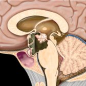
*Sagittal graphic shows a predominantly cystic, partially solid, suprasellar mass with focal rim Ca⁺⁺. Note the small intrasellar component and fluid-fluid level. Craniopharyngiomas are the 90% tumors (90% cystic, 90% Ca⁺⁺, and 90% enhancing).*
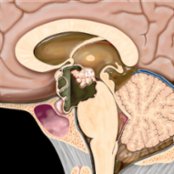
*Sagittal graphic shows a predominantly cystic, partially solid, suprasellar mass with focal rim Ca⁺⁺. Note the small intrasellar component and fluid-fluid level. Craniopharyngiomas are the 90% tumors (90% cystic, 90% Ca⁺⁺, and 90% enhancing).*
 with anterior extension into the sphenoid sinus
  and superior displacement of the optic chiasm.](images/app.statdx.com_image_thumbnail_ff36ded9-e912-42b4-929c-ef63a5e8fa57_size_168_quality_85_f43afdfe_20251014T205219Z.jpg)
*Sagittal T2 MR in a 9 year old with headache and visual changes shows a heterogeneous cystic and solid sellar and suprasellar craniopharyngioma with anterior extension into the sphenoid sinus and superior displacement of the optic chiasm.*
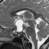
*Sagittal T2 MR in a 9 year old with headache and visual changes shows a heterogeneous cystic and solid sellar and suprasellar craniopharyngioma with anterior extension into the sphenoid sinus and superior displacement of the optic chiasm.*
 with a focal T2-hypointense nodule
  . The T2 imaging mimics a Rathke cleft cyst.](images/app.statdx.com_image_thumbnail_a46de544-a40b-46b9-bd8e-dfba34730496_size_168_quality_85_27db027b_20251014T205219Z.jpg)
*Coronal T2 MR in a 48-year-old man who presented with visual changes shows a cystic suprasellar mass with a focal T2-hypointense nodule . The T2 imaging mimics a Rathke cleft cyst.*
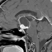
*Sagittal T1 C+ MR in a 45-year-old woman shows a cystic and solid mass with an enhancing portion , which distinguishes this cystic lesion as a craniopharyngioma, not a Rathke cleft cyst. Surgical resection is the primary therapy for this WHO grade 1 neoplasm. However, the recurrence rate at 10 years approaches 20%.*
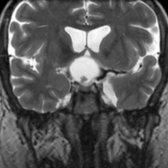
*Coronal T2 MR in a 48-year-old man who presented with visual changes shows a cystic suprasellar mass with a focal T2-hypointense nodule . The T2 imaging mimics a Rathke cleft cyst.*

*Sagittal T1 C+ MR in a 45-year-old woman shows a cystic and solid mass with an enhancing portion , which distinguishes this cystic lesion as a craniopharyngioma, not a Rathke cleft cyst. Surgical resection is the primary therapy for this WHO grade 1 neoplasm. However, the recurrence rate at 10 years approaches 20%.*
 of an adamantinomatous craniopharyngioma. The cystic spaces contain a thick gelatinous material. (Courtesy AFIP.)](images/app.statdx.com_image_thumbnail_8fe443b2-0b89-48cc-a021-94c248ae56c4_size_168_quality_85_275ebdf6_20251014T205219Z.jpg)
*Gross pathologic specimen shows a typical solid and cystic composition of an adamantinomatous craniopharyngioma. The cystic spaces contain a thick gelatinous material. (Courtesy AFIP.)*
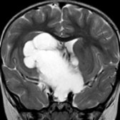
*Coronal T2 MR in a 2 year old with a huge suprasellar mass shows multiple hyperintense cysts . Adamantinomatous craniopharyngiomas typically present between 5-15 years in childhood and between 45-60 years in adults. They are the most common pediatric intracranial tumor of nonglial origin.*
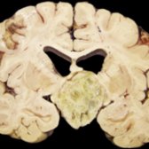
*Gross pathologic specimen shows a typical solid and cystic composition of an adamantinomatous craniopharyngioma. The cystic spaces contain a thick gelatinous material. (Courtesy AFIP.)*

*Coronal T2 MR in a 2 year old with a huge suprasellar mass shows multiple hyperintense cysts . Adamantinomatous craniopharyngiomas typically present between 5-15 years in childhood and between 45-60 years in adults. They are the most common pediatric intracranial tumor of nonglial origin.*
 , and globular Ca⁺⁺. Enhancement was present on postcontrast images.](images/app.statdx.com_image_thumbnail_20fed2a7-ce3d-4ef8-aab8-c4cf436a651f_size_168_quality_85_f9740012_20251014T205220Z.jpg)
*Axial NECT shows classic findings of an adamantinomatous craniopharyngioma. Note the large suprasellar cyst with a fluid-fluid level , rim , and globular Ca⁺⁺. Enhancement was present on postcontrast images.*
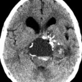
*Axial NECT shows classic findings of an adamantinomatous craniopharyngioma. Note the large suprasellar cyst with a fluid-fluid level , rim , and globular Ca⁺⁺. Enhancement was present on postcontrast images.*
 . (Courtesy R. Hewlett, MD.)](images/app.statdx.com_image_thumbnail_5737449d-e941-4480-bb6c-7a8d8fc2f41a_size_168_quality_85_6ada1f4b_20251014T205219Z.jpg)
*Sagittal gross pathology shows classic adamantinomatous craniopharyngioma with mixed solid, cystic components. The classic machine or "crankcase oil" is present. Note the intrasellar extension . (Courtesy R. Hewlett, MD.)*
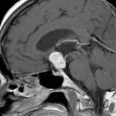
*Sagittal T1 C+ MR in a 45-year-old man with visual changes shows a solid enhancing suprasellar mass . Papillary craniopharyngioma was diagnosed at resection. Papillary craniopharyngiomas are WHO grade 1 tumors; however, they are more commonly solid, noncalcified lesions.*
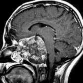
*Sagittal T1WI C+ MR shows a large recurrent craniopharyngioma in the central skull base and nasopharynx with typical heterogeneous cystic and solid morphology. (Courtesy S. Blaser, MD.)*
### Additional Images
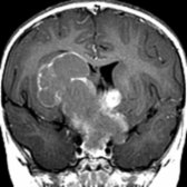
*Axial T1 C+ MR in a child with a huge multilobulated craniopharyngioma shows rim and solid nodular enhancement. Note that the cyst fluid is moderately hyperintense compared to CSF in the lateral ventricles.*
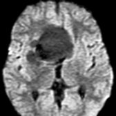
*Axial DWI MR in the same patient with a large craniopharyngioma shows no restriction in the fluid-containing part of the tumor .*
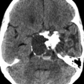
*Axial NECT shows a low-attenuation suprasellar mass with rim and globular Ca⁺⁺. Note the fluid-fluid level formed by intracystic keratin debris .*
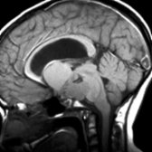
*Sagittal T1WI MR shows a complex predominantly cystic suprasellar mass. Note the T1 shortening within the cyst due to machine oil-like proteinaceous fluid .*
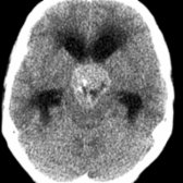
*Axial NECT shows a predominantly solid, minimally calcified , suprasellar craniopharyngioma.*
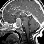
*Sagittal T1 C+ MR shows a principally cystic, sellar/suprasellar mass with rim enhancement .*
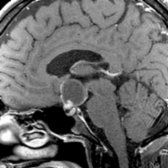
*Sagittal T1 C+ MR in a 48-year-old man with visual changes shows an enhancing nodule , which distinguishes this cystic lesion as a craniopharyngioma, not a Rathke cleft cyst. Surgical resection is the primary therapy for this WHO grade 1 neoplasm; however, the recurrence rate at 10 years approaches 20%.*
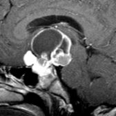
*Sagittal T1WI C+ MR shows a complex cystic suprasellar mass with an enhancing rim and solid components . The cysts contain fluid of different signal intensities. Note the large suprasellar, smaller intrasellar components in this classic craniopharyngioma.*
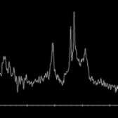
*A short TE (35) H-MRS in a patient with a solid and cystic craniopharyngioma acquired from the center of the cystic portion of the mass shows large lipid-lactate peaks , characteristic of the cholesterol and lipid constituents found in cysts of craniopharyngiomas.*
. (Courtesy R. Hewlett, MD.)](images/app.statdx.com_image_thumbnail_5737449d-e941-4480-bb6c-7a8d8fc2f41a_size_168_quality_85_6ada1f4b_20251014T205219Z.jpg)
*Sagittal gross pathology shows classic adamantinomatous craniopharyngioma with mixed solid, cystic components. The classic machine or "crankcase oil" is present. Note the intrasellar extension . (Courtesy R. Hewlett, MD.)*

*Sagittal T1 C+ MR in a 45-year-old man with visual changes shows a solid enhancing suprasellar mass . Papillary craniopharyngioma was diagnosed at resection. Papillary craniopharyngiomas are WHO grade 1 tumors; however, they are more commonly solid, noncalcified lesions.*

*Sagittal T1WI C+ MR shows a large recurrent craniopharyngioma in the central skull base and nasopharynx with typical heterogeneous cystic and solid morphology. (Courtesy S. Blaser, MD.)*
### Additional Images

*Axial T1 C+ MR in a child with a huge multilobulated craniopharyngioma shows rim and solid nodular enhancement. Note that the cyst fluid is moderately hyperintense compared to CSF in the lateral ventricles.*

*Axial DWI MR in the same patient with a large craniopharyngioma shows no restriction in the fluid-containing part of the tumor .*

*Axial NECT shows a low-attenuation suprasellar mass with rim and globular Ca⁺⁺. Note the fluid-fluid level formed by intracystic keratin debris .*

*Sagittal T1WI MR shows a complex predominantly cystic suprasellar mass. Note the T1 shortening within the cyst due to machine oil-like proteinaceous fluid .*

*Axial NECT shows a predominantly solid, minimally calcified , suprasellar craniopharyngioma.*

*Sagittal T1 C+ MR shows a principally cystic, sellar/suprasellar mass with rim enhancement .*

*Sagittal T1 C+ MR in a 48-year-old man with visual changes shows an enhancing nodule , which distinguishes this cystic lesion as a craniopharyngioma, not a Rathke cleft cyst. Surgical resection is the primary therapy for this WHO grade 1 neoplasm; however, the recurrence rate at 10 years approaches 20%.*

*Sagittal T1WI C+ MR shows a complex cystic suprasellar mass with an enhancing rim and solid components . The cysts contain fluid of different signal intensities. Note the large suprasellar, smaller intrasellar components in this classic craniopharyngioma.*

*A short TE (35) H-MRS in a patient with a solid and cystic craniopharyngioma acquired from the center of the cystic portion of the mass shows large lipid-lactate peaks , characteristic of the cholesterol and lipid constituents found in cysts of craniopharyngiomas.*