---
title: "Craniovertebral Junction Soft Tissue Abnormality"
docid: "3a8dca6c-3452-4cbb-ab83-5e9610d3f0f0"
authors:
- key: "bee1f359-33fb-4cba-9e6b-ed1ca1842439"
value: "Jeffrey S. Ross, MD"
breadcrumbs:
-
name: "Spine"
slug: "spine"
treeNodeId: "b337a156-914a-4696-a77c-af206720fab5"
-
name: "Differential Diagnosis"
slug: "differential-diagnosis"
treeNodeId: "ef5fd925-2033-4f3b-aa7c-640fef9aa956"
-
name: "Craniovertebral Junction"
slug: "craniovertebral-junction"
treeNodeId: "1b500928-e185-4268-95e4-d472fc416f2c"
-
name: "Anatomically Based Differentials"
slug: "anatomically-based-differentials"
treeNodeId: "13201c88-825d-423b-a917-f0e7cce4a599"
-
name: "Craniovertebral Junction Soft Tissue Abnormality"
slug: "craniovertebral-junction-soft-tiss-"
treeNodeId: null
category: "Spine"
documentVersionId: "01ade6cf-e6ab-4b60-b19f-4132e55c4b3a"
imageCount: 25
lastUpdated: "01/18/23"
pageDescription: "Craniovertebral Junction Soft Tissue Abnormality"
pageKeywords: "Spine, Differential Diagnosis, Craniovertebral Junction, Anatomically Based Differentials, Craniovertebral Junction Soft Tissue Abnormality"
pageTitle: "Craniovertebral Junction Soft Tissue Abnormality | STATdx"
enhancedTitle: "Craniovertebral Junction Soft Tissue Abnormality"
type: "DDX"
references: true
breadcrumbs:
- "Spine"
- "Differential Diagnosis"
- "Craniovertebral Junction"
- "Anatomically Based Differentials"
- "Craniovertebral Junction Soft Tissue Abnormality"
---
# ESSENTIAL INFORMATION
- ## Key Differential Diagnosis Issues
- Do intralesion calcifications represent arc-whorl intralesional calcifications (chondrosarcoma) or fragmented destroyed bone (chordoma, metastasis)?
- Does patient have known primary neoplasm (metastasis), myeloma (plasmacytoma), or nasopharyngeal mass (nasopharyngeal carcinoma)?
- ## Helpful Clues for Common Diagnoses
- **Rheumatoid Arthritis**
- Thickened and inflamed synovium called pannus
- Never involves spine without involvement of hands &/or feet
- Odontoid erosions, ligamentous laxity
- C1-C2 instability in 33% of all rheumatoid arthritis (RA) patients
- Neutral, flexion, and extension lateral radiographs performed for evaluation
- High correlation to neurologic symptoms with distance 9 mm or more between C1-C2
- **Retroodontoid Pseudotumor**
- Increased soft tissue dorsal to odontoid secondary to C1-C2 osteoarthritis
- Low-signal mass on T1 and T2 (fibrotic)
- May cause cervicomedullary junction compression
- Usually seen with altered biomechanics of lower cervical spine → surgical/congenital fusion
- Mimics appearance of RA
- Multiple other levels of degenerative disc disease
- **Calcium Pyrophosphate Dihydrate Deposition Disease**
- Pseudogout: Acute, painful episode due to calcium pyrophosphate dihydrate deposition disease (CPPD)
- Crowned dens syndrome: CPPD of atlantooccipital joint causing periodic acute cervicooccipital pain with fever, neck stiffness, and laboratory inflammatory signs
- Calcifications commonly in transverse and alar ligaments, posterior longitudinal ligament, ligamentum flavum
- **Osteomyelitis, C1-C2**
- Infection starts as septic arthritis of C1-C2
- Risk factors include diabetes, drug abuse, endocarditis, immunocompromise
- Soft tissue mass and bone destruction at C1-C2 level
- *Staphylococcus aureus* most common organism in USA
- *Mycobacterium tuberculosis* most common worldwide
- MR shows low T1 signal mass centered at C1-C2 with variable involvement of odontoid and lateral masses at C2
- May show enlarged atlantodental interval
- Epidural mass with thecal sac/cord compression
- Grisel syndrome: Inflammatory, nontraumatic subluxation of C1-C2 following peripharyngeal infection
- **Extramedullary Tumor**
- **Metastases**
- Multiple lesions, bone destruction, systemic primary
- **Lymphoma**
- Large pharyngeal mucosal space mass with associated cervical adenopathy > 50% of time
- Non-Hodgkin lymphoma (NHL) 5x as common as Hodgkin disease in head & neck
- **Nasopharyngeal carcinoma**
- Mass centered in lateral pharyngeal recess of nasopharyngeal with deep extension and cervical adenopathy
- Nodal metastases present in 90% of cases at presentation
- Multiplanar images show invasion of clivus, sphenoid bone and sinus, C1 and C2 bodies
- **Neurofibromatosis type 1**
- Plexiform neurofibroma → diffuse enlargement of major nerve trunks/branches → bulky rope-like ("bag of worms") nerve expansion with adjacent tissue distortion
- Look for kyphoscoliosis ± multiple nerve root tumors, plexiform neurofibroma, dural ectasia/lateral meningocele
- **Schwannoma**
- Hypoglossal or upper cervical roots as site of origin
- Hypoglossal neuropathy results in tongue denervation
- Dumbbell with uniform enhancement
- Larger lesions may show central cystic formation
- **Paraganglioma**
- Multiple black dots ("pepper") in tumor substance indicating high-velocity flow voids from feeding arterial branches
- Jugular foramen (JF) or vagal varieties may present with upper cervical/skull base level mass
- **Chordoma**
- Mass is hyperintense to discs on T2WI with multiple septa
- Destructive, lytic lesion
- May extend into disc, involve 2 or more adjacent vertebrae
- **Chondrosarcoma**
- Lytic mass ± chondroid matrix, cortical disruption, and extension into soft tissues
- Chondroid matrix mineralization of rings and arcs (characteristic)
- **Meningioma**
- Foramen magnum, JF, upper cervical dura locations
- Carotid space → connection to JF above with JF margins showing permeative-sclerotic or hyperostotic changes on bone CT
- Absence of high-velocity flow voids on T1 MR
- T1 C+ MR shows enhancing JF mass
- ## Helpful Clues for Less Common Diagnoses
- **Aneurysm/Vertebral Dissection**
- Multiple etiologies → dissection, posttraumatic, atherosclerotic, iatrogenic, congenital
- **Synovial Cyst**
- Round, central T2-hyperintense mass with low-signal margin
- Associated with dorsal C1-C2 articulation or degenerated facets
- ## Helpful Clues for Rare Diagnoses
- **Neurenteric Cyst**
- Intraspinal cyst + vertebral abnormalities (persistent canal of Kovalevsky, segmentation and fusion anomalies)
## References
# Selected References
1. [Wang HB et al: Cervical myelopathy due to idiopathic retro-odontoid pseudotumor. World Neurosurg.160:e256-60, 2022](http://www.ncbi.nlm.nih.gov/pubmed/?term=34999265%5Bpmid%5D)
1. [Bi WL et al: Skull base tumors: neuropathology and clinical implications. Neurosurgery. 90(3):243-61, 2021](http://www.ncbi.nlm.nih.gov/pubmed/?term=34164689%5Bpmid%5D)
1. [Parperis K et al: Management of calcium pyrophosphate crystal deposition disease: a systematic review. Semin Arthritis Rheum. 51(1):84-94, 2021](http://www.ncbi.nlm.nih.gov/pubmed/?term=33360232%5Bpmid%5D)
1. [Pascart T et al: Treatment of nongout joint deposition diseases: an update. Arthritis. 2014:375202, 2014](http://www.ncbi.nlm.nih.gov/pubmed/?term=24895535%5Bpmid%5D)
1. [Chang EY et al: Frequency of atlantoaxial calcium pyrophosphate dihydrate deposition at CT. Radiology. 269(2):519-24, 2013](http://www.ncbi.nlm.nih.gov/pubmed/?term=23737539%5Bpmid%5D)
1. [Tojo S et al: Factors influencing on retro-odontoid soft-tissue thickness: analysis by magnetic resonance imaging. Spine (Phila Pa 1976). 38(5):401-6, 2013](http://www.ncbi.nlm.nih.gov/pubmed/?term=23448899%5Bpmid%5D)
1. [Di Maio S et al: Current comprehensive management of cranial base chordomas: 10-year meta-analysis of observational studies. J Neurosurg. 115(6):1094-5, 2011](http://www.ncbi.nlm.nih.gov/pubmed/?term=21819197%5Bpmid%5D)
1. [Smith JS et al: Basilar invagination. Neurosurgery. 66(3 Suppl):39-47, 2010](http://www.ncbi.nlm.nih.gov/pubmed/?term=20173526%5Bpmid%5D)
## Images
### Selected Images
 . There is mild compression of medulla by the pannus and obscuration of fat planes.](images/app.statdx.com_image_thumbnail_1e8e97b6-3801-41f7-91f8-a8e00d56c18b_annotated_true_size_900_quality_90_d7c6a53b.jpg)
**Rheumatoid Arthritis**
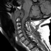
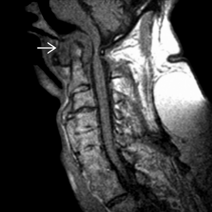
*Sagittal T1WI MR shows rheumatoid arthritis (RA) involving C1-C2 articulation with dens erosion and extensive pannus formation . There is mild compression of medulla by the pannus and obscuration of fat planes.*
**Rheumatoid Arthritis**
*Sagittal T1WI MR shows rheumatoid arthritis (RA) involving C1-C2 articulation with dens erosion and extensive pannus formation . There is mild compression of medulla by the pannus and obscuration of fat planes.*
**Retroodontoid Pseudotumor**
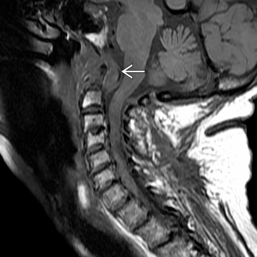
*Sagittal T1WI MR shows C1-C2 degenerative pseudopannus in a patient with diffuse idiopathic skeletal hyperostosis. The odontoid is not eroded, but the atlantodental interval is increased. Cord compression occurs between the degenerative pannus and posterior C1.*
**Rheumatoid Arthritis**
*Sagittal T1WI MR shows rheumatoid arthritis (RA) involving C1-C2 articulation with dens erosion and extensive pannus formation . There is mild compression of medulla by the pannus and obscuration of fat planes.*

**Rheumatoid Arthritis**
*Sagittal T1WI MR shows rheumatoid arthritis (RA) involving C1-C2 articulation with dens erosion and extensive pannus formation . There is mild compression of medulla by the pannus and obscuration of fat planes.*

**Retroodontoid Pseudotumor**
*Sagittal T1WI MR shows C1-C2 degenerative pseudopannus in a patient with diffuse idiopathic skeletal hyperostosis. The odontoid is not eroded, but the atlantodental interval is increased. Cord compression occurs between the degenerative pannus and posterior C1.*
 . No gross erosive changes are present within the odontoid process, as would be present in RA.](images/app.statdx.com_image_thumbnail_b9c089b6-5c25-48d2-a435-b1e13a873488_annotated_true_size_900_quality_90_95c69b21.jpg)
**Calcium Pyrophosphate Dihydrate Deposition Disease**
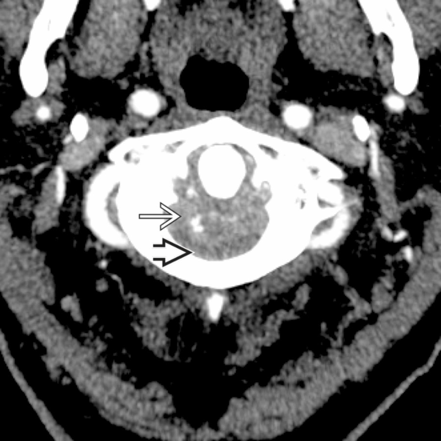
*CTA study shows multiple punctate foci of calcification within a large soft tissue mass posterior to the odontoid process that causes cord compression . No gross erosive changes are present within the odontoid process, as would be present in RA.*
**Calcium Pyrophosphate Dihydrate Deposition Disease**
*CTA study shows multiple punctate foci of calcification within a large soft tissue mass posterior to the odontoid process that causes cord compression . No gross erosive changes are present within the odontoid process, as would be present in RA.*
 .](images/app.statdx.com_image_thumbnail_3eea911f-372e-4f76-a62c-d36e4e54e961_annotated_true_size_900_quality_90_f12897b0.jpg)
**Calcium Pyrophosphate Dihydrate Deposition Disease**
*Sagittal T1WI MR shows a large mass with low T2 signal posterior to the odontoid process with severe cord compression .*
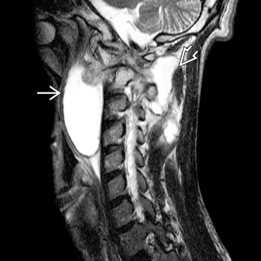
**Osteomyelitis, C1-C2**
*Sagittal T2WI MR shows a large prevertebral abscess spanning C1 to C4 and extension posteriorly involving the interspinous region . These findings are typical for tuberculosis (TB).*
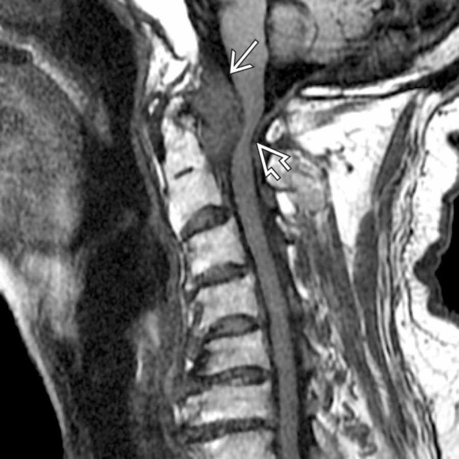
**Calcium Pyrophosphate Dihydrate Deposition Disease**
*Sagittal T1WI MR shows a large mass with low T2 signal posterior to the odontoid process with severe cord compression .*

**Osteomyelitis, C1-C2**
*Sagittal T2WI MR shows a large prevertebral abscess spanning C1 to C4 and extension posteriorly involving the interspinous region . These findings are typical for tuberculosis (TB).*
 and invasion to the pharyngeal mucosal space .](images/app.statdx.com_image_thumbnail_1ead54d2-e7f9-466a-aab9-a6329250bcf1_annotated_true_size_900_quality_90_4865f802.jpg)
**Metastases**
*Axial T1 C+ FS MR shows metastatic lung cancer with extensive extracapsular nodal spread. The mass has ill-defined borders with invasion of the longus capitis muscle and invasion to the pharyngeal mucosal space .*
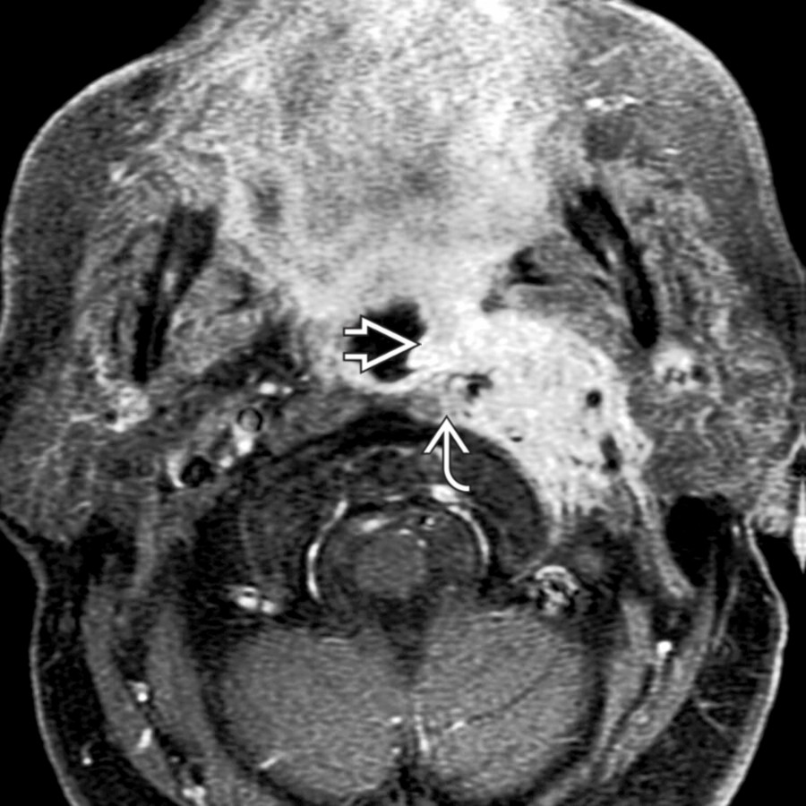
**Metastases**
*Axial T1 C+ FS MR shows metastatic lung cancer with extensive extracapsular nodal spread. The mass has ill-defined borders with invasion of the longus capitis muscle and invasion to the pharyngeal mucosal space .*
 .](images/app.statdx.com_image_thumbnail_483f8d25-4da6-4f41-9f47-66464fb427eb_annotated_true_size_900_quality_90_9711bb56.jpg)
**Metastases**
*Axial CTA shows the classic appearance of thyroid metastasis with a thin, expansile, bony margin with the predominately lytic lesion within the left facet/lamina of C3 .*
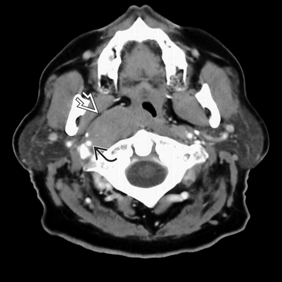
**Lymphoma**
*Axial CECT shows a homogeneous mass in the retropharyngeal space, displacing the parapharyngeal fat anterolaterally and encircling the right internal carotid artery .*
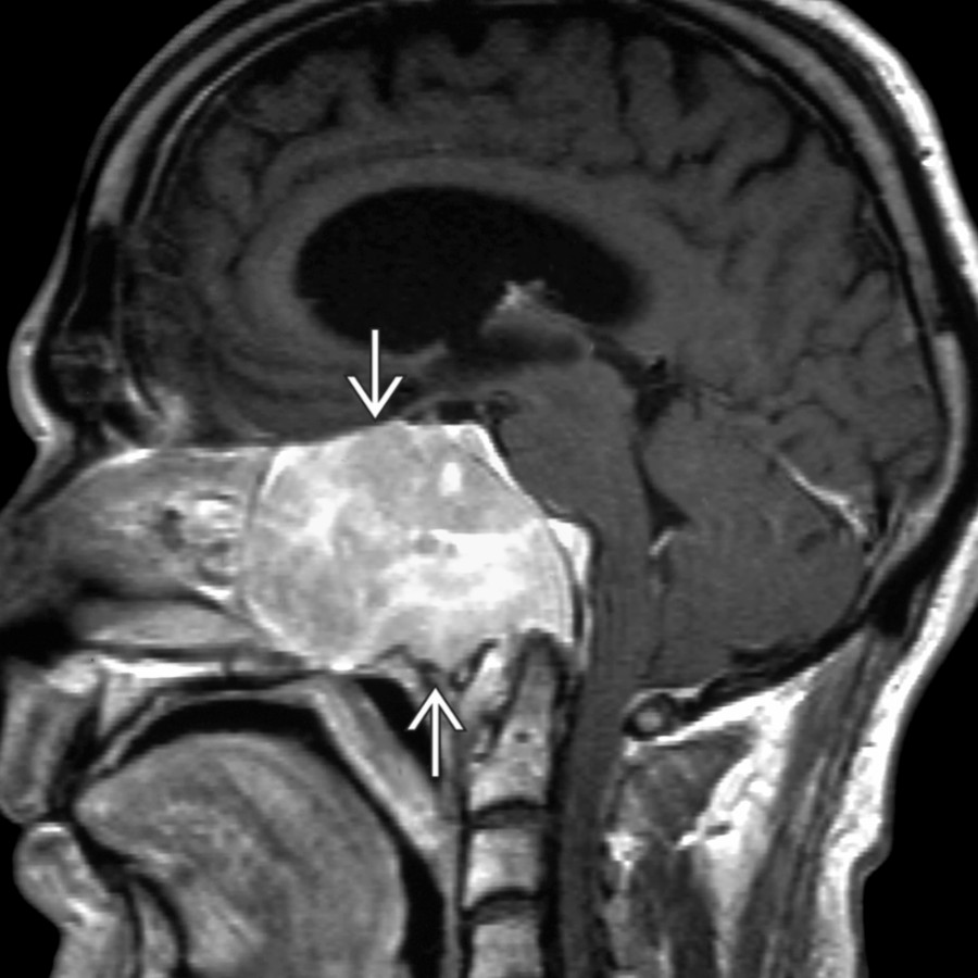
**Plasmacytoma**
*Sagittal T1 C+ MR shows a variant MR case of an unusually large skull base plasmacytoma engulfing the clivus and extending into the nasopharynx and abutting C1-C2.*
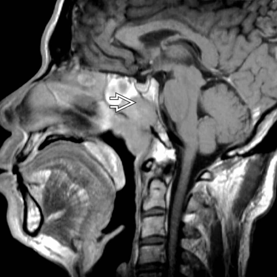
**Nasopharyngeal Carcinoma**
*Sagittal T1WI MR shows a typical case of an aggressive nasopharyngeal squamous cell carcinoma with invasion of the skull base by direct extension .*
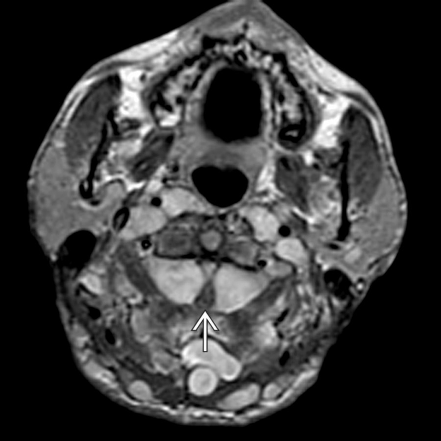
**Neurofibromatosis Type 1**
*Axial T1WI C+ MR shows multiple large neurofibromas within dorsal soft tissues and paravertebral regions. Symmetrical, large intradural lesions compress the cervical cord at the C2 level.*
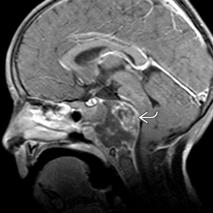
**Chordoma**
*Sagittal T1 C+ MR demonstrates an isointense, expansile mass arising from the clivus . Notice the posterior indentation, or "thumbing," of the pons.*

**Chordoma**
*Sagittal T1WI C+ MR shows a large, heterogeneous-signal mass in the cervical epidural space involving the dorsal aspect of C2-C3 junction and extending laterally with diffuse enhancement.*

**Chondrosarcoma**
*Coronal T1WI MR shows a typical MR case of petrooccipital fissure skull base chondrosarcoma .*

**Meningioma**
*Sagittal T1 C+ MR shows a well-defined, homogeneously enhancing mass with a broad dural margin at the foramen magnum, typical for meningioma. There is compression of the medulla.*

**Aneurysm/Vertebral Dissection**
*Axial CTA shows the CT features of a pseudoaneurysm of the internal carotid artery located below the skull base.*
### Additional Images
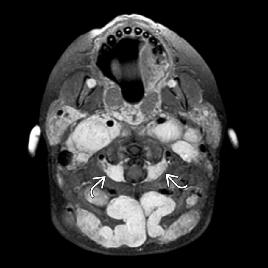
**Neurofibromatosis Type 1**
*Axial T1 C+ FS MR shows the appearance of multiple neurofibromas in a child with neurofibromatosis type 1 with bilateral, symmetric masses involving neural foramen in the carotid sheath and posterior cervical space, consistent with neurofibromas.*
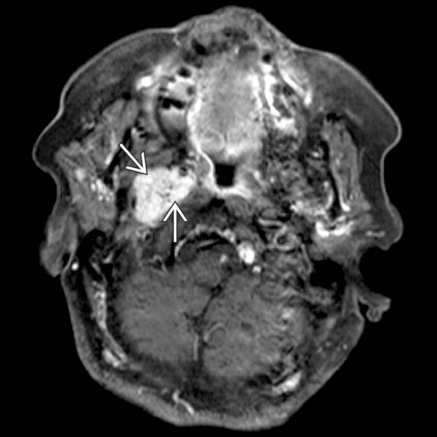
**Paraganglioma**
*Axial T1 C+ FS MR shows the typical imaging appearance of a glomus tumor (vagale paraganglioma): A soft tissue mass below the skull base with small, focal signal voids indicating the presence of high-flow vessels.*

**Meningioma**
*Axial CECT shows a variant case of carotid space meningioma from recurrent intracranial disease with an oval-shaped mass in the right carotid space, medial to the styloid process .*

**Chiari 1 Malformation**
*Sagittal T2WI MR shows a Chiari 1 malformation with inferiorly positioned, peg-shaped tonsils with small, associated cervical syrinx .*

**Chiari 2 Malformation**
*Sagittal T2WI MR shows a typical case of mild Chiari 2 malformation with a small posterior fossa and vermian ectopia to the C2/C3 level . The 4th ventricle is small and elongated.*

**Chiari 2 Malformation**
*Sagittal T2WI MR shows a variant Chiari 2 with syringobulbia. There is a characteristic small posterior fossa and vermian ectopia . There is focal cervicomedullary syrinx .*

**Glioma, Brainstem**
*Sagittal T2WI MR demonstrates an extensive T2-hyperintense mass expanding the medulla and cervical cord from astrocytoma . The tumor causes a septated-appearing neoplastic syrinx in the more caudal cord .*

**Hemangioblastoma, Spinal Cord**
*Sagittal T1WI MR shows well-defined, complex cyst at the cervicomedullary junction due to hemangioblastoma.*

**Meningioma**
*Axial T1 C+ MR shows a well-defined, homogeneously enhancing mass with a broad dural margin at the foramen magnum, typical for meningioma. The left distal vertebral artery is adjacent to the tumor .*
.](images/app.statdx.com_image_thumbnail_483f8d25-4da6-4f41-9f47-66464fb427eb_annotated_true_size_900_quality_90_9711bb56.jpg)
**Metastases**
*Axial CTA shows the classic appearance of thyroid metastasis with a thin, expansile, bony margin with the predominately lytic lesion within the left facet/lamina of C3 .*

**Lymphoma**
*Axial CECT shows a homogeneous mass in the retropharyngeal space, displacing the parapharyngeal fat anterolaterally and encircling the right internal carotid artery .*

**Plasmacytoma**
*Sagittal T1 C+ MR shows a variant MR case of an unusually large skull base plasmacytoma engulfing the clivus and extending into the nasopharynx and abutting C1-C2.*

**Nasopharyngeal Carcinoma**
*Sagittal T1WI MR shows a typical case of an aggressive nasopharyngeal squamous cell carcinoma with invasion of the skull base by direct extension .*

**Neurofibromatosis Type 1**
*Axial T1WI C+ MR shows multiple large neurofibromas within dorsal soft tissues and paravertebral regions. Symmetrical, large intradural lesions compress the cervical cord at the C2 level.*

**Chordoma**
*Sagittal T1 C+ MR demonstrates an isointense, expansile mass arising from the clivus . Notice the posterior indentation, or "thumbing," of the pons.*

**Chordoma**
*Sagittal T1WI C+ MR shows a large, heterogeneous-signal mass in the cervical epidural space involving the dorsal aspect of C2-C3 junction and extending laterally with diffuse enhancement.*

**Chondrosarcoma**
*Coronal T1WI MR shows a typical MR case of petrooccipital fissure skull base chondrosarcoma .*

**Meningioma**
*Sagittal T1 C+ MR shows a well-defined, homogeneously enhancing mass with a broad dural margin at the foramen magnum, typical for meningioma. There is compression of the medulla.*

**Aneurysm/Vertebral Dissection**
*Axial CTA shows the CT features of a pseudoaneurysm of the internal carotid artery located below the skull base.*
### Additional Images

**Neurofibromatosis Type 1**
*Axial T1 C+ FS MR shows the appearance of multiple neurofibromas in a child with neurofibromatosis type 1 with bilateral, symmetric masses involving neural foramen in the carotid sheath and posterior cervical space, consistent with neurofibromas.*

**Paraganglioma**
*Axial T1 C+ FS MR shows the typical imaging appearance of a glomus tumor (vagale paraganglioma): A soft tissue mass below the skull base with small, focal signal voids indicating the presence of high-flow vessels.*

**Meningioma**
*Axial CECT shows a variant case of carotid space meningioma from recurrent intracranial disease with an oval-shaped mass in the right carotid space, medial to the styloid process .*

**Chiari 1 Malformation**
*Sagittal T2WI MR shows a Chiari 1 malformation with inferiorly positioned, peg-shaped tonsils with small, associated cervical syrinx .*

**Chiari 2 Malformation**
*Sagittal T2WI MR shows a typical case of mild Chiari 2 malformation with a small posterior fossa and vermian ectopia to the C2/C3 level . The 4th ventricle is small and elongated.*

**Chiari 2 Malformation**
*Sagittal T2WI MR shows a variant Chiari 2 with syringobulbia. There is a characteristic small posterior fossa and vermian ectopia . There is focal cervicomedullary syrinx .*

**Glioma, Brainstem**
*Sagittal T2WI MR demonstrates an extensive T2-hyperintense mass expanding the medulla and cervical cord from astrocytoma . The tumor causes a septated-appearing neoplastic syrinx in the more caudal cord .*

**Hemangioblastoma, Spinal Cord**
*Sagittal T1WI MR shows well-defined, complex cyst at the cervicomedullary junction due to hemangioblastoma.*

**Meningioma**
*Axial T1 C+ MR shows a well-defined, homogeneously enhancing mass with a broad dural margin at the foramen magnum, typical for meningioma. The left distal vertebral artery is adjacent to the tumor .*