---
title: "Cyst With Nodule"
docid: "6cb71737-f574-4121-a8fb-02eeada9f9f7"
authors:
- key: "4b6589b0-9b8d-4467-8a90-01a0a59742fc"
value: "Troy A. Hutchins, MD"
- key: "8d5254e9-8dda-478b-8f08-bdee97a32c79"
value: "Karen L. Salzman, MD, FACR"
breadcrumbs:
-
name: "Brain"
slug: "brain"
treeNodeId: "6d8829f1-14d7-45af-8675-255189aa526a"
-
name: "Differential Diagnosis"
slug: "differential-diagnosis"
treeNodeId: "a7fdd139-664e-4bb8-8d18-400e4733ff60"
-
name: "Brain Parenchyma, General"
slug: "brain-parenchyma-general"
treeNodeId: "e79be97b-28c0-4023-be87-334c0579d35d"
-
name: "Generic Imaging Patterns"
slug: "generic-imaging-patterns"
treeNodeId: "66ab9cf6-74ad-42b7-a40a-4b6224edaa25"
-
name: "Cyst With Nodule"
slug: "cyst-with-nodule"
treeNodeId: null
category: "Brain"
documentVersionId: "a8a6b610-83a2-461f-bc34-b34af327c00c"
imageCount: 35
lastUpdated: "03/14/23"
pageDescription: "Cyst With Nodule"
pageKeywords: "Brain, Differential Diagnosis, Brain Parenchyma, General, Generic Imaging Patterns, Cyst With Nodule"
pageTitle: "Cyst With Nodule | STATdx"
enhancedTitle: "Cyst With Nodule"
type: "DDX"
references: true
breadcrumbs:
- "Brain"
- "Differential Diagnosis"
- "Brain Parenchyma, General"
- "Generic Imaging Patterns"
- "Cyst With Nodule"
---
# ESSENTIAL INFORMATION
- ## Key Differential Diagnosis Issues
- Cystic lesions with solid nodular components can be divided into 2 categories
- Lesions that typically demonstrate cyst with nodule morphology
- Neurocysticercosis (NCC), pilocytic astrocytoma, ganglioglioma, hemangioblastoma, pleomorphic xanthoastrocytoma (PXA), desmoplastic infantile ganglioglioma (DIG), intraparenchymal schwannoma
- Lesions that may demonstrate cyst with nodule morphology
- Metastases, glioblastoma (GBM), abscess, toxoplasmosis, parasites, dysplastic neuroepithelial tumor (DNET), thrombosed arteriovenous malformation (AVM), supratentorial ependymoma
- Although metastases, abscesses, & GBMs do not classically present as "cysts with nodules," they are included because of their overall prevalence
- Statistically, atypical form of these common diseases may be more likely than some of other "classic" cysts with nodule lesions
- ## Helpful Clues for Common Diagnoses
- **Neurocysticercosis**
- Intracranial parasitic infection caused by pork tapeworm *Taenia solium*
- Cyst with "dot" inside representing scolex
- Imaging appearance varies with stage; increased enhancement & edema when organism dies (inflammatory host response)
- Location: Convexity subarachnoid space > > cisterns > parenchyma > ventricles
- Lesions may be at different stages in same patient
- **Pilocytic Astrocytoma**
- Cerebellar cystic mass with mural nodule in child; rarely supratentorial
- T1 C+ MR: Nodule shows intense but heterogeneous enhancement
- Cyst wall may show enhancement
- T1 & T2 MR: Cyst content iso- to hyperintense to CSF
- Most common brain tumor in children
- **Ganglioglioma**
- Cortically based, slow-growing, enhancing mass in older child or young adult
- Circumscribed cyst with mural nodule most common
- May be solid and appear well circumscribed
- Often expands cortex; calcification common
- Most common tumor to cause temporal lobe epilepsy
- Cortical dysplasia is commonly associated
- **Hemangioblastoma**
- Vascular neoplasm of uncertain etiology
- Parenchymal posterior fossa cyst with nodule mass in adult
- T1 C+ MR: Nodule abuts pial surface & shows intense, homogeneous enhancement
- Prominent flow voids may be seen
- Multiple in von Hippel-Lindau syndrome (VHL) (25-40% of hemangioblastomas)
- ## Helpful Clues for Less Common Diagnoses
- **Metastases, Parenchymal**
- Discrete, gray-white interface mass(es) with adjacent vasogenic edema
- Multiplicity, history of primary malignancy helpful if present
- Solitary metastasis may mimic GBM
- Often known history of primary neoplasm
- **Glioblastoma, IDH-Wildtype**
- Malignant white matter mass with central necrosis
- Predilection to spread across midline along corpus callosum; "butterfly glioma"
- T1 C+ MR: Thick, irregular, nodular, enhancing margins
- T2/FLAIR MR: Surrounding hyperintensity & mass effect reflect edema + infiltrative tumor
- **Pleomorphic Xanthoastrocytoma**
- Cortically based cyst + nodule ± involvement of adjacent meninges
- T1 C+ MR
- Enhancing nodule
- Thickening, enhancement of adjacent meninges
- 70% have dural tail
- Temporal lobe predominance; young adult
- Maybe associated with cortical dysplasia
- **Abscess**
- T2 MR: Hypointense rim with surrounding edema classic
- T1 C+ MR: Enhancing capsule thinnest at ventricular side
- DWI MR: Cystic component bright (diffusion restriction)
- SWI MR: Dual rim sign (hypointense outside, hyperintense inside )
- **Opportunistic Infection, AIDS, Toxoplasmosis**
- Caused by parasite *Toxoplasma gondii*
- Toxoplasmosis: Ring-enhancing lesion containing eccentric nodule = eccentric target sign specific but not sensitive
- Location: Basal ganglia > hemispheres
- Clinical: Immunocompromised patient
- **Parasites, Miscellaneous**
- Multiple enhancing lesions typical
- May mimic brain tumor
- Travel history critical
- **Dysplastic Neuroepithelial Tumor**
- Bubbly, wedge-shaped, cortically based mass "points" toward lateral ventricle
- T2 MR: Very hyperintense; nodular, septate; no surrounding edema
- FLAIR MR: Hyperintense ring sign
- Thin rim of well-defined peritumoral hyperintensity separating it from surrounding normal brain
- T1 C+ MR: No to minimal enhancement; may be nodular
- Temporal lobe predominance
- ## Helpful Clues for Rare Diagnoses
- **Desmoplastic Infantile Ganglioglioma**
- Supratentorial cystic/nodular mass with dominance of cyst
- Cortically based nodule with intense enhancement & dural tail
- May be massive
- Peak age: 3-6 months
- **Schwannoma, Intraparenchymal**
- Only 1-2% of schwannomas are parenchymal
- Cyst with strongly enhancing nodule
- **Arteriovenous Malformation**
- When hemorrhagic with partial or complete thrombosis, may present as cyst with nodule
- Blood breakdown products of various ages; fluid-fluid levels
- **Ependymoma, Supratentorial**
- 40% of supratentorial ependymomas are extraventricular
- Large, complex, mixed solid/cystic mass
- Calcification, intratumoral hemorrhage common
- Moderate but inhomogeneous enhancement
- **Meningioma (Cystic)**
- Meningioma with intraparenchymal cyst may mimic cyst + nodule mass
- **Rosette-Forming Glioneuronal Tumor**
- Rare, slowly growing benign tumor of young adults
- 4th ventricle most common site > cerebellum
- Mixed solid-cystic appearance, variable Ca⁺⁺, hemorrhage
- May show cyst with nodule configuration
- **Papillary Glioneuronal Tumor**
- Temporal lobe predilection
- Parenchymal mass with solid, cystic, or cyst/mural nodule architecture
- May show calcification
- Imaging may be indistinguishable from ganglioglioma
- ## Alternative Differential Approaches
- By location
- Posterior fossa: Pilocytic astrocytoma, hemangioblastoma, metastasis, Rosette-forming glioneuronal tumor
- Temporal lobe: Ganglioglioma, PXA, DNET, papillary glioneuronal tumor
- Gray-white junction: Metastases, abscess
- Hemispheric: NCC, metastases, GBM, infections, DIG, AVM, supratentorial ependymoma
- Patient age
- Child & young adult: Pilocytic astrocytoma, ganglioglioma, PXA, DNET
- Adult: Hemangioblastoma, GBM, metastases
- Any age: NCC, abscess, other infections
- Multiple lesions
- Metastases (50-55%), NCC (50-70%), hemangioblastoma (VHL), abscesses (septic emboli), toxoplasmosis, parasites
## References
# Selected References
1. [Sotoudeh H et al: Radiomics for differentiation of the posterior fossa pilocytic astrocytoma versus hemangioblastomas in adults. A pilot study. Clin Imaging. 93:26-30, 2022](http://www.ncbi.nlm.nih.gov/pubmed/?term=36370592%5Bpmid%5D)
1. [Medhi G et al: Imaging features of rosette-forming glioneuronal tumours (RGNTs): a series of seven cases. Eur Radiol. 26(1):262-70, 2016](http://www.ncbi.nlm.nih.gov/pubmed/?term=26017735%5Bpmid%5D)
1. [Carangelo B et al: Papillary glioneuronal tumor: case report and review of literature. G Chir. 36(2):63-9, 2015](http://www.ncbi.nlm.nih.gov/pubmed/?term=26017104%5Bpmid%5D)
1. [Raz E et al: Cyst with a mural nodule tumor of the brain. Cancer Imaging. 12:237-44, 2012](http://www.ncbi.nlm.nih.gov/pubmed/?term=22935908%5Bpmid%5D)
1. [Kumar GG et al: Eccentric target sign in cerebral toxoplasmosis: neuropathological correlate to the imaging feature. J Magn Reson Imaging. 31(6):1469-72, 2010](http://www.ncbi.nlm.nih.gov/pubmed/?term=20512900%5Bpmid%5D)
1. [Smirniotopoulos JG et al: Patterns of contrast enhancement in the brain and meninges. Radiographics. Mar-Apr;27(2):525-51, 2007](http://www.ncbi.nlm.nih.gov/pubmed/?term=17374867%5Bpmid%5D)
## Images
### Selected Images
 with an eccentric nodule (scolex)
  . Additional lesions have surrounding edema
  . Edema and enhancement vary with the stage of neurocysticercosis and host response.](images/app.statdx.com_image_thumbnail_e25e1c8a-0345-4b38-89ff-714d2a60ffcc_size_168_quality_85_5bce5347_20251014T195847Z.jpg)
**Neurocysticercosis**
*Axial FLAIR MR demonstrates a neurocysticercosis cyst with an eccentric nodule (scolex) . Additional lesions have surrounding edema . Edema and enhancement vary with the stage of neurocysticercosis and host response.*

**Neurocysticercosis**
*Axial FLAIR MR demonstrates a neurocysticercosis cyst with an eccentric nodule (scolex) . Additional lesions have surrounding edema . Edema and enhancement vary with the stage of neurocysticercosis and host response.*

**Pilocytic Astrocytoma**
*Axial T1 C+ SPGR MR shows a pilocytic astrocytoma with enhancing, eccentric nodule . There is cyst wall enhancement (present in ~ 50% of cases). Note obstructive hydrocephalus with dilation of superior 4th ventricle secondary to mass effect.*

**Ganglioglioma**
*Coronal T1 C+ MR in a young adult patient with epilepsy shows a cystic and solid mass in the temporal lobe with intense enhancement of the solid mural nodule . Gangliogliomas are the most common tumor to cause temporal lobe epilepsy.*

**Hemangioblastoma**
*Coronal T1 C+ MR in an adult patient reveals a cystic mass with an intensely enhancing mural nodule in the posterior fossa. Additional enhancing nodules are noted in this patient with von Hippel-Lindau.*

**Metastases, Parenchymal**
*Coronal T1 C+ MR in a patient with primary malignancy demonstrates a cystic and solid, enhancing, nodular mass with the surrounding vasogenic edema . Multiple additional lesions and clinical history can help with diagnosis.*

**Glioblastoma, IDH-Wildtype**
*Axial T1 C+ FS MR shows an infiltrative left frontal lobe glioblastoma with cystic and heterogeneously enhancing, solid, nodular components. Glioblastomas have a predilection for crossing midline through the corpus callosum.*
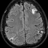
**Neurocysticercosis**
*Axial FLAIR MR demonstrates a neurocysticercosis cyst with an eccentric nodule (scolex) . Additional lesions have surrounding edema . Edema and enhancement vary with the stage of neurocysticercosis and host response.*

**Neurocysticercosis**
*Axial FLAIR MR demonstrates a neurocysticercosis cyst with an eccentric nodule (scolex) . Additional lesions have surrounding edema . Edema and enhancement vary with the stage of neurocysticercosis and host response.*

**Pilocytic Astrocytoma**
*Axial T1 C+ SPGR MR shows a pilocytic astrocytoma with enhancing, eccentric nodule . There is cyst wall enhancement (present in ~ 50% of cases). Note obstructive hydrocephalus with dilation of superior 4th ventricle secondary to mass effect.*

**Ganglioglioma**
*Coronal T1 C+ MR in a young adult patient with epilepsy shows a cystic and solid mass in the temporal lobe with intense enhancement of the solid mural nodule . Gangliogliomas are the most common tumor to cause temporal lobe epilepsy.*

**Hemangioblastoma**
*Coronal T1 C+ MR in an adult patient reveals a cystic mass with an intensely enhancing mural nodule in the posterior fossa. Additional enhancing nodules are noted in this patient with von Hippel-Lindau.*

**Metastases, Parenchymal**
*Coronal T1 C+ MR in a patient with primary malignancy demonstrates a cystic and solid, enhancing, nodular mass with the surrounding vasogenic edema . Multiple additional lesions and clinical history can help with diagnosis.*

**Glioblastoma, IDH-Wildtype**
*Axial T1 C+ FS MR shows an infiltrative left frontal lobe glioblastoma with cystic and heterogeneously enhancing, solid, nodular components. Glioblastomas have a predilection for crossing midline through the corpus callosum.*
 with an enhancing nodule
  in a young adult. Enhancement & thickening of the adjacent dura
  help diagnose PXA & differentiate from a ganglioglioma.](images/app.statdx.com_image_thumbnail_5191f79e-6206-4dc0-a3a9-1eadd8da6b52_size_168_quality_85_34d41e2d_20251014T195847Z.jpg)
**Pleomorphic Xanthoastrocytoma**
*Coronal T1 C+ MR shows a cortically based left temporal lobe cystic mass with an enhancing nodule in a young adult. Enhancement & thickening of the adjacent dura help diagnose PXA & differentiate from a ganglioglioma.*

**Abscess**
*Sagittal T1 C+ MR in a patient treated for sinusitis who presented with headache, fever, and seizures shows a large, ring-enhancing abscess with an eccentric nodule . DWI (not shown) showed characteristic diffusion restriction in the central nonenhancing component.*

**Opportunistic Infection, AIDS, Toxoplasmosis**
*Axial T1 C+ FS MR in an immunocompromised patient shows a ring-enhancing lesion with peripheral enhancing nodule . This eccentric target sign is specific but not sensitive for toxoplasmosis. Note the numerous additional nodular and ring-enhancing lesions and surrounding edema .*

**Parasites, Miscellaneous**
*Axial CECT demonstrates a ring-enhancing lesion with an associated nodule and surrounding vasogenic edema. Multiple punctate lesions are also apparent in this patient with amebic encephalitis.*

**Dysplastic Neuroepithelial Tumor**
*Axial T1 C+ MR in a patient with epilepsy shows a cortically based, bubbly, cystic mass with an eccentric, enhancing nodule . Faint, nodular enhancement can be seen in 20% of DNETs. FLAIR images often show a well-defined, hyperintense ring surrounding the mass.*

**Desmoplastic Infantile Ganglioglioma**
*Coronal T1 C+ MR shows a large cyst with a cortically based, intensely enhancing mural nodule in an infant. Note the adjacent dural thickening & enhancement , typical of desmoplastic infantile ganglioglioma.*

**Schwannoma, Intraparenchymal**
*Axial T1 C+ MR shows a cystic parenchymal mass with an intensely enhancing mural nodule in the right occipital lobe. Although ganglioglioma was the preoperative diagnosis, schwannoma was found on pathology.*

**Ependymoma, Supratentorial**
*Axial T2 MR shows a recurrent supratentorial ependymoma with a cyst and nodule architecture. 40% of supratentorial ependymomas are extraventricular. They are commonly associated with calcification and hemorrhage.*
### Additional Images

**Neurocysticercosis**
*Axial FLAIR MR demonstrates a classic neurocysticercosis cyst with an eccentric nodule (scolex) in the right parietal lobe with surrounding vasogenic edema. The enhancement and edema varies with the stage of neurocysticercosis and host response.*

**Pleomorphic Xanthoastrocytoma**
*Coronal T1 C+ MR shows a cortically based left temporal lobe cystic mass with an enhancing nodule in a young adult. Enhancement & thickening of the adjacent dura help diagnose PXA & differentiate from a ganglioglioma.*

**Abscess**
*Sagittal T1 C+ MR in a patient treated for sinusitis who presented with headache, fever, and seizures shows a large, ring-enhancing abscess with an eccentric nodule . DWI (not shown) showed characteristic diffusion restriction in the central nonenhancing component.*

**Opportunistic Infection, AIDS, Toxoplasmosis**
*Axial T1 C+ FS MR in an immunocompromised patient shows a ring-enhancing lesion with peripheral enhancing nodule . This eccentric target sign is specific but not sensitive for toxoplasmosis. Note the numerous additional nodular and ring-enhancing lesions and surrounding edema .*

**Parasites, Miscellaneous**
*Axial CECT demonstrates a ring-enhancing lesion with an associated nodule and surrounding vasogenic edema. Multiple punctate lesions are also apparent in this patient with amebic encephalitis.*

**Dysplastic Neuroepithelial Tumor**
*Axial T1 C+ MR in a patient with epilepsy shows a cortically based, bubbly, cystic mass with an eccentric, enhancing nodule . Faint, nodular enhancement can be seen in 20% of DNETs. FLAIR images often show a well-defined, hyperintense ring surrounding the mass.*

**Desmoplastic Infantile Ganglioglioma**
*Coronal T1 C+ MR shows a large cyst with a cortically based, intensely enhancing mural nodule in an infant. Note the adjacent dural thickening & enhancement , typical of desmoplastic infantile ganglioglioma.*

**Schwannoma, Intraparenchymal**
*Axial T1 C+ MR shows a cystic parenchymal mass with an intensely enhancing mural nodule in the right occipital lobe. Although ganglioglioma was the preoperative diagnosis, schwannoma was found on pathology.*

**Ependymoma, Supratentorial**
*Axial T2 MR shows a recurrent supratentorial ependymoma with a cyst and nodule architecture. 40% of supratentorial ependymomas are extraventricular. They are commonly associated with calcification and hemorrhage.*
### Additional Images

**Neurocysticercosis**
*Axial FLAIR MR demonstrates a classic neurocysticercosis cyst with an eccentric nodule (scolex) in the right parietal lobe with surrounding vasogenic edema. The enhancement and edema varies with the stage of neurocysticercosis and host response.*
 & left lateral ventricular "cyst with dot." The "dot" or scolex may be T1 hyperintense . Edema & enhancement vary with stage & host response.](3d10372e-9f99-4e2a-b41f-3be239a24680)
**Neurocysticercosis**
*Axial T1WI MR shows a frontal & left lateral ventricular "cyst with dot." The "dot" or scolex may be T1 hyperintense . Edema & enhancement vary with stage & host response.*

**Pilocytic Astrocytoma**
*Axial T1 C+ MR shows a classic cerebellar pilocytic astrocytoma. There is well-defined enhancement of the cyst wall with an enhancing eccentric nodule . Note the mass effect on the 4th ventricle with obstructive hydrocephalus.*

**Pilocytic Astrocytoma**
*Axial T1 C+ MR shows a cystic mass with an intense, heterogeneously enhancing mural nodule in the posterior fossa of a child. Note associated temporal horn dilatation related to the tumor.*

**Ganglioglioma**
*Coronal T1 C+ MR in a child with epilepsy demonstrates a cystic and solid mass in the temporal lobe with intense enhancement of the solid mural nodule . Gangliogliomas commonly cause temporal lobe epilepsy.*

**Ganglioglioma**
*Coronal T1 C+ MR shows a circumscribed, cystic and solid mass in the temporal lobe with intense enhancement of the solid mural nodule . Note cortical location and lack of significant mass effect and edema. Gangliogliomas commonly cause temporal lobe epilepsy.*

**Hemangioblastoma**
*Axial CECT in a middle-aged woman shows a cystic mass with an intensely enhancing mural nodule in the posterior fossa. Note extensive perilesional edema with mass effect and obstructive hydrocephalus. Histopathology revealed a hemangioblastoma.*

**Neurocysticercosis**
*Axial T1WI MR shows a frontal & left lateral ventricular "cyst with dot." The "dot" or scolex may be T1 hyperintense . Edema & enhancement vary with stage & host response.*

**Pilocytic Astrocytoma**
*Axial T1 C+ MR shows a classic cerebellar pilocytic astrocytoma. There is well-defined enhancement of the cyst wall with an enhancing eccentric nodule . Note the mass effect on the 4th ventricle with obstructive hydrocephalus.*

**Pilocytic Astrocytoma**
*Axial T1 C+ MR shows a cystic mass with an intense, heterogeneously enhancing mural nodule in the posterior fossa of a child. Note associated temporal horn dilatation related to the tumor.*

**Ganglioglioma**
*Coronal T1 C+ MR in a child with epilepsy demonstrates a cystic and solid mass in the temporal lobe with intense enhancement of the solid mural nodule . Gangliogliomas commonly cause temporal lobe epilepsy.*

**Ganglioglioma**
*Coronal T1 C+ MR shows a circumscribed, cystic and solid mass in the temporal lobe with intense enhancement of the solid mural nodule . Note cortical location and lack of significant mass effect and edema. Gangliogliomas commonly cause temporal lobe epilepsy.*

**Hemangioblastoma**
*Axial CECT in a middle-aged woman shows a cystic mass with an intensely enhancing mural nodule in the posterior fossa. Note extensive perilesional edema with mass effect and obstructive hydrocephalus. Histopathology revealed a hemangioblastoma.*
 in the posterior fossa of an adult. The nodule typically abuts the pial surface.](442c4f93-fd1d-448e-8a5e-d892fb5d31f6)
**Hemangioblastoma**
*Sagittal T1 C+ MR shows a cystic mass with an intensely and homogeneously enhancing mural nodule in the posterior fossa of an adult. The nodule typically abuts the pial surface.*

**Metastases, Parenchymal**
*Axial T1 C+ MR in a patient with breast carcinoma shows multiple metastatic lesions in the posterior fossa with a few showing a cyst and nodule morphology.*

**Metastases, Parenchymal**
*Coronal T1 C+ MR shows a cystic mass with a large, enhancing nodule in the cerebellar hemisphere with rim enhancement . This is an atypical appearance for a metastasis. History of primary malignancy & presence of other lesions are helpful for diagnosis.*

**Metastases, Parenchymal**
*Axial T1 C+ FS MR shows a "cystic" mass with nodular enhancement in this patient with colon cancer. Multiple lesions and clinical history are helpful for diagnosis.*

**Metastases, Parenchymal**
*Coronal T1 C+ MR shows an irregularly enhancing perisylvian cystic & solid mass in this patient with multiple lesions related to metastatic disease. Note the surrounding vasogenic edema in the frontal lobe.*

**Glioblastoma, IDH-Wildtype**
*Coronal T1 C+ MR shows a large, cystic mass with rim enhancement and an intensely enhancing, eccentric nodule . Note the mass effect with mild subfalcine herniation. Glioblastoma was the final pathology in this case.*

**Glioblastoma, IDH-Wildtype**
*Axial CECT shows a heterogeneous mass with irregular peripheral enhancement containing a nodular component . Aggressive features help diagnose this malignant tumor.*

**Abscess**
*Axial T1 C+ FS MR demonstrates a ring-enhancing lesion with a small, enhancing mural nodule . DWI MR (not shown) showed characteristic diffusion restriction in the central nonenhancing component.*

**Opportunistic Infection, AIDS, Toxoplasmosis**
*Sagittal T1 C+ MR in an HIV-positive patient shows a ring-enhancing lesion in the parietal lobe with marked perilesional edema. Note the target appearance with central nodule , typical of toxoplasmosis.*

**Opportunistic Infection, AIDS, Toxoplasmosis**
*Coronal T1 C+ MR shows basal ganglia, thalamic, & parenchymal ring-enhancing lesions in an immunocompromised patient. Note the target appearance with a central nodule in the right temporal lobe lesion.*

**Dysplastic Neuroepithelial Tumor**
*Axial T2WI MR shows a wedge-shaped, extremely hyperintense, cortically based, bubbly mass that "points" toward the lateral ventricle. Faint nodular enhancement can be seen in 20% of cases.*

**Dysplastic Neuroepithelial Tumor**
*Axial T1 C+ MR shows a left temporal lobe mass with a small focus of mild enhancement within the bubbly, cystic mass. Faint nodular enhancement can be seen in 20% of DNETs. Lesions are typically T2 hyperintense & may erode the adjacent calvarium, as in this case.*

**Ependymoma, Supratentorial**
*Axial T2 MR shows a recurrent supratentorial ependymoma with a cyst and nodule architecture. NECT showed calcifications with the solid component. Note the old parietal craniotomy.*

**Arteriovenous Malformation**
*Axial CECT shows a mixed-density, cystic and solid lesion with rim enhancement . There is a fluid-fluid level within 1 of the cysts , representing hemorrhage in this partially thrombosed arteriovenous malformation.*
in the posterior fossa of an adult. The nodule typically abuts the pial surface.](442c4f93-fd1d-448e-8a5e-d892fb5d31f6)
**Hemangioblastoma**
*Sagittal T1 C+ MR shows a cystic mass with an intensely and homogeneously enhancing mural nodule in the posterior fossa of an adult. The nodule typically abuts the pial surface.*

**Metastases, Parenchymal**
*Axial T1 C+ MR in a patient with breast carcinoma shows multiple metastatic lesions in the posterior fossa with a few showing a cyst and nodule morphology.*

**Metastases, Parenchymal**
*Coronal T1 C+ MR shows a cystic mass with a large, enhancing nodule in the cerebellar hemisphere with rim enhancement . This is an atypical appearance for a metastasis. History of primary malignancy & presence of other lesions are helpful for diagnosis.*

**Metastases, Parenchymal**
*Axial T1 C+ FS MR shows a "cystic" mass with nodular enhancement in this patient with colon cancer. Multiple lesions and clinical history are helpful for diagnosis.*

**Metastases, Parenchymal**
*Coronal T1 C+ MR shows an irregularly enhancing perisylvian cystic & solid mass in this patient with multiple lesions related to metastatic disease. Note the surrounding vasogenic edema in the frontal lobe.*

**Glioblastoma, IDH-Wildtype**
*Coronal T1 C+ MR shows a large, cystic mass with rim enhancement and an intensely enhancing, eccentric nodule . Note the mass effect with mild subfalcine herniation. Glioblastoma was the final pathology in this case.*

**Glioblastoma, IDH-Wildtype**
*Axial CECT shows a heterogeneous mass with irregular peripheral enhancement containing a nodular component . Aggressive features help diagnose this malignant tumor.*

**Abscess**
*Axial T1 C+ FS MR demonstrates a ring-enhancing lesion with a small, enhancing mural nodule . DWI MR (not shown) showed characteristic diffusion restriction in the central nonenhancing component.*

**Opportunistic Infection, AIDS, Toxoplasmosis**
*Sagittal T1 C+ MR in an HIV-positive patient shows a ring-enhancing lesion in the parietal lobe with marked perilesional edema. Note the target appearance with central nodule , typical of toxoplasmosis.*

**Opportunistic Infection, AIDS, Toxoplasmosis**
*Coronal T1 C+ MR shows basal ganglia, thalamic, & parenchymal ring-enhancing lesions in an immunocompromised patient. Note the target appearance with a central nodule in the right temporal lobe lesion.*

**Dysplastic Neuroepithelial Tumor**
*Axial T2WI MR shows a wedge-shaped, extremely hyperintense, cortically based, bubbly mass that "points" toward the lateral ventricle. Faint nodular enhancement can be seen in 20% of cases.*

**Dysplastic Neuroepithelial Tumor**
*Axial T1 C+ MR shows a left temporal lobe mass with a small focus of mild enhancement within the bubbly, cystic mass. Faint nodular enhancement can be seen in 20% of DNETs. Lesions are typically T2 hyperintense & may erode the adjacent calvarium, as in this case.*

**Ependymoma, Supratentorial**
*Axial T2 MR shows a recurrent supratentorial ependymoma with a cyst and nodule architecture. NECT showed calcifications with the solid component. Note the old parietal craniotomy.*

**Arteriovenous Malformation**
*Axial CECT shows a mixed-density, cystic and solid lesion with rim enhancement . There is a fluid-fluid level within 1 of the cysts , representing hemorrhage in this partially thrombosed arteriovenous malformation.*