---
title: "Cystic CPA Mass"
docid: "6c60db6d-8093-4df5-8cbb-c6f6570ae167"
authors:
- key: "07a2c087-6202-49e7-870b-7aa162d18f06"
value: "Bronwyn E. Hamilton, MD"
breadcrumbs:
-
name: "Head and Neck"
slug: "head-and-neck"
treeNodeId: "ed24ed8c-5d57-4629-879b-447b82d2973d"
-
name: "Differential Diagnosis"
slug: "differential-diagnosis"
treeNodeId: "40d68862-8975-4dde-ac2b-ebc43ab0fb5c"
-
name: "CPA-IAC and Posterior Fossa"
slug: "cpa-iac-and-posterior-fossa"
treeNodeId: "c590eedb-4a3b-4158-a04f-ad880564c992"
-
name: "Generic Imaging Patterns"
slug: "generic-imaging-patterns"
treeNodeId: "ba996846-af9d-4714-b15b-84315b9ad282"
-
name: "Cystic CPA Mass"
slug: "cystic-cpa-mass"
treeNodeId: null
category: "Head and Neck"
cmeTopicId: "75974523-991b-4e4d-90c7-5a70d21f2598"
documentVersionId: "dd13cf85-d7fc-40b3-b753-b3231a135c6b"
imageCount: 23
lastUpdated: "02/09/24"
pageDescription: "Cystic CPA Mass"
pageKeywords: "Head and Neck, Differential Diagnosis, CPA-IAC and Posterior Fossa, Generic Imaging Patterns, Cystic CPA Mass"
pageTitle: "Cystic CPA Mass | STATdx"
enhancedTitle: "Cystic CPA Mass"
type: "DDX"
references: true
breadcrumbs:
- "Head and Neck"
- "Differential Diagnosis"
- "CPA-IAC and Posterior Fossa"
- "Generic Imaging Patterns"
- "Cystic CPA Mass"
---
# ESSENTIAL INFORMATION
- ## Key Differential Diagnosis Issues
- This differential diagnosis is constructed around lesions of cerebellopontine angle (CPA) that may have "cystic" imaging features
- Typically cystic lesions: Epidermoid, arachnoid, & neurenteric cysts; neurocysticercosis & large endolymphatic sac anomaly
- Many solid CPA tumors may have either intramural cysts, necrosis, or extramural cysts as typical or variant MR imaging manifestation
- Schwannoma: Vestibular, facial nerve, trigeminal, or jugular foramen schwannoma with intramural or extramural cysts can all be found in CPA area
- Hemangioblastoma: Cerebellar cystic & solid tumor that may project into CPA cistern
- Endolymphatic sac tumor: CPA involved if large
- Cystic meningioma
- Idealized imaging protocol in evaluating cystic CPA masses
- T1 C+ fat-saturated MR is gold standard
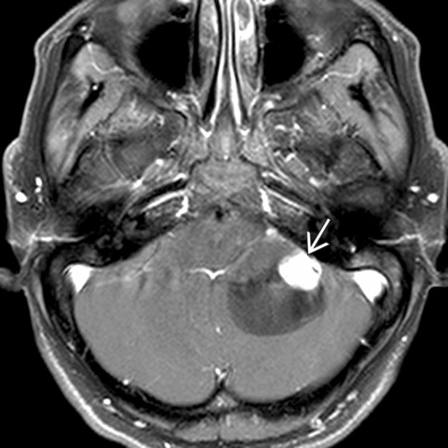
- Contrast helps differentiate solid from cystic components of tumors, such as vestibular or facial nerve schwannoma & hemangioblastoma
- DWI for possible epidermoid cyst (restricted diffusion)
- T2 thin-section, high-resolution MR
- Also sorts out solid and cystic components of lesions
- Helps with cranial nerve & vascular anatomy
- ## Helpful Clues for Common Diagnoses
- **Epidermoid Cyst**
- Key facts
- Definition: Congenital rest of epithelial tissue in CPA
- Imaging findings
- Insinuating or scalloping brainstem margin
- T1 MR: Iso- to slightly hyperintense relative to CSF
- T1 C+ MR: Typically nonenhancing cyst; may be hard to see
- Minimal marginal cyst enhancement in 25%
- Soft tissue enhancement present in rare cases of malignant degeneration to squamous cell carcinoma; PET avidity can help confirm suspicion
- T2 MR: High-signal, well-circumscribed CPA mass
- FLAIR: Lack of complete fluid suppression
- DWI: Restricted diffusion (high signal) makes diagnosis
- **Arachnoid Cyst**
- Key facts
- Definition: Congenital lesion resulting from failure of embryonic meninges to merge results in cyst between split arachnoid membrane
- Imaging findings
- Fills cistern with rounded margins
- T1 C+ MR: No enhancement
- FLAIR: Lesion attenuates (black like CSF)
- DWI: Isointense to CSF (no restriction)
- ## Helpful Clues for Less Common Diagnoses
- **Vestibular Schwannoma W****ith Intramural Cyst(s)**
- Key facts
- Definition: Vestibular schwannoma may have either intramural or extramural cysts associated
- Imaging findings
- Solid CPA-IAC mass with intramural cysts
- Bone CT: Larger lesions flare medial IAC component
- T1 C+ MR: Enhancing solid tumor component ± intramural cysts (common) ± extramural cyst (rare)
- Microhemorrhages on T2* GRE or SWI favor schwannoma over meningioma
- **Hemangioblastoma**
- Key facts
- Definition: Benign tumor composed of stromal cells in small blood vessels of CNS
- Adult with intraaxial posterior fossa mass abutting pia
- Associated with von Hippel Lindau (VHL) in 25-40%
- Supratentorial hemangioblastomas more likely VHL related
- Imaging findings
- Cerebellar cystic & solid tumor
- T1 C+ MR: 60% of tumors with solid enhancing & cystic components (40% solid only)
- **Large Endolymphatic Sac Anomaly (IP-II)**
- Key facts
- Bilateral congenital sensorineural hearing loss that appears in child with cascading hearing loss pattern
- Most common congenital imaging abnormality
- Recommendation to avoid contact sports
- Imaging findings
- Bone CT: Enlarged bony vestibular aqueduct
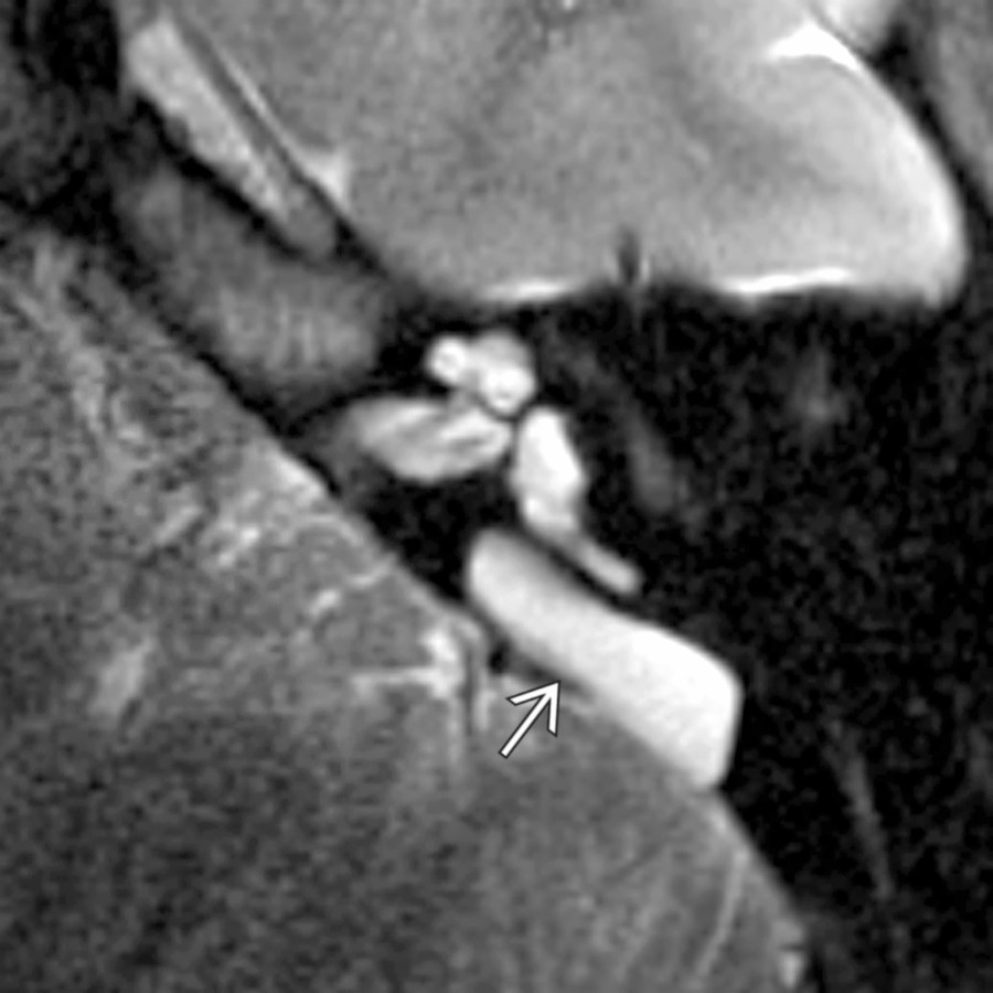
- T2 high-resolution MR: Enlarged endolymphatic sac & duct + mild cochlear malformation (modiolar deficiency, deficient apical septation, & scalar chamber asymmetry)
- **Neurocysticercosis**
- Key facts
- Definition: Intracranial infection caused by pork tapeworm (*Taenia solium*)
- Imaging findings
- Cysts with "dots" inside
- Appearance varies with stage
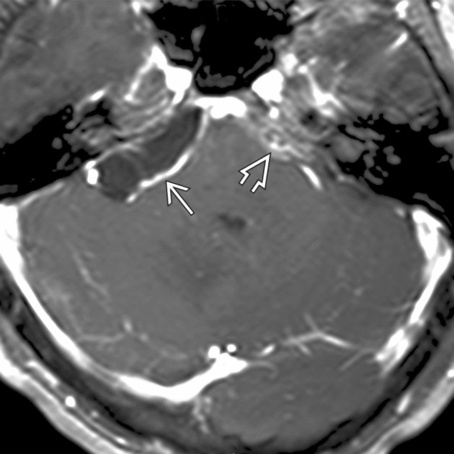
- T1 C+ MR: Cysts with enhancing thin or thick wall
- May be associated with hydrocephalus
- ## Helpful Clues for Rare Diagnoses
- **Vestibular Schwannoma W****ith Extramural Cyst**
- Key facts
- Vestibular schwannoma with extramural cyst
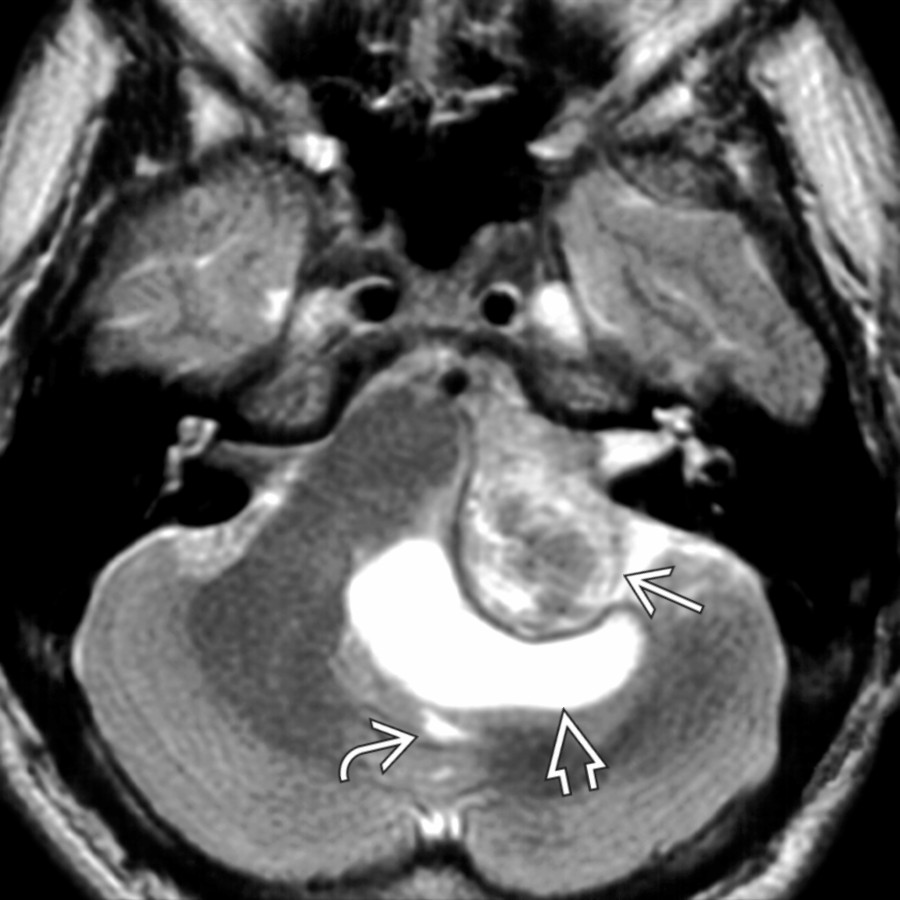
- Cyst may be "trapped CSF" or actual arachnoid cyst
- Neurootologists call extramural cyst "herald cyst"
- Imaging findings
- CPA-IAC mass with extramural cyst
- Bone CT: Large lesions flare IAC medial component
- T1 C+ MR: Enhancing solid tumor component ± extramural cyst
- Microhemorrhages on T2* GRE or SWI favor schwannoma over meningioma
- **Facial Nerve Schwannoma in CPA-IAC W****ith Cyst**
- Key facts
- Rare CPA-IAC mass with labyrinthine tail involving labyrinthine segment of facial nerve canal
- Often presents with hearing loss before facial nerve symptoms
- Imaging findings
- Bone CT: Labyrinthine segment of facial nerve may be enlarged
- T1 C+ MR: Enhancing tubular mass in CPA-IAC & labyrinthine segment of facial nerve; intramural or extramural cyst visible
- **Schwannoma, Trigeminal, Skull Base**
- Key facts
- Most common presentation is ipsilateral facial pain
- Imaging findings
- CPA cystic enhancing mass with tail extending toward or into Meckel cave is characteristic
- Look for denervation of masticator space muscles
- **Neurenteric****C****yst**
- Key facts
- Arises at time of notochordal development during transitory existence of neurenteric canal
- Benign endodermal lesion of CNS
- Often presents as incidental rounded to ovoid mass in prepontine cistern
- Imaging findings
- MR shows intermediate- to high-signal T1 prepontine "cystic" mass
- **Jugular Foramen Schwannoma W****ith Intramural Cyst**
- Key facts
- Presents with mixture of 9-12 cranial neuropathy
- Imaging findings
- Bone CT: Enlarged, sharply marginated jugular foramen
- T1 MR: Iso-g to hyperintense compared to CSF
- T1 C+ MR: Enhancing mass with intramural cysts arising from jugular foramen
- Intramural cysts in 25% of jugular foramen schwannomas
- Other MR findings: Mass projects superomedially into CPA cistern, often with brainstem compression
- **Cystic Metastasis in CPA**
- Key facts
- Usually in patients with known primary cancer
- Imaging findings
- T1 C+ MR: Enhancing mass with cystic component may mimic schwannoma or cystic meningioma
- Likely to exhibit rapid growth pattern and more symptoms, such as facial nerve palsy, than benign tumors like schwannoma
- **Endolymphatic Sac Tumor**
- Key facts
- Sporadic or associated with VHL
- Bilateral tumors &/or concurrent hemangioblastoma indicates VHL
- Imaging findings
- Bone CT: Permeative destruction of posterior petrous temporal bone centered at vestibular aqueduct
- T1 MR: Hyperintense foci due to internal hemorrhage are characteristic
- T1 C+ MR: Partly cystic, heterogenous enhancing mass
- DOTATATE scan may show mild uptake due to somatostatin receptor type 2A expression in tumor vasculature not tumor cells
- **Meningioma, Cystic**
- T1 C+ MR: Rare, heterogeneous, partly cystic enhancing mass
- Rare meningioma variant that mimics schwannoma; most are clear cell subtype
## References
# Selected References
1. [Ali NE et al: Natural history of cystic vestibular schwannomas. Ann Otol Rhinol Laryngol. 132(7):795-9, 2023](http://www.ncbi.nlm.nih.gov/pubmed/?term=35993287%5Bpmid%5D)
1. [Adachi S et al: Unusual imaging characteristics of cystic meningioma in cerebellopontine angle. Neuroradiol J. 35(6):777-9, 2022](http://www.ncbi.nlm.nih.gov/pubmed/?term=35503008%5Bpmid%5D)
1. [Geng Y et al: Endolymphatic sac tumour: exploring the role of CT and MRI features in the diagnosis of 22 cases. Clin Radiol. 77(8):e592-8, 2022](http://www.ncbi.nlm.nih.gov/pubmed/?term=35643739%5Bpmid%5D)
1. [Talukdar R et al: Endolymphatic sac tumor: single-institution series of seven cases with updated review of literature. Eur Arch Otorhinolaryngol. 279(5):2591-8, 2022](http://www.ncbi.nlm.nih.gov/pubmed/?term=34410472%5Bpmid%5D)
1. [Parlak S et al: 3 Tesla MR imaging of the large endolymphatic duct and sac anomaly with audiological correlation. Eur J Radiol. 145:110064, 2021](http://www.ncbi.nlm.nih.gov/pubmed/?term=34839211%5Bpmid%5D)
1. [Saigal G et al: Utility of microhemorrhage as a diagnostic tool in distinguishing vestibular schwannomas from other cerebellopontine angle (CPA) Tumors. Indian J Otolaryngol Head Neck Surg. 73(3):321-6, 2021](http://www.ncbi.nlm.nih.gov/pubmed/?term=34471620%5Bpmid%5D)
1. [Lou R et al: 68Ga-DOTATATE uptake in an endolymphatic sac tumor: radiologic-pathologic correlation. Clin Nucl Med. 45(7):563-5, 2020](http://www.ncbi.nlm.nih.gov/pubmed/?term=32433163%5Bpmid%5D)
1. [Connor SEJ et al: Is CT or MRI the optimal imaging investigation for the diagnosis of large vestibular aqueduct syndrome and large endolymphatic sac anomaly? Eur Arch Otorhinolaryngol. 276(3):693-702, 2019](http://www.ncbi.nlm.nih.gov/pubmed/?term=30635710%5Bpmid%5D)
1. [Eliezer M et al: Clinical and radiological characteristics of malignant tumors located to the cerebellopontine angle and/or internal acoustic meatus. Otol Neurotol. 40(9):1237-45, 2019](http://www.ncbi.nlm.nih.gov/pubmed/?term=31469787%5Bpmid%5D)
1. [Le H et al: Clinicoradiologic characteristics of endolymphatic sac tumors. Eur Arch Otorhinolaryngol. 276(10):2705-14, 2019](http://www.ncbi.nlm.nih.gov/pubmed/?term=31197530%5Bpmid%5D)
1. [Touska P et al: Temporal bone tumors: an imaging update. Neuroimaging Clin N Am. 29(1):145-72, 2019](http://www.ncbi.nlm.nih.gov/pubmed/?term=30466638%5Bpmid%5D)
1. [Schnack DT et al: Sporadic endolymphatic sac tumor-a very rare cause of hearing loss, tinnitus, and dizziness. J Int Adv Otol. 13(2):289-91, 2017](http://www.ncbi.nlm.nih.gov/pubmed/?term=28716765%5Bpmid%5D)
1. [Agarwal A: Intracranial trigeminal schwannoma. Neuroradiol J. 28(1):36-41, 2015](http://www.ncbi.nlm.nih.gov/pubmed/?term=25924170%5Bpmid%5D)
1. [Dispenza F et al: Imaging of vestibular schwannoma with prevalent cystic component: cystic vestibular schwannoma. Otol Neurotol. 30(5):681-2, 2009](http://www.ncbi.nlm.nih.gov/pubmed/?term=18716565%5Bpmid%5D)
1. [Piccirillo E et al: Cystic vestibular schwannoma: classification, management, and facial nerve outcomes. Otol Neurotol. 30(6):826-34, 2009](http://www.ncbi.nlm.nih.gov/pubmed/?term=19704364%5Bpmid%5D)
1. [Zhang L et al: Trigeminal schwannomas: a report of 42 cases and review of the relevant surgical approaches. Clin Neurol Neurosurg. 111(3):261-9, 2009](http://www.ncbi.nlm.nih.gov/pubmed/?term=19081670%5Bpmid%5D)
1. [Bonneville F et al: Imaging of cerebellopontine angle lesions: an update. Part 1: enhancing extra-axial lesions. Eur Radiol. 17(10):2472-82, 2007](http://www.ncbi.nlm.nih.gov/pubmed/?term=17562049%5Bpmid%5D)
1. [Bonneville F et al: Imaging of cerebellopontine angle lesions: an update. Part 2: intra-axial lesions, skull base lesions that may invade the CPA region, and non-enhancing extra-axial lesions. Eur Radiol. 17(11):2908-20, 2007](http://www.ncbi.nlm.nih.gov/pubmed/?term=17569053%5Bpmid%5D)
1. [Kiliçkesmez O: Endolymphatic sac tumor in a patient with von Hippel-Lindau disease: MR imaging findings. Diagn Interv Radiol. 12(1):14-6, 2006](http://www.ncbi.nlm.nih.gov/pubmed/?term=16538578%5Bpmid%5D)
1. [Patel NP et al: The radiologic diagnosis of endolymphatic sac tumors. Laryngoscope. 116(1):40-6, 2006](http://www.ncbi.nlm.nih.gov/pubmed/?term=16481807%5Bpmid%5D)
1. [Preece MT et al: Intracranial neurenteric cysts: imaging and pathology spectrum. AJNR Am J Neuroradiol. 27(6):1211-6, 2006](http://www.ncbi.nlm.nih.gov/pubmed/?term=16775266%5Bpmid%5D)
1. [Nelson MD Jr et al: A different approach to cysts of the posterior fossa. Pediatr Radiol. 34(9):720-32, 2004](http://www.ncbi.nlm.nih.gov/pubmed/?term=15316692%5Bpmid%5D)
1. [Bonneville F et al: Unusual lesions of the cerebellopontine angle: a segmental approach. Radiographics. 21(2):419-38, 2001](http://www.ncbi.nlm.nih.gov/pubmed/?term=11259705%5Bpmid%5D)
1. [Davidson HC et al: MR evaluation of vestibulocochlear anomalies associated with large endolymphatic duct and sac. AJNR Am J Neuroradiol. 20(8):1435-41, 1999](http://www.ncbi.nlm.nih.gov/pubmed/?term=10512225%5Bpmid%5D)
1. [Koeller KK et al: Congenital cystic masses of the neck: radiologic-pathologic correlation. Radiographics. 19(1):121-46; quiz 152-3, 1999](http://www.ncbi.nlm.nih.gov/pubmed/?term=9925396%5Bpmid%5D)
1. [Lau KY et al: MRI demonstration of subarachnoid neurocysticercosis simulating metastatic disease. Neuroradiology. 40(11):724-6, 1998](http://www.ncbi.nlm.nih.gov/pubmed/?term=9860122%5Bpmid%5D)
1. [Mukherji SK et al: Papillary endolymphatic sac tumors: CT, MR imaging, and angiographic findings in 20 patients. Radiology. 202(3):801-8, 1997](http://www.ncbi.nlm.nih.gov/pubmed/?term=9051037%5Bpmid%5D)
1. [Tong KA et al: Large vestibular aqueduct syndrome: a genetic disease? AJR Am J Roentgenol. 168(4):1097-101, 1997](http://www.ncbi.nlm.nih.gov/pubmed/?term=9124122%5Bpmid%5D)
1. [Friedman DP et al: Vascular neoplasms and malformations, ischemia, and hemorrhage affecting the spinal cord: MR imaging findings. AJR Am J Roentgenol. 162(3):685-92, 1994](http://www.ncbi.nlm.nih.gov/pubmed/?term=8109522%5Bpmid%5D)
1. [Kollias SS et al: Cystic malformations of the posterior fossa: differential diagnosis clarified through embryologic analysis. Radiographics. 13(6):1211-31, 1993](http://www.ncbi.nlm.nih.gov/pubmed/?term=8031352%5Bpmid%5D)
1. [Smirniotopoulos JG et al: Cerebellopontine angle masses: radiologic-pathologic correlation. Radiographics. 13(5):1131-47, 1993](http://www.ncbi.nlm.nih.gov/pubmed/?term=8210595%5Bpmid%5D)
## Images
### Selected Images
 and along the right cerebellar hemisphere
  . DWI MR sequence would show restricted diffusion. FLAIR would show lack of complete fluid suppression.](images/app.statdx.com_image_thumbnail_9c20ab07-d905-4fe9-825e-f669857ed535_annotated_true_size_900_quality_90_e351d8febf9476ec73cfce123b434851a06e0145.jpg)
**Epidermoid Cyst**
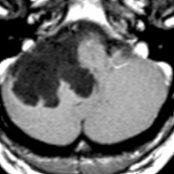
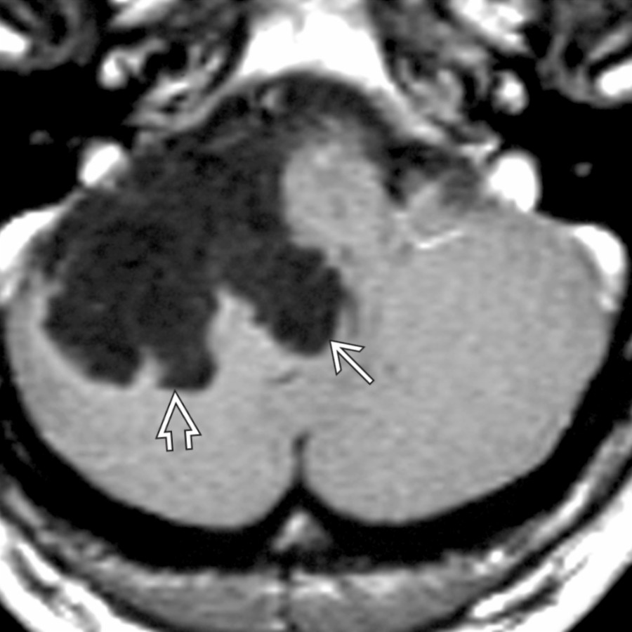
*Axial T1 C+ MR reveals a low signal intensity, nonenhancing epidermoid cyst that insinuates into the foramen of Luschka and along the right cerebellar hemisphere . DWI MR sequence would show restricted diffusion. FLAIR would show lack of complete fluid suppression.*
**Epidermoid Cyst**
*Axial T1 C+ MR reveals a low signal intensity, nonenhancing epidermoid cyst that insinuates into the foramen of Luschka and along the right cerebellar hemisphere . DWI MR sequence would show restricted diffusion. FLAIR would show lack of complete fluid suppression.*
**Arachnoid Cyst**
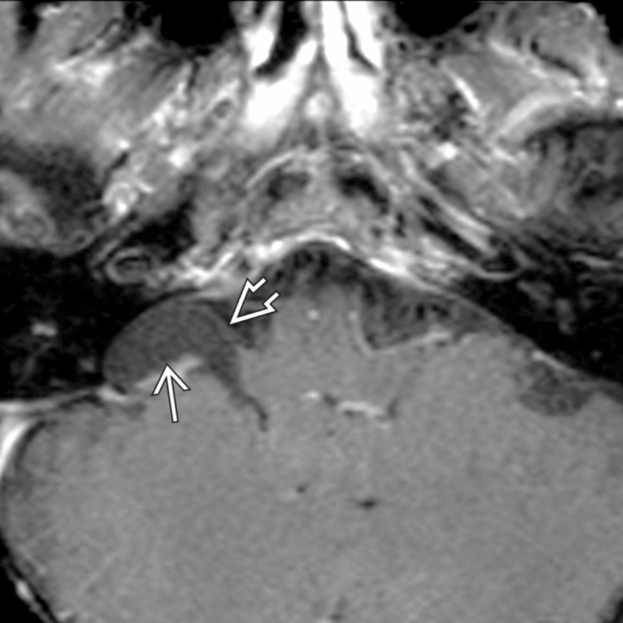
*Axial T1 C+ FS MR demonstrates a right cerebellopontine angle (CPA) cistern arachnoid cyst displacing the proximal facial and vestibulocochlear nerves anteriorly .*
**Vestibular Schwannoma With Intramural Cyst(s)**
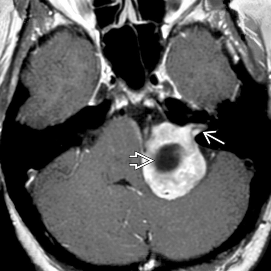
*Axial T1 C+ MR shows a large enhancing vestibular schwannoma projecting from the internal auditory canal (IAC) into the CPA cistern. The tumor has a large intramural cyst that compresses the brainstem and cerebellum.*
**Epidermoid Cyst**
*Axial T1 C+ MR reveals a low signal intensity, nonenhancing epidermoid cyst that insinuates into the foramen of Luschka and along the right cerebellar hemisphere . DWI MR sequence would show restricted diffusion. FLAIR would show lack of complete fluid suppression.*

**Epidermoid Cyst**
*Axial T1 C+ MR reveals a low signal intensity, nonenhancing epidermoid cyst that insinuates into the foramen of Luschka and along the right cerebellar hemisphere . DWI MR sequence would show restricted diffusion. FLAIR would show lack of complete fluid suppression.*

**Arachnoid Cyst**
*Axial T1 C+ FS MR demonstrates a right cerebellopontine angle (CPA) cistern arachnoid cyst displacing the proximal facial and vestibulocochlear nerves anteriorly .*

**Vestibular Schwannoma With Intramural Cyst(s)**
*Axial T1 C+ MR shows a large enhancing vestibular schwannoma projecting from the internal auditory canal (IAC) into the CPA cistern. The tumor has a large intramural cyst that compresses the brainstem and cerebellum.*
 .](images/app.statdx.com_image_thumbnail_c0eea3bd-14f3-436b-bd9e-2818f22f0815_annotated_true_size_900_quality_90_a453d30064cbb01de279d0559dfd07ad5bee5732.jpg)
**Vestibular Schwannoma With Intramural Cyst(s)**
*Axial T2 MR shows a heterogeneous CPA mass extending into the left internal auditory canal. Note numerous large intramural tumor cysts .*
**Hemangioblastoma**
*Axial T1 C+ FS MR shows a superficial, intracerebellar mixed cystic-solid hemangioblastoma projecting into the left CPA cistern area. The solid nodule is avidly enhancing.*
**Large Endolymphatic Sac Anomaly (IP-II)**
*Axial T2WI MR shows a large endolymphatic sac within the posterior wall of the temporal bone. CT (not shown) would reveal a large bony vestibular aqueduct in this patient with large endolymphatic sac anomaly.*
**Neurocysticercosis**
*Axial T1 C+ FS MR demonstrates a cystic mass in the right CPA cistern with a peripherally enhancing wall . Adjacent enhancing, thickened meninges are also seen.*
**Vestibular Schwannoma With Extramural Cyst**
*Axial T2WI MR shows a vestibular schwannoma projecting from the IAC into the CPA cistern. An associated (extramural) arachnoid cyst is visible compressing the brainstem and 4th ventricle .*
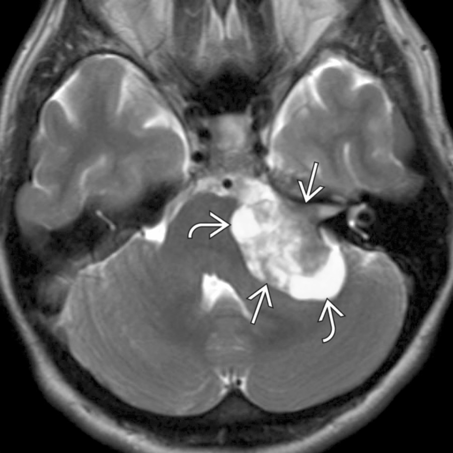
**Vestibular Schwannoma With Intramural Cyst(s)**
*Axial T2 MR shows a heterogeneous CPA mass extending into the left internal auditory canal. Note numerous large intramural tumor cysts .*

**Hemangioblastoma**
*Axial T1 C+ FS MR shows a superficial, intracerebellar mixed cystic-solid hemangioblastoma projecting into the left CPA cistern area. The solid nodule is avidly enhancing.*

**Large Endolymphatic Sac Anomaly (IP-II)**
*Axial T2WI MR shows a large endolymphatic sac within the posterior wall of the temporal bone. CT (not shown) would reveal a large bony vestibular aqueduct in this patient with large endolymphatic sac anomaly.*

**Neurocysticercosis**
*Axial T1 C+ FS MR demonstrates a cystic mass in the right CPA cistern with a peripherally enhancing wall . Adjacent enhancing, thickened meninges are also seen.*

**Vestibular Schwannoma With Extramural Cyst**
*Axial T2WI MR shows a vestibular schwannoma projecting from the IAC into the CPA cistern. An associated (extramural) arachnoid cyst is visible compressing the brainstem and 4th ventricle .*
 from CNVII denervation.](images/app.statdx.com_image_thumbnail_fd005ccd-437a-4a14-9656-afa375f84039_annotated_true_size_900_quality_90_ed15f34e2656f0d3eb999951c956965f618988d6.jpg)
**Facial Nerve Schwannoma in CPA-IAC With Cyst**
*Axial T1 C+ MR reveals an enhancing CPA mass , with intramural cysts, which projected into the IAC but did not involve the labyrinthine segment. Note atrophy of the right platysma muscle from CNVII denervation.*
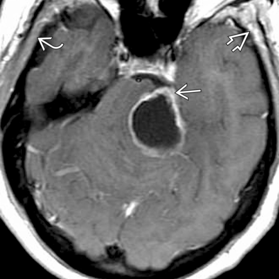
**Schwannoma, Trigeminal, Skull Base**
*Axial T1 C+ MR demonstrates a large CPA cystic mass "pointing" toward the Meckel cave , providing a clue to the origin of this mass. Note chronic denervation changes of volume loss and fatty infiltration in the left suprazygomatic masticator space compared to the right , partly seen here.*

**Neurenteric Cyst**
*Axial T1WI MR shows a small mass anterior to the pontomedullary junction . The neurenteric cyst is well delineated, demonstrating nearly isointense signal to brain, and does not enhance significantly.*
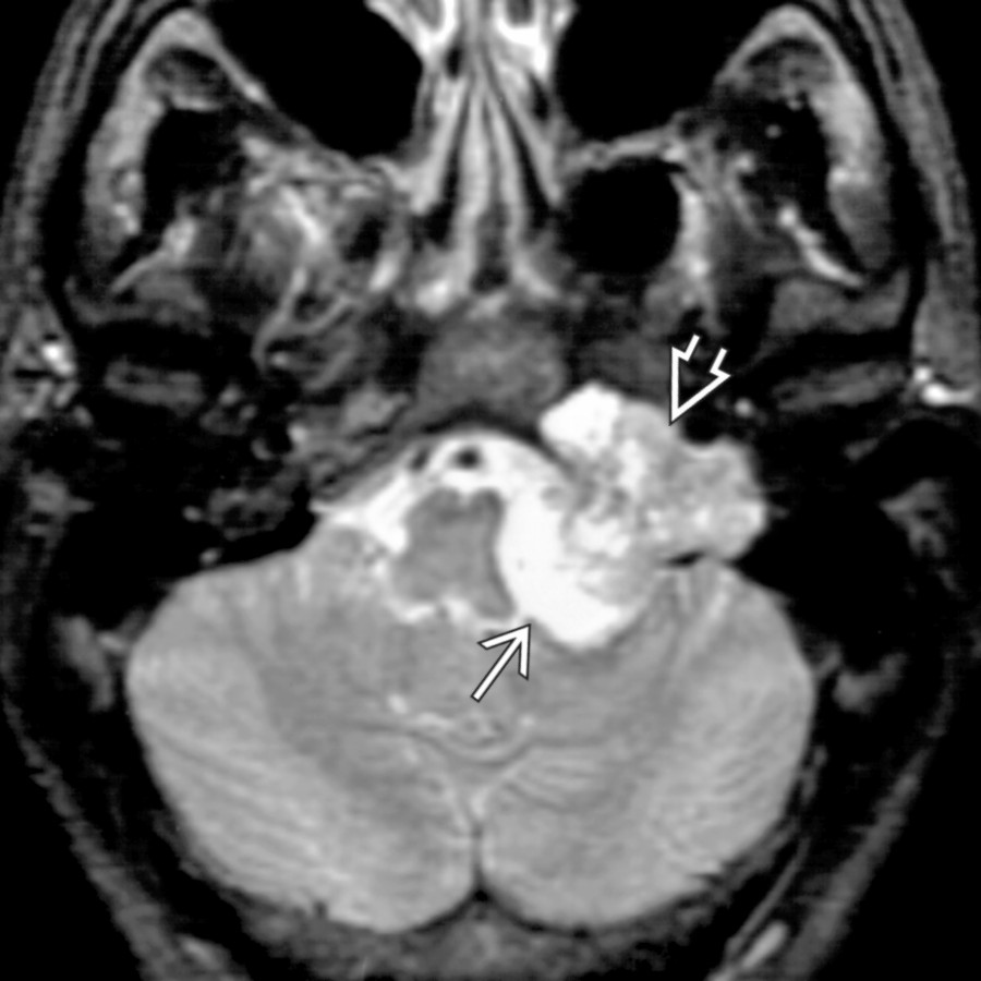
**Jugular Foramen Schwannoma With Intramural Cyst**
*Axial T2 FS MR reveals a large, sharply marginated lesion expanding the left jugular foramen . The high-signal mass projects medially into the low CPA cistern where it compresses the brainstem .*
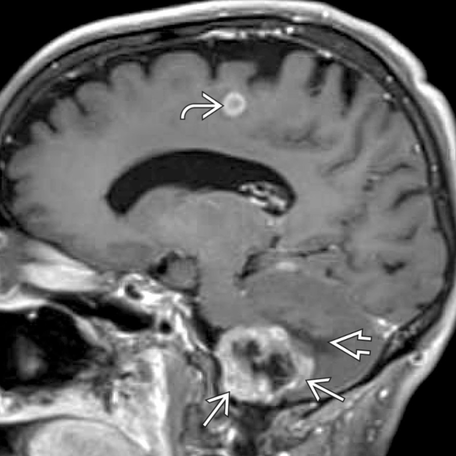
**Cystic Metastasis in CPA**
*Parasagittal T1 C+ FS MR shows a heterogeneous, cystic enhancing mass in the right CPA . Adjacent brain edema is noted . Although the mass might mimic a cystic schwannoma, the presence of a 2nd enhancing mass and a clinical history of lung cancer helps establish diagnosis of metastasis.*
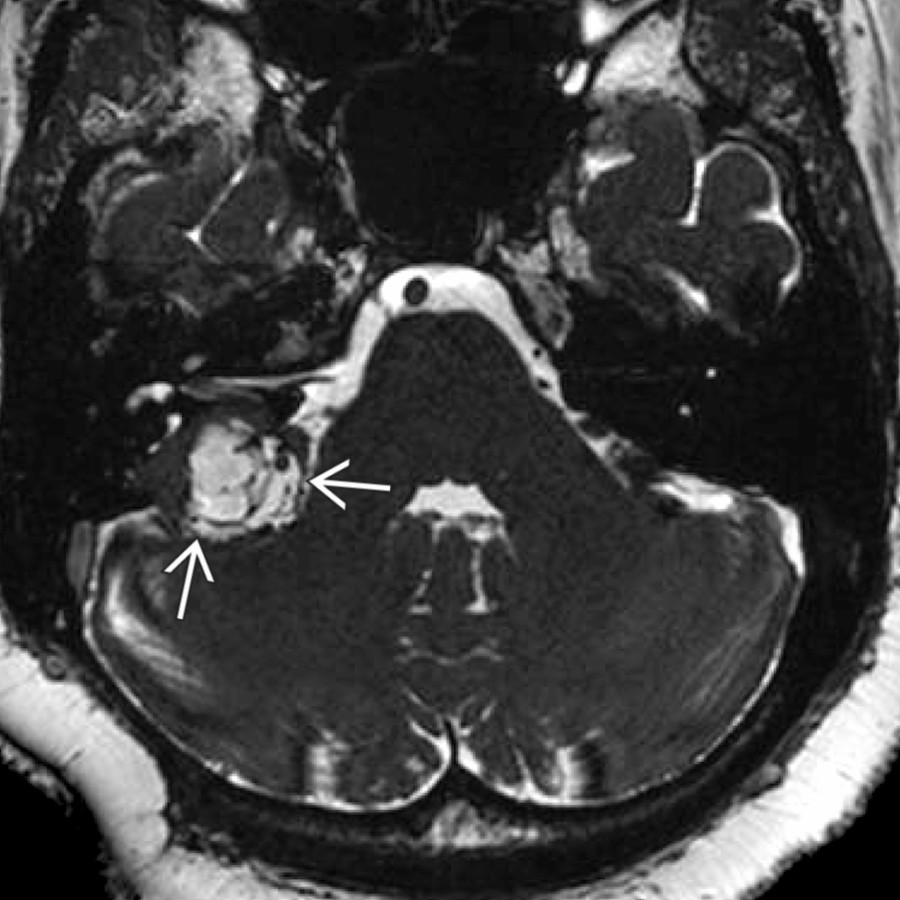
**Endolymphatic Sac Tumor**
*Axial high-resolution T2 MR shows a heterogeneous cystic CPA mass . The key to diagnosis is identifying the mass origin from the posterior temporal bone in the expected location of the endolymphatic sac.*
### Additional Images
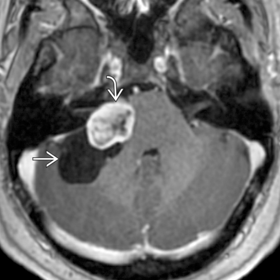
**Vestibular Schwannoma With Extramural Cyst**
*Axial T1 C+ MR shows an atypical heterogeneously enhancing left vestibular schwannoma with an associated arachnoid cyst .*
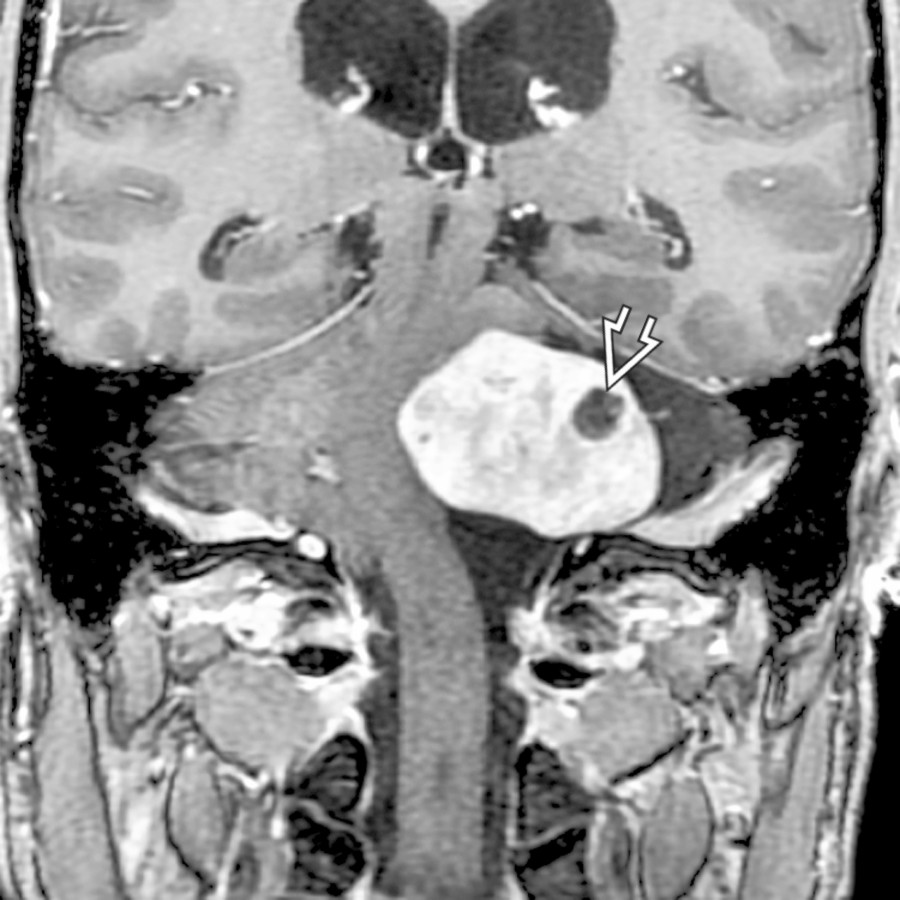
**Jugular Foramen Schwannoma With Intramural Cyst**
*Coronal T1 C+ MR shows variant MR case of a jugular foramen schwannoma with intramural cyst and very large cisternal component. The jugular foramen connection is seen on the next image.*

**Jugular Foramen Schwannoma With Intramural Cyst**
*Coronal T1 C+ MR reveals an enhancing schwannoma in the jugular foramen . The tumor projects superomedially to fill the CPA cistern and compress the brainstem.*
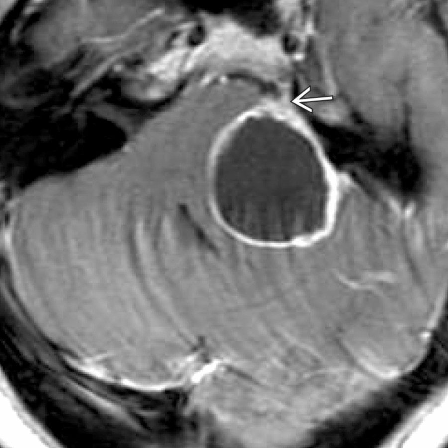
**Schwannoma, Trigeminal, Skull Base**
*Axial T1 C+ MR in a patient presenting with left facial pain demonstrates a peripherally enhancing cystic mass in the left CPA with a vector of spread extending towards Meckel cave , that was the clue to its origin.*
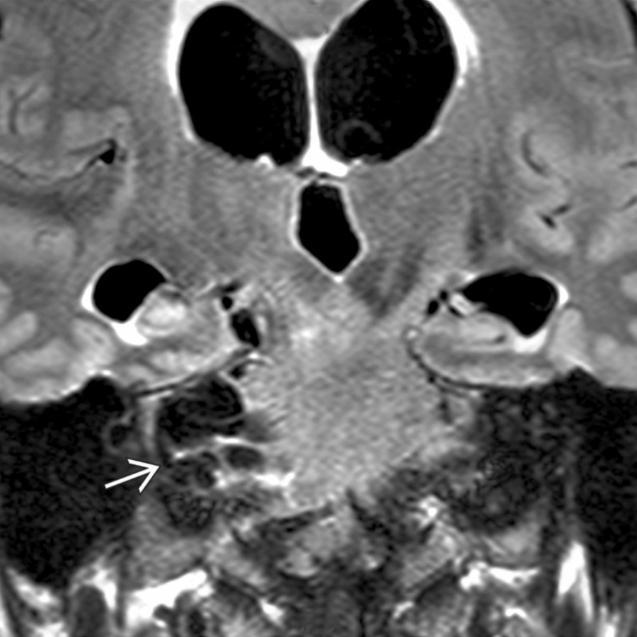
**Neurocysticercosis**
*Coronal T1 C+ FS MR shows multiple cysts in the right CPA cistern causing mass effect on the brainstem. Secondary hydrocephalus is present.*
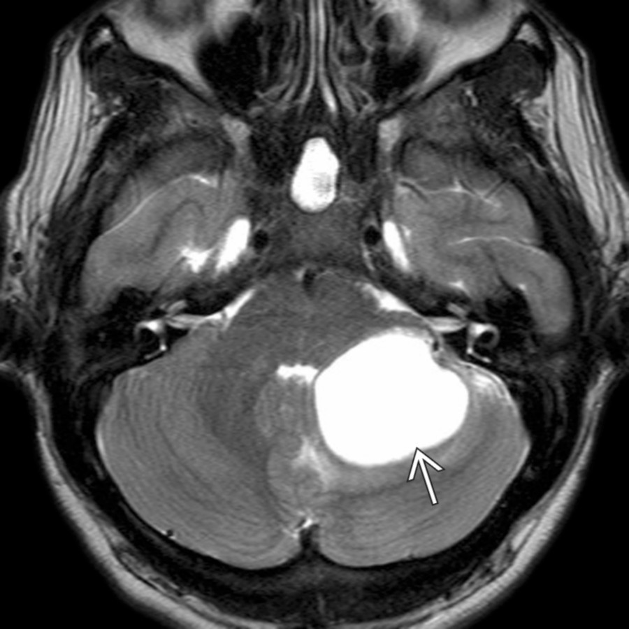
**Hemangioblastoma**
*Axial T2WI MR reveals an intracerebellar high signal hemangioblastoma projecting into the CPA cistern area. Contrast is required to define enhancing nodule if present.*
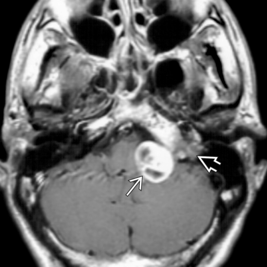
**Jugular Foramen Schwannoma With Intramural Cyst**
*Axial T1 C+ MR demonstrates an ovoid enhancing mass in the low CPA cistern . Multiple intramural cysts suggest the diagnosis of schwannoma. Extension into the jugular foramen is evident.*
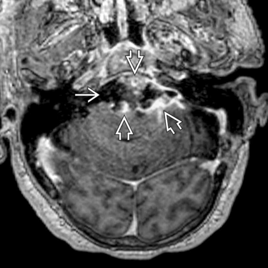
**Epidermoid Cyst**
*Axial T1 C+ MR shows nodular soft tissue enhancement along the margins of a cystic prepontine and left CPA epidermoid cyst , due to a rare complication: Malignant degeneration into squamous cell carcinoma.*
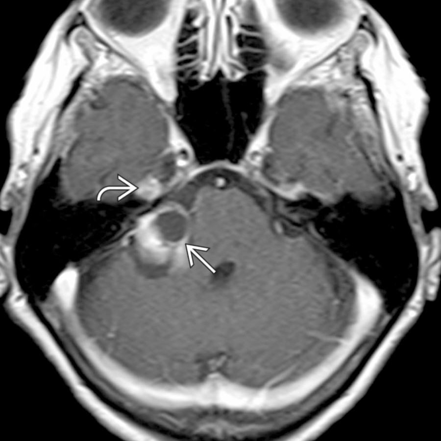
**Meningioma, Cystic**
*Axial T1 C+ MR shows a partly cystic, partly solid mass in the right CPA and posterior Meckel cave . Biopsy supported meningioma, which rarely may have cystic morphologies that mimic schwannoma.*
from CNVII denervation.](images/app.statdx.com_image_thumbnail_fd005ccd-437a-4a14-9656-afa375f84039_annotated_true_size_900_quality_90_ed15f34e2656f0d3eb999951c956965f618988d6.jpg)
**Facial Nerve Schwannoma in CPA-IAC With Cyst**
*Axial T1 C+ MR reveals an enhancing CPA mass , with intramural cysts, which projected into the IAC but did not involve the labyrinthine segment. Note atrophy of the right platysma muscle from CNVII denervation.*

**Schwannoma, Trigeminal, Skull Base**
*Axial T1 C+ MR demonstrates a large CPA cystic mass "pointing" toward the Meckel cave , providing a clue to the origin of this mass. Note chronic denervation changes of volume loss and fatty infiltration in the left suprazygomatic masticator space compared to the right , partly seen here.*

**Neurenteric Cyst**
*Axial T1WI MR shows a small mass anterior to the pontomedullary junction . The neurenteric cyst is well delineated, demonstrating nearly isointense signal to brain, and does not enhance significantly.*

**Jugular Foramen Schwannoma With Intramural Cyst**
*Axial T2 FS MR reveals a large, sharply marginated lesion expanding the left jugular foramen . The high-signal mass projects medially into the low CPA cistern where it compresses the brainstem .*

**Cystic Metastasis in CPA**
*Parasagittal T1 C+ FS MR shows a heterogeneous, cystic enhancing mass in the right CPA . Adjacent brain edema is noted . Although the mass might mimic a cystic schwannoma, the presence of a 2nd enhancing mass and a clinical history of lung cancer helps establish diagnosis of metastasis.*

**Endolymphatic Sac Tumor**
*Axial high-resolution T2 MR shows a heterogeneous cystic CPA mass . The key to diagnosis is identifying the mass origin from the posterior temporal bone in the expected location of the endolymphatic sac.*
### Additional Images

**Vestibular Schwannoma With Extramural Cyst**
*Axial T1 C+ MR shows an atypical heterogeneously enhancing left vestibular schwannoma with an associated arachnoid cyst .*

**Jugular Foramen Schwannoma With Intramural Cyst**
*Coronal T1 C+ MR shows variant MR case of a jugular foramen schwannoma with intramural cyst and very large cisternal component. The jugular foramen connection is seen on the next image.*

**Jugular Foramen Schwannoma With Intramural Cyst**
*Coronal T1 C+ MR reveals an enhancing schwannoma in the jugular foramen . The tumor projects superomedially to fill the CPA cistern and compress the brainstem.*

**Schwannoma, Trigeminal, Skull Base**
*Axial T1 C+ MR in a patient presenting with left facial pain demonstrates a peripherally enhancing cystic mass in the left CPA with a vector of spread extending towards Meckel cave , that was the clue to its origin.*

**Neurocysticercosis**
*Coronal T1 C+ FS MR shows multiple cysts in the right CPA cistern causing mass effect on the brainstem. Secondary hydrocephalus is present.*

**Hemangioblastoma**
*Axial T2WI MR reveals an intracerebellar high signal hemangioblastoma projecting into the CPA cistern area. Contrast is required to define enhancing nodule if present.*

**Jugular Foramen Schwannoma With Intramural Cyst**
*Axial T1 C+ MR demonstrates an ovoid enhancing mass in the low CPA cistern . Multiple intramural cysts suggest the diagnosis of schwannoma. Extension into the jugular foramen is evident.*

**Epidermoid Cyst**
*Axial T1 C+ MR shows nodular soft tissue enhancement along the margins of a cystic prepontine and left CPA epidermoid cyst , due to a rare complication: Malignant degeneration into squamous cell carcinoma.*

**Meningioma, Cystic**
*Axial T1 C+ MR shows a partly cystic, partly solid mass in the right CPA and posterior Meckel cave . Biopsy supported meningioma, which rarely may have cystic morphologies that mimic schwannoma.*