---
title: "Dementia With Lewy Bodies"
docid: "e8e46d1d-46d2-4e5a-880f-f025a84c5871"
authors:
- key: "1fa14dfd-71ea-4960-908e-e720313bc63a"
value: "Santhosh Gaddikeri, MD"
- key: "a25c450b-3d34-4f64-bba3-cc0834813df6"
value: "Miral D. Jhaveri, MD, MBA"
breadcrumbs:
-
name: "Brain"
slug: "brain"
treeNodeId: "6d8829f1-14d7-45af-8675-255189aa526a"
-
name: "Diagnosis"
slug: "diagnosis"
treeNodeId: "51c00394-446e-4a38-94af-d3b1d14d34e8"
-
name: "Pathology-Based Diagnoses"
slug: "pathology-based-diagnoses"
treeNodeId: "d9d3a8ed-f21b-4831-8c77-591a3500ef77"
-
name: "Acquired Toxic/Metabolic/Degenerative Disorders"
slug: "acquired-toxicmetabolicdegenerativ-"
treeNodeId: "ba3cfeaf-64d9-4117-91e8-d2ce58783fc5"
-
name: "Dementias and Degenerative Disorders"
slug: "dementias-and-degenerative-disorde-"
treeNodeId: "6381104d-7a4c-4be5-bb19-3cd90837d547"
-
name: "Dementia With Lewy Bodies"
slug: "dementia-with-lewy-bodies"
treeNodeId: null
category: "Brain"
cmeTopicId: "3a0ceb4e-585c-4343-b8c3-30199a551c37"
documentVersionId: "8e3dd335-218e-43f3-9635-d2a11eb0a445"
imageCount: 8
lastUpdated: "08/10/20"
pageDescription: "Dementia With Lewy Bodies"
pageKeywords: "Brain, Diagnosis, Pathology-Based Diagnoses, Acquired Toxic/Metabolic/Degenerative Disorders, Dementias and Degenerative Disorders, Dementia With Lewy Bodies"
pageTitle: "Dementia With Lewy Bodies | STATdx"
enhancedTitle: "Dementia With Lewy Bodies"
type: "DX"
references: true
breadcrumbs:
- "Brain"
- "Diagnosis"
- "Pathology-Based Diagnoses"
- "Acquired Toxic/Metabolic/Degenerative Disorders"
- "Dementias and Degenerative Disorders"
- "Dementia With Lewy Bodies"
---
# KEY FACTS
- ## Terminology
- Progressive neurodegenerative dementia
- Parkinsonism, visual hallucinations prominent
- Caused by abnormal accumulation of α-synuclein protein
- ## Imaging
- MR may differentiate Alzheimer disease (AD) from dementia with Lewy bodies (DLB)
- PET, SPECT most useful for DLB diagnosis
- Voxel-based morphometry
- Relatively preserved hippocampal/medial temporal lobe volume in DLB vs. AD
- ↓ volume of hypothalamus, substantia innominata, & putamen in DLB vs. AD
- FDG PET
- ↓ in glucose metabolism in occipital cortex, especially primary visual cortex
- F-18 fluorodopa-PET: ↓ striatal dopamine uptake in DLB vs. AD
- SPECT: Occipital lobe hypoperfusion, especially visual cortex
- 123 FP-CIT SPECT: ↓ uptake in striatum in DLB vs. AD
- ## Top Differential Diagnoses
- Parkinson disease-associated dementia (PDD)
- Similar clinical, pathological, imaging features with DLB
- AD
- Frontotemporal lobar degeneration (FTLD)
- Vascular dementia
- ## Pathology
- Pathologic aggregation of α-synuclein protein in neurites (LB)
- ## Diagnostic Checklist
- Unlike AD, medial temporal lobe atrophy not prominent feature
# TERMINOLOGY
- ## Abbreviations
- Dementia with Lewy bodies (DLB)
- ## Definitions
- Neurodegenerative dementia characterized by cognitive fluctuations, visual hallucinations, & motor parkinsonism
- Caused by pathologic aggregation of α-synuclein protein in neurites (LB)
# IMAGING
- ## General Features
- ### Best diagnostic clue
- MR may differentiate Alzheimer disease (AD) from DLB
- PET, SPECT most useful for DLB diagnosis
- ## Imaging Recommendations
- ### Best imaging tool
- PET or SPECT
- ## MR Findings
- ### T1WI
- Mild generalized atrophy
- ### T2WI
- Nonspecific white matter (WM) hyperintensities
- ### MRS
- ↓ WM NAA/Cr in DLB vs. healthy controls (HC)
- ↑ Cho/Cr ratios in DLB vs. HC
- Normal levels of NAA/Cr & myoinositol in DLB vs. AD
- Voxel-based morphometry
- Relatively preserved hippocampal/medial temporal lobe volume in DLB vs. AD
- ↓ volume of hypothalamus, substantia innominata, & putamen in DLB vs. AD
- ↓ gray matter in temporal, parietal, & occipital regions vs. HC
- DTI
- ↑ mean diffusivity in amygdala
- ↓ fractional anisotropy in pons & left thalamus vs. AD
- ↓ fractional anisotropy in inferior longitudinal fasciculus & inferior occipitofrontal fasciculi vs. HC
- ## Nuclear Medicine Findings
- ### PET
- FDG PET: ↓ glucose metabolism in occipital cortex & visual association cortex with relative preservation of posterior cingulate
- F-18 fluorodopa-PET: ↓ striatal dopamine uptake in DLB vs. AD
- ### MIBG scintigraphy
- ↓ myocardial uptake in DLB due to ↓ postganglionic sympathetic cardiac innervation
- SPECT
- Occipital lobe hypoperfusion, especially visual cortex
- 123 FP-CIT SPECT: Visualize DAT (dopamine transporter) loss
- ↓ uptake in striatum in DLB
# DIFFERENTIAL DIAGNOSIS
- [Parkinson Disease-Associated Dementia](/document/parkinson-disease/0bc3188a-935b-416d-b1a0-25b2d52c6399)
- Dementia typically develops at least 12 months after onset of initial parkinsonian symptoms
- Similar clinical, pathologic, imaging features to DLB
- Less pronounced atrophy in temporal, occipital, & parietal lobes vs. DLB
- [Alzheimer Disease](/document/alzheimer-disease/f71f5cf5-b1af-4c6d-b145-b4c10eec7b58)
- Parietal/temporal cortical atrophy
- Disproportionate hippocampal volume loss
- Amyloid uptake of cerebral cortex in PiB-PET
- More severe, faster rate of progression than DLB
- [Frontotemporal Lobar Degeneration](/document/frontotemporal-lobar-degeneration/49510d0e-acf7-45cb-9eb1-53f8193b0b6d)
- Asymmetric frontal, anterior temporal lobar atrophy
- Behavioral variant: Both frontal lobes atrophic
- Semantic variant: Asymmetric anterior temporal lobe atrophy
- [Vascular Dementia](/document/vascular-dementia/f59dab57-c511-4369-8fcc-592421a4b8d1)
- 2nd most common dementia (15-30%)
- WM & deep gray lacunae
- Infarcts of different ages
- Hyperintense lesions on T2WI, hypodense areas on CT, & focal atrophy suggestive of chronic infarcts
# PATHOLOGY
- ## General Features
- ### Etiology
- Accumulation of α-synuclein protein (LB)
- LB, neuronal loss in substantia nigra → dopamine depletion
- Loss of cholinergic neurons in nucleus basalis of Meynert
- → cognitive impairment, visual hallucinations
- ### Genetics
- Majority of DLB is sporadic; some are familial
- α-synuclein gene mutation on chromosome 4 (*A53T*, *E46K* mutation)
- Similar inheritance, similar genetic risk for PD
- Other genes associated with DLB include *SNCA*, *APP*, *PSEN1*/*PSEN2*, *MAPT*, *GBA*, & *APOE*
- ## Staging, Grading, & Classification
- 3 major forms: Brainstem dominant, limbic/transitional, diffuse neocortical
- ## Gross Pathologic & Surgical Features
- Nonspecific & overlap with other neurodegenerative dementias
- Cortical atrophy is less than AD
- Atrophy affects frontal, temporal, & parietal lobes, relative sparing of occipital lobes
- Amygdala & cingulate gyri can show severe atrophy
- ## Microscopic Features
- LB in substantia nigra, neocortex, limbic system
- α-synuclein protein aggregates: Pale eosinophilic inclusions
- α-synuclein
- Physiologic function: Synaptic transmission, neuroprotective effect
- Predominantly expressed in neurons
- Lewy neurites in hippocampus, amygdala, brainstem nuclei
- Neuronal loss in substantia nigra, locus ceruleus, nucleus basalis of Meynert, dorsal raphe nuclei
- Relative preservation of cortical neurons
- Superficial microvacuolation of cerebral cortex, especially temporal cortex in severe cases
- 80% have associated AD-like pathology
- Neuritic/diffuse plaques or neurofibrillary tangles
# CLINICAL ISSUES
- ## Presentation
- ### Most common signs/symptoms
- Cognitive fluctuations, visual hallucinations, parkinsonism
- Dysautonomia & sleep disorders
- Clinical criteria for DLB diagnosis
- Core clinical features
- Fluctuating cognition with pronounced variations in attention & alertness
- Recurrent visual hallucinations (typically well formed & detailed)
- REM sleep behavior disorder (May precede cognitive decline)
- 1 or more spontaneous cardinal features of parkinsonism (bradykinesia, rest tremor, rigidity)
- Supportive clinical features
- Severe sensitivity to antipsychotic agents
- Postural instability, repeated falls
- Syncope or other transient episodes of unresponsiveness
- Severe autonomic dysfunction (constipation, orthostatic hypotension, urinary incontinence)
- Hypersomnia, hyposmia, hallucinations in other modalities, systematized delusions
- Apathy, anxiety, & depression
- Indicative biomarkers
- ↓ dopamine transporter uptake in basal ganglia by SPECT or PET
- Abnormal (low-uptake) I-123-MIBG myocardial scintigraphy
- Polysomnographic confirmation of REM sleep without atonia
- Supportive biomarkers
- Relative preservation of medial temporal lobe structures on CT/MR
- Generalized low uptake on SPECT/PET perfusion/metabolism scan with ↓ occipital activity ± cingulate island sign on FDG PET imaging
- Prominent posterior slow-wave activity on EEG with periodic fluctuations in pre-alpha/theta range
- Probable DLB
- ≥ 2 core clinical features of DLB with or without indicative biomarkers; **or**
- Only 1 core clinical feature but with ≥ 1 indicative biomarkers
- Probable DLB should not be diagnosed on basis of biomarkers alone
- Possible DLB
- Only 1 core clinical feature of DLB with no indicative biomarker evidence; **or**
- ≥ 1 indicative biomarkers but no core clinical features
- DLB is less likely
- Presence of any other physical illness or brain disorder, including cerebrovascular disease, sufficient to account in part or in total for clinical picture
- If parkinsonian features are only core clinical feature & appear for 1st time at stage of severe dementia
- ## Demographics
- ### Age
- 55-85 years; age is only risk factor
- Average at presentation is 75 years
- ### Ethnicity
- LB formation more common in African Americans than Caucasians, but clinical diagnosis of DLB is not significantly different
- ### Sex
- M:F = 4:1
- ### Epidemiology
- 5% of general population & 30% of dementia cases
- 2nd most common neurodegenerative dementia (after AD)
- Incidence rate of 0.1% per year in general population & 3.2% for new dementia cases
- ## Natural History & Prognosis
- Average survival after diagnosis < 8 years
- ## Treatment
- ### Options, risks, complications
- No disease-modifying treatments for DLB
- Symptomatic, targeted toward specific disease manifestations
- Cholinesterase inhibitor for cognitive features
- DLB responds better to cholinesterase inhibitor than AD
- Treatment against hallucination should be conservative due to neuroleptic hypersensitivity of DLB
# DIAGNOSTIC CHECKLIST
- ## Image Interpretation Pearls
- No characteristic features on standard MR
- Clinical dementia + no/relatively mild medial temporal lobe atrophy
- Unlike AD, medial temporal lobe atrophy is not prominent
f336b865-74c1-456e-9130-6db7efb9e7b3
## References
# Selected References
1. [Colloby SJ et al: Cortical thinning in dementia with Lewy bodies and Parkinson disease dementia. Aust N Z J Psychiatry. 4867419885165, 2019](http://www.ncbi.nlm.nih.gov/pubmed/?term=31696728%5Bpmid%5D)
1. [Gupta V et al: Metabolic imaging patterns in posterior cortical atrophy and Lewy body dementia. Nucl Med Commun. 40(12):1275-82, 2019](http://www.ncbi.nlm.nih.gov/pubmed/?term=31633646%5Bpmid%5D)
1. [Yamada M et al: Diagnostic criteria for dementia with lewy bodies: updates and future directions. J Mov Disord. ePub, 2019](http://www.ncbi.nlm.nih.gov/pubmed/?term=31694357%5Bpmid%5D)
1. [Orimo S: [Differential diagnosis of dementia with lewy bodies.] Brain Nerve. 67(4):413-25, 2015](http://www.ncbi.nlm.nih.gov/pubmed/?term=25846590%5Bpmid%5D)
1. [Bertelson JA et al: Neuroimaging of dementia. Neurol Clin. 32(1):59-93, 2014](http://www.ncbi.nlm.nih.gov/pubmed/?term=24287385%5Bpmid%5D)
1. [Broski SM et al: Structural and functional imaging in parkinsonian syndromes. Radiographics. 34(5):1273-92, 2014](http://www.ncbi.nlm.nih.gov/pubmed/?term=25208280%5Bpmid%5D)
1. [Mak E et al: Neuroimaging characteristics of dementia with Lewy bodies. Alzheimers Res Ther. 6(2):18, 2014](http://www.ncbi.nlm.nih.gov/pubmed/?term=25031634%5Bpmid%5D)
1. [Peraza LR et al: fMRI resting state networks and their association with cognitive fluctuations in dementia with Lewy bodies. Neuroimage Clin. 4:558-65, 2014](http://www.ncbi.nlm.nih.gov/pubmed/?term=24818081%5Bpmid%5D)
1. [Bhogal P et al: The common dementias: a pictorial review. Eur Radiol. 23(12):3405-17, 2013](http://www.ncbi.nlm.nih.gov/pubmed/?term=24081643%5Bpmid%5D)
1. [Mortimer AM et al: Neuroimaging in dementia: a practical guide. Pract Neurol. 13(2):92-103, 2013](http://www.ncbi.nlm.nih.gov/pubmed/?term=23468560%5Bpmid%5D)
1. [Burton EJ et al: Medial temporal lobe atrophy on MRI differentiates Alzheimer's disease from dementia with Lewy bodies and vascular cognitive impairment: a prospective study with pathological verification of diagnosis. Brain. 132(Pt 1):195-203, 2009](http://www.ncbi.nlm.nih.gov/pubmed/?term=19022858%5Bpmid%5D)
1. [Ota M et al: Degeneration of dementia with Lewy bodies measured by diffusion tensor imaging. NMR Biomed. 22(3):280-4, 2009](http://www.ncbi.nlm.nih.gov/pubmed/?term=19009555%5Bpmid%5D)
1. [Watson R et al: Magnetic resonance imaging in lewy body dementias. Dement Geriatr Cogn Disord. 28(6):493-506, 2009](http://www.ncbi.nlm.nih.gov/pubmed/?term=19996594%5Bpmid%5D)
1. [Edison P et al: Amyloid load in Parkinson's disease dementia and Lewy body dementia measured with [11C]PIB positron emission tomography. J Neurol Neurosurg Psychiatry. 79(12):1331-8, 2008](http://www.ncbi.nlm.nih.gov/pubmed/?term=18653550%5Bpmid%5D)
1. [Perneczky R et al: Cerebral metabolic dysfunction in patients with dementia with Lewy bodies and visual hallucinations. Dement Geriatr Cogn Disord. 25(6):531-8, 2008](http://www.ncbi.nlm.nih.gov/pubmed/?term=18477846%5Bpmid%5D)
1. [Schmidt SL et al: Value of combining activated brain FDG-PET and cardiac MIBG for the differential diagnosis of dementia: differentiation of dementia with Lewy bodies and Alzheimer disease when the diagnoses based on clinical and neuroimaging criteria are difficult. Clin Nucl Med. 33(6):398-401, 2008](http://www.ncbi.nlm.nih.gov/pubmed/?term=18496445%5Bpmid%5D)
1. [McKeith I et al: Sensitivity and specificity of dopamine transporter imaging with 123I-FP-CIT SPECT in dementia with Lewy bodies: a phase III, multicentre study. Lancet Neurol. 6(4):305-13, 2007](http://www.ncbi.nlm.nih.gov/pubmed/?term=17362834%5Bpmid%5D)
1. [Seppi K et al: Dementia with Lewy bodies and Parkinson disease with dementia: can MRI make the difference? Neurology. 69(8):717-8, 2007](http://www.ncbi.nlm.nih.gov/pubmed/?term=17709701%5Bpmid%5D)
1. [Whitwell JL et al: Focal atrophy in dementia with Lewy bodies on MRI: a distinct pattern from Alzheimer's disease. Brain. 130(Pt 3):708-19, 2007](http://www.ncbi.nlm.nih.gov/pubmed/?term=17267521%5Bpmid%5D)
1. [Burton EJ et al: Progression of white matter hyperintensities in Alzheimer disease, dementia with lewy bodies, and Parkinson disease dementia: a comparison with normal aging. Am J Geriatr Psychiatry. 14(10):842-9, 2006](http://www.ncbi.nlm.nih.gov/pubmed/?term=17001024%5Bpmid%5D)
## Images
### Selected Images
 & precuneus
  . Note moderate ↓ metabolic activity in right occipital lobe
  & precuneus
  & relative sparing of bilateral posterior cingulate gyri
  .](images/app.statdx.com_image_thumbnail_d2ff700c-f618-401b-94df-595198c45827_size_168_quality_85_36e94475_20251014T193405Z.jpg)
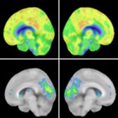
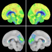
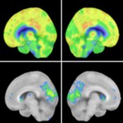
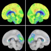
*Right & left medial sagittal FDG PET source & 3DSSP images of a 74-year-old man with dementia with Lewy bodies (DLB) presenting with visual hallucinations show severe ↓ metabolic activity in left visual cortex & precuneus . Note moderate ↓ metabolic activity in right occipital lobe & precuneus & relative sparing of bilateral posterior cingulate gyri .*
*Right & left medial sagittal FDG PET source & 3DSSP images of a 74-year-old man with dementia with Lewy bodies (DLB) presenting with visual hallucinations show severe ↓ metabolic activity in left visual cortex & precuneus . Note moderate ↓ metabolic activity in right occipital lobe & precuneus & relative sparing of bilateral posterior cingulate gyri .*
*Right & left medial sagittal FDG PET source & 3DSSP images of a 74-year-old man with dementia with Lewy bodies (DLB) presenting with visual hallucinations show severe ↓ metabolic activity in left visual cortex & precuneus . Note moderate ↓ metabolic activity in right occipital lobe & precuneus & relative sparing of bilateral posterior cingulate gyri .*
*Right & left medial sagittal FDG PET source & 3DSSP images of a 74-year-old man with dementia with Lewy bodies (DLB) presenting with visual hallucinations show severe ↓ metabolic activity in left visual cortex & precuneus . Note moderate ↓ metabolic activity in right occipital lobe & precuneus & relative sparing of bilateral posterior cingulate gyri .*
*Right & left medial sagittal FDG PET source & 3DSSP images of a 74-year-old man with dementia with Lewy bodies (DLB) presenting with visual hallucinations show severe ↓ metabolic activity in left visual cortex & precuneus . Note moderate ↓ metabolic activity in right occipital lobe & precuneus & relative sparing of bilateral posterior cingulate gyri .*
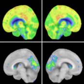
*Right & left medial sagittal FDG PET source & 3DSSP images of a 74-year-old man with dementia with Lewy bodies (DLB) presenting with visual hallucinations show severe ↓ metabolic activity in left visual cortex & precuneus . Note moderate ↓ metabolic activity in right occipital lobe & precuneus & relative sparing of bilateral posterior cingulate gyri .*
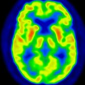
*Axial PET of the same patient shows preserved metabolism in frontal & temporal lobes. (Courtesy S Behnia, MD.)*
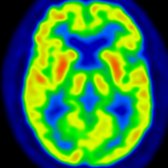
*Axial PET of the same patient shows preserved metabolism in frontal & temporal lobes. (Courtesy S Behnia, MD.)*

*Axial T2WI MR in a patient with DLB shows nonspecific diffuse cortical atrophy. Conventional MR findings are frequently nonspecific in DLB.*

*Axial T2WI MR in a patient with DLB shows nonspecific diffuse cortical atrophy. Conventional MR findings are frequently nonspecific in DLB.*
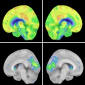
*Right & left medial sagittal FDG PET source & 3DSSP images of a 74-year-old man with dementia with Lewy bodies (DLB) presenting with visual hallucinations show severe ↓ metabolic activity in left visual cortex & precuneus . Note moderate ↓ metabolic activity in right occipital lobe & precuneus & relative sparing of bilateral posterior cingulate gyri .*

*Right & left medial sagittal FDG PET source & 3DSSP images of a 74-year-old man with dementia with Lewy bodies (DLB) presenting with visual hallucinations show severe ↓ metabolic activity in left visual cortex & precuneus . Note moderate ↓ metabolic activity in right occipital lobe & precuneus & relative sparing of bilateral posterior cingulate gyri .*

*Right & left medial sagittal FDG PET source & 3DSSP images of a 74-year-old man with dementia with Lewy bodies (DLB) presenting with visual hallucinations show severe ↓ metabolic activity in left visual cortex & precuneus . Note moderate ↓ metabolic activity in right occipital lobe & precuneus & relative sparing of bilateral posterior cingulate gyri .*

*Right & left medial sagittal FDG PET source & 3DSSP images of a 74-year-old man with dementia with Lewy bodies (DLB) presenting with visual hallucinations show severe ↓ metabolic activity in left visual cortex & precuneus . Note moderate ↓ metabolic activity in right occipital lobe & precuneus & relative sparing of bilateral posterior cingulate gyri .*

*Right & left medial sagittal FDG PET source & 3DSSP images of a 74-year-old man with dementia with Lewy bodies (DLB) presenting with visual hallucinations show severe ↓ metabolic activity in left visual cortex & precuneus . Note moderate ↓ metabolic activity in right occipital lobe & precuneus & relative sparing of bilateral posterior cingulate gyri .*

*Right & left medial sagittal FDG PET source & 3DSSP images of a 74-year-old man with dementia with Lewy bodies (DLB) presenting with visual hallucinations show severe ↓ metabolic activity in left visual cortex & precuneus . Note moderate ↓ metabolic activity in right occipital lobe & precuneus & relative sparing of bilateral posterior cingulate gyri .*

*Axial PET of the same patient shows preserved metabolism in frontal & temporal lobes. (Courtesy S Behnia, MD.)*

*Axial PET of the same patient shows preserved metabolism in frontal & temporal lobes. (Courtesy S Behnia, MD.)*

*Axial T2WI MR in a patient with DLB shows nonspecific diffuse cortical atrophy. Conventional MR findings are frequently nonspecific in DLB.*

*Axial T2WI MR in a patient with DLB shows nonspecific diffuse cortical atrophy. Conventional MR findings are frequently nonspecific in DLB.*
 . Using DAT imaging, it is not possible to distinguish DLB from atypical parkinsonian syndromes like MSA, PSP, & CBD.](images/app.statdx.com_image_thumbnail_d5cf7988-1211-4bec-888b-b72a8bdf8139_size_168_quality_85_22e550ce_20251014T193405Z.jpg)
*123FP-CIT-SPECT, DAT imaging (dopamine transporter) shows normal symmetric uptake in the striatum of a healthy control (HC). In DLB, there is marked ↓ update in the putamen & mild in the caudate nuclei . Using DAT imaging, it is not possible to distinguish DLB from atypical parkinsonian syndromes like MSA, PSP, & CBD.*
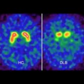
*123FP-CIT-SPECT, DAT imaging (dopamine transporter) shows normal symmetric uptake in the striatum of a healthy control (HC). In DLB, there is marked ↓ update in the putamen & mild in the caudate nuclei . Using DAT imaging, it is not possible to distinguish DLB from atypical parkinsonian syndromes like MSA, PSP, & CBD.*
### Additional Images
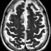
*Axial T2WI MR in a patient with cognitive decline, visual hallucination, & parkinsonism shows diffuse cortical atrophy consistent with DLB.*
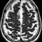
*Axial T2WI MR in a patient with cognitive decline, visual hallucination, & parkinsonism shows diffuse cortical atrophy consistent with DLB.*

*Axial T2WI MR in the same patient exhibits mild atrophy of the medial temporal lobes.*

*Axial T2WI MR in the same patient exhibits mild atrophy of the medial temporal lobes.*
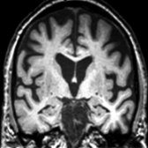
*Coronal T1WI MR in a patient with DLB shows prominent frontal lobe volume loss with relative sparing of hippocampal volume. (Courtesy M.J. Firbank, MD & J.T. O'Brien, MD.)*
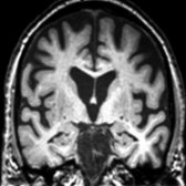
*Coronal T1WI MR in a patient with DLB shows prominent frontal lobe volume loss with relative sparing of hippocampal volume. (Courtesy M.J. Firbank, MD & J.T. O'Brien, MD.)*
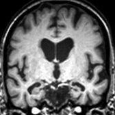
*Coronal T1WI MR in a patient with Alzheimer disease (AD) shows marked hippocampal volume loss & relative sparing of frontal lobes. (Courtesy M. J. Firbank, MD & J. T. O'Brien, MD.)*
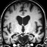
*Coronal T1WI MR in a patient with Alzheimer disease (AD) shows marked hippocampal volume loss & relative sparing of frontal lobes. (Courtesy M. J. Firbank, MD & J. T. O'Brien, MD.)*
. Using DAT imaging, it is not possible to distinguish DLB from atypical parkinsonian syndromes like MSA, PSP, & CBD.](images/app.statdx.com_image_thumbnail_d5cf7988-1211-4bec-888b-b72a8bdf8139_size_168_quality_85_22e550ce_20251014T193405Z.jpg)
*123FP-CIT-SPECT, DAT imaging (dopamine transporter) shows normal symmetric uptake in the striatum of a healthy control (HC). In DLB, there is marked ↓ update in the putamen & mild in the caudate nuclei . Using DAT imaging, it is not possible to distinguish DLB from atypical parkinsonian syndromes like MSA, PSP, & CBD.*

*123FP-CIT-SPECT, DAT imaging (dopamine transporter) shows normal symmetric uptake in the striatum of a healthy control (HC). In DLB, there is marked ↓ update in the putamen & mild in the caudate nuclei . Using DAT imaging, it is not possible to distinguish DLB from atypical parkinsonian syndromes like MSA, PSP, & CBD.*
### Additional Images

*Axial T2WI MR in a patient with cognitive decline, visual hallucination, & parkinsonism shows diffuse cortical atrophy consistent with DLB.*

*Axial T2WI MR in a patient with cognitive decline, visual hallucination, & parkinsonism shows diffuse cortical atrophy consistent with DLB.*

*Axial T2WI MR in the same patient exhibits mild atrophy of the medial temporal lobes.*

*Axial T2WI MR in the same patient exhibits mild atrophy of the medial temporal lobes.*

*Coronal T1WI MR in a patient with DLB shows prominent frontal lobe volume loss with relative sparing of hippocampal volume. (Courtesy M.J. Firbank, MD & J.T. O'Brien, MD.)*

*Coronal T1WI MR in a patient with DLB shows prominent frontal lobe volume loss with relative sparing of hippocampal volume. (Courtesy M.J. Firbank, MD & J.T. O'Brien, MD.)*

*Coronal T1WI MR in a patient with Alzheimer disease (AD) shows marked hippocampal volume loss & relative sparing of frontal lobes. (Courtesy M. J. Firbank, MD & J. T. O'Brien, MD.)*

*Coronal T1WI MR in a patient with Alzheimer disease (AD) shows marked hippocampal volume loss & relative sparing of frontal lobes. (Courtesy M. J. Firbank, MD & J. T. O'Brien, MD.)*