---
title: "Demyelinating Diseases"
docid: "e3ba880e-d924-4594-a6f4-c21c5f1f0ae7"
authors:
- key: "838e1722-2479-4fbd-a5fe-d965980a1a2c"
value: "Blaise V. Jones, MD"
breadcrumbs:
-
name: "Pediatrics"
slug: "pediatrics"
treeNodeId: "a915965c-d436-44cf-ae65-2f22e7246ea4"
-
name: "Diagnosis"
slug: "diagnosis"
treeNodeId: "2b5cea64-a083-489e-ac0c-ec14ba059026"
-
name: "Brain"
slug: "brain"
treeNodeId: "95caa0da-bc4f-4103-8551-f58d6e415781"
-
name: "Metabolic, Infectious, and Inflammatory Disorders"
slug: "metabolic-infectious-and-inflammat-"
treeNodeId: "ed178695-cb39-40bc-981d-d0031c835a98"
-
name: "Demyelinating Diseases"
slug: "demyelinating-diseases"
treeNodeId: null
category: "Pediatrics"
cmeTopicId: "273337e9-da4e-4caa-8a31-95fa5c11a1b9"
documentVersionId: "5557de59-59d3-4508-b2dd-6913988c047d"
imageCount: 23
lastUpdated: "11/04/21"
pageDescription: "Demyelinating Diseases"
pageKeywords: "Pediatrics, Diagnosis, Brain, Metabolic, Infectious, and Inflammatory Disorders, Demyelinating Diseases"
pageTitle: "Demyelinating Diseases | STATdx"
enhancedTitle: "Demyelinating Diseases"
type: "DX"
references: true
breadcrumbs:
- "Pediatrics"
- "Diagnosis"
- "Brain"
- "Metabolic, Infectious, and Inflammatory Disorders"
- "Demyelinating Diseases"
---
# KEY FACTS
- ## Diagnostic Checklist
- Multiple sclerosis (MS)
- Demyelinating disease characterized by multiple lesions disseminated in time & space
- Brain lesions: Multiple T2- & FLAIR MR hyperintense foci, typically small (5-10 mm), ovoid, discrete, periventricular, & perpendicular to ventricular margins
- Optic neuritis (ON): Unilateral, short segment, intraorbital; myelitis: < 2 vertebrae in length, < 50% of cross-sectional area, typically peripheral
- Acute disseminated encephalomyelitis (ADEM)
- Acute demyelinating disease with encephalopathy, without NMOSD or anti-MOG associated antibodies
- Characteristically arises subsequent to infection (viral respiratory) or vaccination
- Brain: Ill-defined, larger T2-/FLAIR hyperintense lesions
- Neuromyelitis optica spectrum disorders (NMOSD)
- Inflammatory CNS disease caused by antibodies to aquaporin-4 (AQP-4) on astrocytic end feet
- ON & transverse myelitis predominate clinically
- Brain: Commonly periventricular but parallel to ependymal lining; area postrema involvement is classic
- ON: Bilateral, posterior predominant (including chiasm)
- Myelitis: Longitudinally extensive transverse myelitis (LETM) (> 3 vertebrae), > 50% of cord cross section, central
- Anti-MOG syndromes
- Acute demyelinating disease caused by antibodies to myelin oligodendrocyte glycoprotein (MOG)
- Extensive clinical overlap with ADEM & NMOSD; encephalopathy in younger patients, ON in older
- Brain: Similar to ADEM
- ON: Bilateral anterior predominant (including optic disc) with perineural enhancement
- Myelitis: LETM vs. short segment; conus involvement
- Lyme disease
- 11% develop neurologic manifestations
- May be accompanied by ON or other CN inflammation
# TERMINOLOGY
- ## Definitions
- Acquired demyelinating processes characterized by inflammation
- Multiple sclerosis (MS)
- Demyelinating disease characterized by multiple lesions disseminated in time & space
- Acute disseminated encephalomyelitis (ADEM)
- Acute demyelinating disease with encephalopathy, without NMOSD or anti-MOG-associated antibodies
- Neuromyelitis optica spectrum disorders (NMOSD)
- Inflammatory CNS disease caused by antibodies to aquaporin-4 (AQP-4) on astrocytic end feet
- Clinically characterized by optic neuritis & transverse myelitis
- Antimyelin oligodendrocyte glycoprotein (MOG) syndromes
- Acute demyelinating disease caused by antibodies to MOG
- Extensive clinical overlap with ADEM & NMOSD
- Lyme disease
- CNS inflammation associated with *Borrelia burgdorferi* infection
# IMAGING
- ## General Features
- MS
- Brain: Multiple T2- & FLAIR MR hyperintense lesions, typically small (5-10 mm), ovoid, discrete
- > 85% are periventricular: Callosal involvement, hemispheric white matter; perpendicular to ventricle margin in perivenular distribution
- Variable enhancement: Presumed to reflect active demyelination
- Nodular, diffuse, or ring-like
- Can be mass-like: Tumefactive MS
- Diffusion restriction in acute lesions
- Diffusely abnormal ADC values
- "Black holes" (due to axonal destruction) on T1 are much more likely to be seen in MS than ADEM
- Optic neuritis (ON): Unilateral, short length, intraorbital
- Myelitis: < 2 vertebral lengths, < 50% of cord cross section, cervical > thoracic
- ADEM
- Brain: Ill-defined larger T2-/FLAIR hyperintense lesions
- Lesions are more likely to be diffuse & bilateral
- Frequent brainstem & thalamic involvement
- ON: Less common; myelitis: Less common
- NMOSD
- Brain: May have extensive lesions
- Commonly periventricular but parallel
- Dorsal brainstem (especially area postrema)
- ON: Bilateral long segment
- Posterior predominant, including chiasm
- Myelitis: Longitudinally extensive transverse myelitis (LETM) (> 3 segments), typically central & > 50% circumference
- Anti-MOG syndromes
- Brain: Similar in appearance to ADEM
- Much less likely to involve corpus callosum
- ON: Bilateral long length with perineural enhancement
- Anterior predominant, including optic disc
- Myelitis: LETM vs. short segment; conus often affected
- Lyme disease
- Presents as meningoencephalitis
- May be accompanied by ON or other cranial nerve inflammation; Bell palsy is characteristic
- ## Imaging Recommendations
- ### Best imaging tool
- MR
- ### Protocol advice
- FLAIR MR imaging for detection
- Postcontrast FLAIR may ↑ detection of meningeal disease
- Fat-saturated, high-resolution postcontrast orbital MR for assessment of ON
- Spine imaging with contrast & axial T2-weighted sequences
# DIFFERENTIAL DIAGNOSIS
- [Posterior Reversible Encephalopathy Syndrome](/document/acute-hypertensive-encephalopathy--/efc6f9c2-dad9-4eb8-bad2-421bfaf1ec57)
- Subcortical vasogenic edema associated with hypertension
- [Viral Encephalitis](/document/acute-encephalitis/a45f63bb-c25b-481d-a001-9c520c58060b)
- Widely variable, but often affects white matter & deep gray nuclei
- [Autoimmune-Mediated Vasculitis](/document/miscellaneous-vasculitis/5a4d4cbd-67e3-4722-8a44-8d411cbb98f0)
- Enhancing lesions spare callososeptal interface
- Reported in COVID-19 infection
- Beaded angiogram appearance
- [Leukodystrophies](/document/leukodystrophies/f4ff3738-131c-46bf-be71-1811f2c1776c)
- Patterns vary by metabolic defect
- Metachromatic leukodystrophy, Alexander disease, X-linked adrenoleukodystrophy
- ## Toxin-Induced Brain Injury
- Carbon monoxide or methanol poisoning
- Bilateral symmetric basal ganglia lesions
- Accompanying subcortical lesions in methanol poisoning
# PATHOLOGY
- ## General Features
- ### Etiology
- MS
- Possibly viral-incited autoimmune reaction in genetically susceptible individuals
- No "trigger" identified
- Activated T cells attack myelinated axons
- B cells, antibodies, macrophages, & microglia all contribute to lesions
- Cox-2, iNOS may cause excitotoxic death of oligodendrocytes
- ADEM
- Autoimmune-mediated demyelination
- Characteristically arises subsequent to infection (viral respiratory) or vaccination
- NMOSD
- Antibodies to aquaporin-4 (AQP-4) channels on astrocytic end feet
- Technically not demyelinating
- AQP-4 is dispersed throughout CNS
- Highly expressed in optic nerves & spinal cord
- AQP-4 antibodies are more highly expressed in peripheral blood than CSF
- Anti-MOG syndromes: Antibodies to MOG
- MOG is CNS specific protein expressed on outer surface of myelin sheath
- Lyme disease
- Caused by spirochete *Borrelia burgdorferi*
- Tick-borne disease; deer tick (Ixodes scapularis) or Western black-legged tick (Ixodes pacificus)
- ## Staging, Grading, & Classification
- Major clinical subtypes of MS
- Relapsing-remitting**** (85% initial presentation)
- Primary-progressive****, a.k.a. chronic progressive (5-10%)
- Progressive from start
- Secondary-progressive****, a.k.a. relapsing progressive
- By 10 years 50% & by 25 years 90% of relapsing-remitting patients enter secondary-progressive phase
- Progressive-relapsing****
- Rare; defined as progressive disease with clear acute relapses ± full recovery
- Periods between relapses are characterized by continuing disease progression
- Clinically isolated syndrome (CIS): Single episode > 24 hours; vast majority progress to MS after number of years
- MS variants/subtypes
- Malignant****: Younger patients, febrile prodrome, clinically fulminant, death in months
- Schilder ****type ("diffuse sclerosis"): Extensive, confluent, asymmetric demyelination in bilateral supra-/infratentorial parenchyma
- Baló ****type ("concentric sclerosis"): Large lesions with alternating zones of demyelinated/myelinated white matter
- ## Gross Pathologic & Surgical Features
- Acute MS: Poorly delineated, yellowish-white, periventricular plaques
- Chronic MS: Gray, granular, well-demarcated plaques ± generalized volume loss
- ## Microscopic Features
- MS
- Perivenous demyelination & oligodendrocyte loss
- Active: Foamy macrophages with myelin fragments, lipids; reactive astrocytes + perivascular inflammation; some are hypercellular with atypical reactive astrocytes & mitoses (mimics tumor)
- Chronic: Marked loss of myelin & oligodendrocytes; dense astrogliosis; minimal/no perivascular inflammation
- Axonal transection
- CSF positive for oligoclonal bands
# CLINICAL ISSUES
- ## Presentation
- ### Most common signs/symptoms
- MS
- Variable
- Initially impaired/double vision of acute ON (50% with positive MR develop MS)
- Weakness, numbness, tingling, gait disturbances
- ↓ sphincter control, blindness, paralysis, dementia
- Cranial nerve palsies; usually multiple, 1-5% isolated (CNV & VI are most common)
- Spinal cord symptoms in 80%
- ADEM
- Cranial nerve palsies, encephalopathy, headache 2 days to 4 weeks after prodrome
- Seizures in 10-35%
- Monophasic illness
- Can recur in small percentage of cases (controversial)
- NMO
- Rapid onset of vision loss
- Subsequent spinal cord symptoms, paralysis
- Lyme disease
- Stereotypical expanding rash around tick bite: Erythema chronicum migrans
- Infrequently recognized
- Bell palsy, meningitis, arthralgias
- ## Demographics
- Estimated 2,500,000 have MS worldwide
- MS most often occurs in temperate climates
- Most common disabling CNS disease of young adults: 1 in 1,000 in Western world
- 3-5% of MS is diagnosed before age 15 years
- 20% of childhood MS is initially diagnosed as ADEM
- 1/3 of acute demyelinating disease in children is anti-MOG
- ## Natural History & Prognosis
- MS: 45% of MS patients are not severely affected & are nearly normal
- > 80% with "probable" MS & positive MR progress to clinically definite MS
- ADEM: Characteristically monophasic
- Recurrence suggests anti-MOG
- Anti-MOG syndromes are more frequently seen in young
- Encephalopathy is more common in younger patients, ON in older
- > 90% of NMOSD in pediatrics have relapsing disease
- Lyme disease: 11% develop neurologic manifestations
- ## Treatment
- MS & NMOSD are both treated with immune-modulating therapy
- ADEM is typically treated with high-dose steroids
- Alternative treatments include IVIg & plasma exchange
- Anti-MOG syndromes respond quickly to steroid & IVIg treatment
- Lyme is treated with antibiotics
- No evidence that antibiotic therapy alters natural history
# DIAGNOSTIC CHECKLIST
- ## Image Interpretation Pearls
- 95% with definite MS clinically have positive MR
12c5cb99-14a6-4793-9a7d-a6f1c3511f06
## References
# Selected References
1. [Chhabda S et al: Relapsing demyelinating syndromes in children: a practical review of neuroradiological mimics. Front Neurol. 11:627, 2020](http://www.ncbi.nlm.nih.gov/pubmed/?term=32849169%5Bpmid%5D)
1. [Padilha IG et al: Pediatric multiple sclerosis: from clinical basis to imaging spectrum and differential diagnosis. Pediatr Radiol. 50(6):776-92, 2020](http://www.ncbi.nlm.nih.gov/pubmed/?term=31925460%5Bpmid%5D)
1. [Bulut E et al: Brain MRI findings in pediatric-onset neuromyelitis optica spectrum disorder: challenges in differentiation from acute disseminated encephalomyelitis. AJNR Am J Neuroradiol. 40(4):726-31, 2019](http://www.ncbi.nlm.nih.gov/pubmed/?term=30846436%5Bpmid%5D)
1. [Galardi MM et al: Differential diagnosis of pediatric multiple sclerosis. Children (Basel). 6(6), 2019](http://www.ncbi.nlm.nih.gov/pubmed/?term=31163654%5Bpmid%5D)
1. [Lana-Peixoto MA et al: Neuromyelitis optica spectrum disorder and anti-MOG syndromes. Biomedicines. 7(2), 2019](http://www.ncbi.nlm.nih.gov/pubmed/?term=31212763%5Bpmid%5D)
1. [Troxell RM et al: Atypical pediatric demyelinating diseases of the central nervous system. Curr Neurol Neurosci Rep. 19(12):95, 2019](http://www.ncbi.nlm.nih.gov/pubmed/?term=31773416%5Bpmid%5D)
1. [Reich DS et al: Multiple sclerosis. N Engl J Med. 378(2):169-80, 2018](http://www.ncbi.nlm.nih.gov/pubmed/?term=29320652%5Bpmid%5D)
1. [Berzero G et al: Diagnosis and therapy of acute disseminated encephalomyelitis and its variants. Expert Rev Neurother. 16(1):83-101, 2016](http://www.ncbi.nlm.nih.gov/pubmed/?term=26620160%5Bpmid%5D)
1. [Faguy K: Multiple sclerosis: an update. Radiol Technol. 87(5):529-50, 2016](http://www.ncbi.nlm.nih.gov/pubmed/?term=27146176%5Bpmid%5D)
1. [Filippi M et al: MRI criteria for the diagnosis of multiple sclerosis: MAGNIMS consensus guidelines. Lancet Neurol. 15(3):292-303, 2016](http://www.ncbi.nlm.nih.gov/pubmed/?term=26822746%5Bpmid%5D)
1. [Borchers AT et al: Lyme disease: a rigorous review of diagnostic criteria and treatment. J Autoimmun. 57:82-115, 2015](http://www.ncbi.nlm.nih.gov/pubmed/?term=25451629%5Bpmid%5D)
1. [Koelman DL et al: Acute disseminated encephalomyelitis: current controversies in diagnosis and outcome. J Neurol. 262(9):2013-24, 2015](http://www.ncbi.nlm.nih.gov/pubmed/?term=25761377%5Bpmid%5D)
1. [Wingerchuk DM et al: International consensus diagnostic criteria for neuromyelitis optica spectrum disorders. Neurology. 85(2):177-89, 2015](http://www.ncbi.nlm.nih.gov/pubmed/?term=26092914%5Bpmid%5D)
1. [Flanagan EP et al: Neuromyelitis optica spectrum disorders. Curr Neurol Neurosci Rep. 14(9):483, 2014](http://www.ncbi.nlm.nih.gov/pubmed/?term=25027264%5Bpmid%5D)
1. [Ketelslegers IA et al: A comparison of MRI criteria for diagnosing pediatric ADEM and MS. Neurology. 74(18):1412-5, 2010](http://www.ncbi.nlm.nih.gov/pubmed/?term=20335562%5Bpmid%5D)
1. [VanLandingham M et al: An uncommon illness with a rare presentation: neurosurgical management of ADEM with tumefactive demyelination in children. Childs Nerv Syst. 26(5):655-61, 2010](http://www.ncbi.nlm.nih.gov/pubmed/?term=19949803%5Bpmid%5D)
1. [Calabrese M et al: Cortical lesions in primary progressive multiple sclerosis: a 2-year longitudinal MR study. Neurology. 72(15):1330-6, 2009](http://www.ncbi.nlm.nih.gov/pubmed/?term=19365054%5Bpmid%5D)
1. [Callen DJ et al: Role of MRI in the differentiation of ADEM from MS in children. Neurology. 72(11):968-73, 2009](http://www.ncbi.nlm.nih.gov/pubmed/?term=19038851%5Bpmid%5D)
1. [Filippi M et al: Conventional MRI in multiple sclerosis. J Neuroimaging. 17 Suppl 1:3S-9S, 2007](http://www.ncbi.nlm.nih.gov/pubmed/?term=17425730%5Bpmid%5D)
1. [Janardhan V et al: Multiple sclerosis: hyperintense lesions in the brain on nonenhanced T1-weighted MR images evidenced as areas of T1 shortening. Radiology. 244(3):823-31, 2007](http://www.ncbi.nlm.nih.gov/pubmed/?term=17690319%5Bpmid%5D)
1. [Traboulsee AL et al: The role of MRI in the diagnosis of multiple sclerosis. Adv Neurol. 98:125-46, 2006](http://www.ncbi.nlm.nih.gov/pubmed/?term=16400831%5Bpmid%5D)
1. [Polman CH et al: Diagnostic criteria for multiple sclerosis: 2005 revisions to the "McDonald Criteria". Ann Neurol. 58(6):840-6, 2005](http://www.ncbi.nlm.nih.gov/pubmed/?term=16283615%5Bpmid%5D)
## Images
### Selected Images
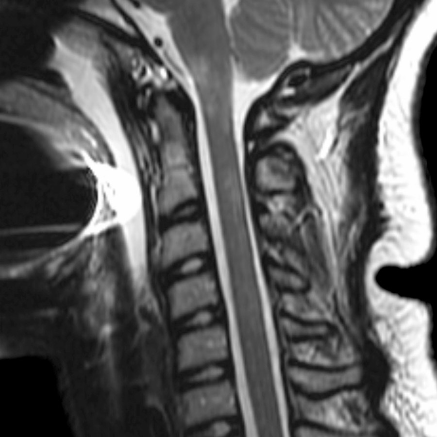
*Sagittal T2 MR in a 9-year-old with optic neuritis shows multiple ill-defined hyperintensities in the medulla & cervical cord. Subsequent serum testing revealed antibodies to aquaporin 4, confirming a diagnosis of neuromyelitis optica spectrum disorders (NMOSD).*
*Sagittal T2 MR in a 9-year-old with optic neuritis shows multiple ill-defined hyperintensities in the medulla & cervical cord. Subsequent serum testing revealed antibodies to aquaporin 4, confirming a diagnosis of neuromyelitis optica spectrum disorders (NMOSD).*
 in this 9-year-old with vision loss. Clinical features were suggestive of NMOSD, but CSF analysis confirmed anti-myelin oligodendrocyte glycoprotein (MOG) disease.](images/app.statdx.com_image_thumbnail_0644d2a0-6cc0-4d76-aa55-0dc02b971f87_annotated_true_size_900_quality_90_b85e5c5c_20251018T152401Z.jpg)
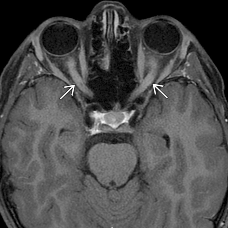
*Axial T1 C+ FS MR through the orbits shows diffuse bilateral optic nerve enhancement in this 9-year-old with vision loss. Clinical features were suggestive of NMOSD, but CSF analysis confirmed anti-myelin oligodendrocyte glycoprotein (MOG) disease.*
*Axial T1 C+ FS MR through the orbits shows diffuse bilateral optic nerve enhancement in this 9-year-old with vision loss. Clinical features were suggestive of NMOSD, but CSF analysis confirmed anti-myelin oligodendrocyte glycoprotein (MOG) disease.*
 .](images/app.statdx.com_image_thumbnail_be1040b7-52b7-4198-85c6-7c794b869ca0_annotated_true_size_900_quality_90_40619fe9_20251018T152401Z.jpg)
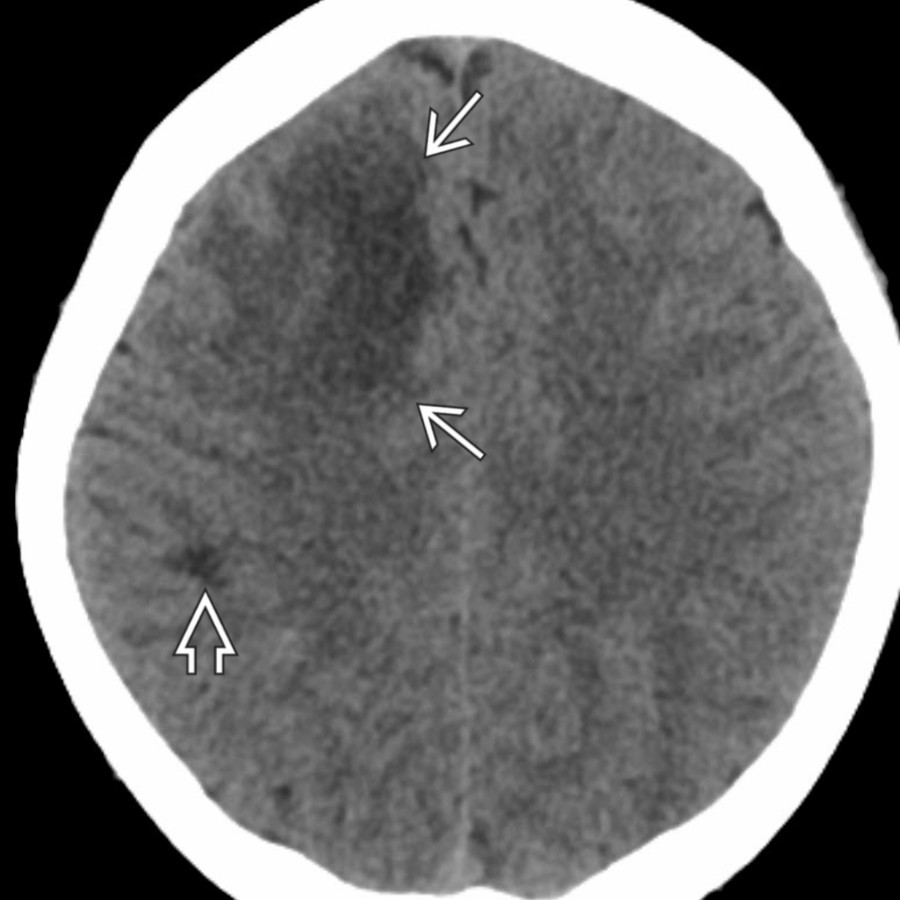
*Axial NECT in a 16-year-old with progressive left-sided weakness after minor trauma shows a large, low-attenuation white matter lesion in the anterior right frontal lobe & a smaller one near the right motor strip .*
*Axial NECT in a 16-year-old with progressive left-sided weakness after minor trauma shows a large, low-attenuation white matter lesion in the anterior right frontal lobe & a smaller one near the right motor strip .*
 . This open ring appearance can help distinguish tumefactive MS from abscess or neoplasm (which more typically have complete ring enhancement).](images/app.statdx.com_image_thumbnail_f6c3b95c-7708-493e-adf2-d530a89cf1b3_annotated_true_size_900_quality_90_9839b7e5_20251018T152401Z.jpg)
*Sagittal T1 C+ MR in the same patient shows the borders of the large lesion nearest to the cortex to be nonenhancing as compared to the other margins . This open ring appearance can help distinguish tumefactive MS from abscess or neoplasm (which more typically have complete ring enhancement).*
### Additional Images
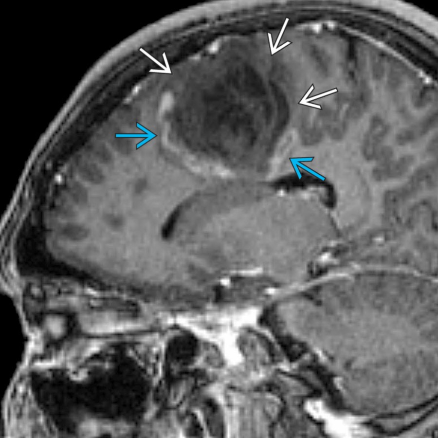
*Sagittal T1 C+ MR in the same patient shows the borders of the large lesion nearest to the cortex to be nonenhancing as compared to the other margins . This open ring appearance can help distinguish tumefactive MS from abscess or neoplasm (which more typically have complete ring enhancement).*
### Additional Images
 at the callososeptal interface along penetrating venules.](images/app.statdx.com_image_thumbnail_5982d841-bf3c-48bf-981b-2b113c170f77_annotated_true_size_900_quality_90_2f597c2e_20251018T152401Z.jpg)
*Sagittal graphic illustrates MS plaques involving the corpus callosum, pons, & spinal cord. Note the characteristic perpendicular orientation of the lesions at the callososeptal interface along penetrating venules.*
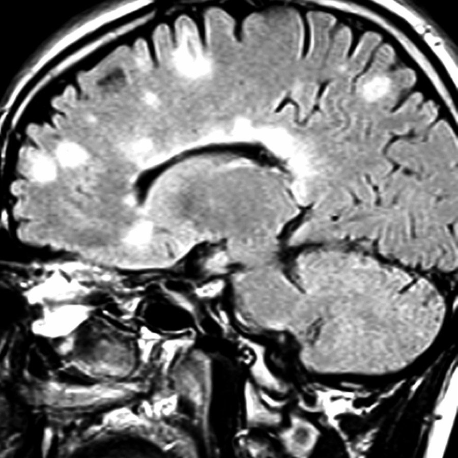
*Sagittal FLAIR MR shows numerous MS plaques with typical perpendicular orientation at the callososeptal interface along penetrating venules ("Dawson fingers") as well as in the subcortical white matter.*

*Sagittal FLAIR MR shows perpendicular callosal/pericallosal MS plaques with hyperintense rims & hypointense centers (with corresponding hypointensities also demonstrated on T1 as "black holes," not shown). Note an additional posterior fossa lesion .*
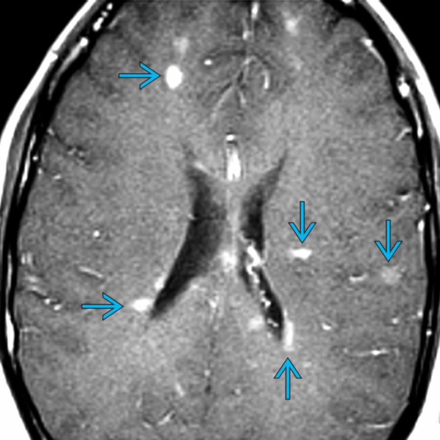
*Axial T1 C+ MR demonstrates multiple nodular, enhancing multiple sclerosis plaques . Note the common periventricular location with perpendicular orientation as well as the involvement of subcortical white matter.*

*Axial FLAIR MR shows confluent multiple sclerosis plaques in commonly seen periventricular locations.*
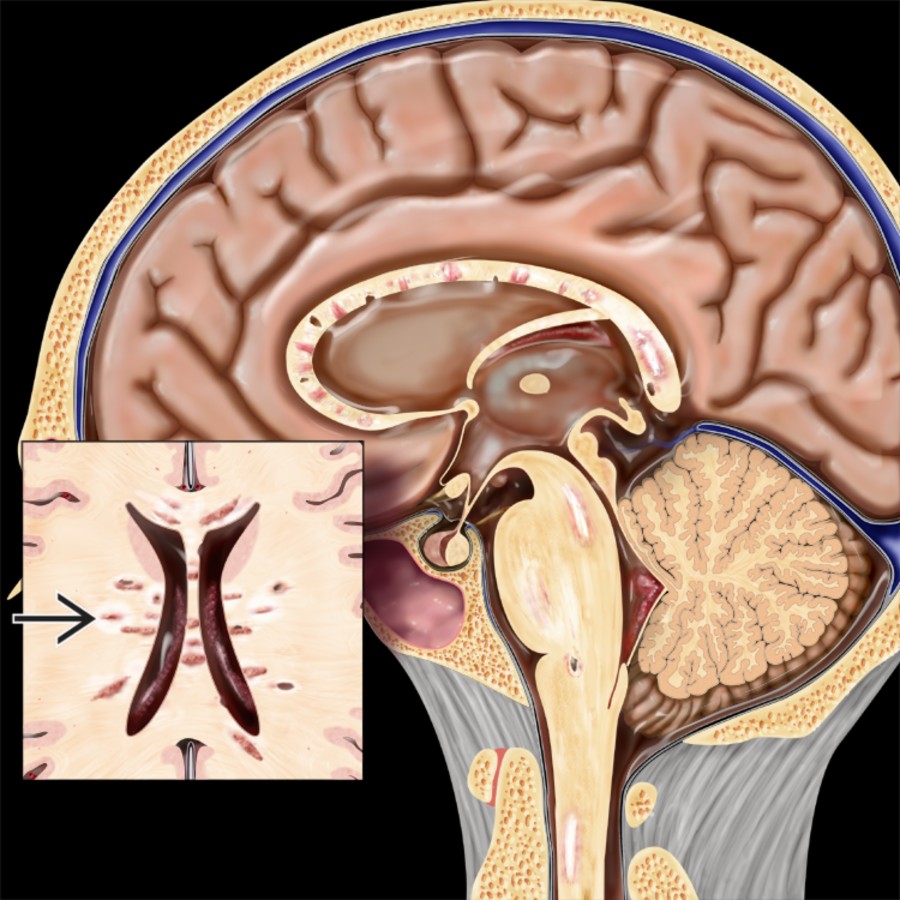
*Sagittal graphic illustrates MS plaques involving the corpus callosum, pons, & spinal cord. Note the characteristic perpendicular orientation of the lesions at the callososeptal interface along penetrating venules.*

*Sagittal FLAIR MR shows numerous MS plaques with typical perpendicular orientation at the callososeptal interface along penetrating venules ("Dawson fingers") as well as in the subcortical white matter.*

*Sagittal FLAIR MR shows perpendicular callosal/pericallosal MS plaques with hyperintense rims & hypointense centers (with corresponding hypointensities also demonstrated on T1 as "black holes," not shown). Note an additional posterior fossa lesion .*

*Axial T1 C+ MR demonstrates multiple nodular, enhancing multiple sclerosis plaques . Note the common periventricular location with perpendicular orientation as well as the involvement of subcortical white matter.*

*Axial FLAIR MR shows confluent multiple sclerosis plaques in commonly seen periventricular locations.*
 , basal ganglia , & insula . Involvement of the deep nuclei is a relatively common feature of acute disseminated encephalomyelitis.](0e99452e-0b58-4f4c-bfac-641ed551772f)
*Axial FLAIR MR in a 9-year-old patient with altered mental status & hyperreflexia shows ill-defined, hyperintense lesions in the thalami , basal ganglia , & insula . Involvement of the deep nuclei is a relatively common feature of acute disseminated encephalomyelitis.*

*Axial FLAIR MR shows large lesions in the thalamus & basal ganglia in this 16-year-old with a headache & weakness 2 weeks after a viral illness. Acute disseminated encephalomyelitis will frequently affect deep gray matter structures.*

*Coronal FLAIR MR in a 12-year-old patient with neuromyelitis optica & bladder dysfunction shows large lesions extending across the corpus callosum & left cerebral peduncle .*

*Axial NECT in a 14-year-old patient with vomiting shows a nonspecific, low-attenuation lesion in the left posterior frontal subcortical white matter.*

*Axial FLAIR MR in the same patient acquired the next day shows several ovoid MS plaques . Active lesions will also show contrast enhancement & restricted diffusion.*

*Axial FLAIR MR in a 14-year-old with MS shows multiple ovoid lesions oriented perpendicular to the long axis of the lateral ventricles with hazy ↑ signal intensity in the white matter between them.*

*Axial T2 MR in a 17-year-old with Baló concentric sclerosis .*

*Sagittal T1 C+ FS MR shows an enhancing MS lesion in the dorsal aspect of the cervical cord . Approximately 2/3 of spinal cord MS lesions are found in the cervical cord. Typical features include a dorsal intramedullary lesion spanning < 2 vertebral segments in length.*

*Axial FLAIR MR shows numerous peripheral white matter & cortical lesions that exhibited robust contrast enhancement (not shown) in an 18-year-old woman with malignant (Marburg) MS. The patient presented with a 2-week history of behavioral changes & leg pain & died 3 weeks after presentation. The autopsy showed typical demyelinating pathology.*

*Axial T1 C+ FS MR in a patient with MS shows ring-enhancing masses of active demyelination. The rings of enhancement are incomplete with each ring defect pointing towards an adjacent cortex.*

*Coronal T1 C+ MR shows a superficial hypointense mass in the left parasagittal posterior frontal region with a peripheral crescent of incomplete or "horseshoe" enhancement . This enhancement pattern is classic for tumefactive demyelinating disease, most commonly MS.*

*Axial FLAIR MR in a 9-year-old patient with altered mental status & hyperreflexia shows ill-defined, hyperintense lesions in the thalami , basal ganglia , & insula . Involvement of the deep nuclei is a relatively common feature of acute disseminated encephalomyelitis.*

*Axial FLAIR MR shows large lesions in the thalamus & basal ganglia in this 16-year-old with a headache & weakness 2 weeks after a viral illness. Acute disseminated encephalomyelitis will frequently affect deep gray matter structures.*

*Coronal FLAIR MR in a 12-year-old patient with neuromyelitis optica & bladder dysfunction shows large lesions extending across the corpus callosum & left cerebral peduncle .*

*Axial NECT in a 14-year-old patient with vomiting shows a nonspecific, low-attenuation lesion in the left posterior frontal subcortical white matter.*

*Axial FLAIR MR in the same patient acquired the next day shows several ovoid MS plaques . Active lesions will also show contrast enhancement & restricted diffusion.*

*Axial FLAIR MR in a 14-year-old with MS shows multiple ovoid lesions oriented perpendicular to the long axis of the lateral ventricles with hazy ↑ signal intensity in the white matter between them.*

*Axial T2 MR in a 17-year-old with Baló concentric sclerosis .*

*Sagittal T1 C+ FS MR shows an enhancing MS lesion in the dorsal aspect of the cervical cord . Approximately 2/3 of spinal cord MS lesions are found in the cervical cord. Typical features include a dorsal intramedullary lesion spanning < 2 vertebral segments in length.*

*Axial FLAIR MR shows numerous peripheral white matter & cortical lesions that exhibited robust contrast enhancement (not shown) in an 18-year-old woman with malignant (Marburg) MS. The patient presented with a 2-week history of behavioral changes & leg pain & died 3 weeks after presentation. The autopsy showed typical demyelinating pathology.*

*Axial T1 C+ FS MR in a patient with MS shows ring-enhancing masses of active demyelination. The rings of enhancement are incomplete with each ring defect pointing towards an adjacent cortex.*

*Coronal T1 C+ MR shows a superficial hypointense mass in the left parasagittal posterior frontal region with a peripheral crescent of incomplete or "horseshoe" enhancement . This enhancement pattern is classic for tumefactive demyelinating disease, most commonly MS.*
 with extensive surrounding white matter edema . Note that the imaging features present in this case could also be seen with neoplasm.](29decf6b-f0d7-43f9-a4bf-788ed632fd56)
*Axial FLAIR MR shows a case of proven tumefactive MS with extensive surrounding white matter edema . Note that the imaging features present in this case could also be seen with neoplasm.*

*Long TE MRS in a case of tumefactive MS reveals elevated choline , ↓ NAA , & a lactate doublet . These MRS findings could be consistent with acute demyelination & probably reflect a combination of membrane disruption, neuronal loss or dysfunction, & inflammation. Note that the MRS findings in MS are not specific. The spectral pattern of demyelination & low-grade neoplasms can be similar & should therefore be interpreted cautiously.*

*Axial T1 C+ MR shows numerous enhancing MS plaques that were present throughout the infratentorial & supratentorial brain. MS lesions may show homogeneous enhancement but may also exhibit ring or incomplete ring patterns of enhancement.*
with extensive surrounding white matter edema . Note that the imaging features present in this case could also be seen with neoplasm.](29decf6b-f0d7-43f9-a4bf-788ed632fd56)
*Axial FLAIR MR shows a case of proven tumefactive MS with extensive surrounding white matter edema . Note that the imaging features present in this case could also be seen with neoplasm.*

*Long TE MRS in a case of tumefactive MS reveals elevated choline , ↓ NAA , & a lactate doublet . These MRS findings could be consistent with acute demyelination & probably reflect a combination of membrane disruption, neuronal loss or dysfunction, & inflammation. Note that the MRS findings in MS are not specific. The spectral pattern of demyelination & low-grade neoplasms can be similar & should therefore be interpreted cautiously.*

*Axial T1 C+ MR shows numerous enhancing MS plaques that were present throughout the infratentorial & supratentorial brain. MS lesions may show homogeneous enhancement but may also exhibit ring or incomplete ring patterns of enhancement.*