---
title: "Dilated Aorta"
docid: "9daee273-f1e9-4cf9-a979-8990a9b82e40"
authors:
- key: "df804626-c042-4296-96e3-836a6da50fd6"
value: "Gregory Kicska, MD, PhD"
breadcrumbs:
-
name: "Cardiac"
slug: "cardiac"
treeNodeId: "fa90100b-619c-430e-8074-b5b9789bab39"
-
name: "Differential Diagnosis"
slug: "differential-diagnosis"
treeNodeId: "952326a0-b3ea-4a21-aa7a-d796cc9325ed"
-
name: "Dilated Aorta"
slug: "dilated-aorta"
treeNodeId: null
category: "Cardiac"
documentVersionId: "a4834ace-8f60-4350-a339-72988d153f26"
imageCount: 15
lastUpdated: "03/17/22"
pageDescription: "Dilated Aorta"
pageKeywords: "Cardiac, Differential Diagnosis, Dilated Aorta"
pageTitle: "Dilated Aorta | STATdx"
enhancedTitle: "Dilated Aorta"
type: "DDX"
breadcrumbs:
- "Cardiac"
- "Differential Diagnosis"
- "Dilated Aorta"
---
# ESSENTIAL INFORMATION
- ## Key Differential Diagnosis Issues
- Pathology indicated by outer diameter measurements
- Measurements providing high specificity for pathology
- Ascending > 4.5 cm
- Proximal descending > 3.2 cm
- Ascending:descending ratio > 1.5:1
- Isthmus:hiatus ratio > 1.4:1
- Aorta should taper throughout course; focal distal diameter increase of > 50% is abnormal
- Morphology
- Saccular (false aneurysm): Dissection, mycotic, posttraumatic, penetrating atherosclerotic ulcer (PAU)
- Fusiform (true aneurysm): Atherosclerosis, valvular disease
- Location
- Ascending aorta: Valvular pathology, dissection, connective tissue disease, syphilis
- Descending aorta: Dissection, PAU, atherosclerotic, mycotic, posttraumatic
- Distance of aneurysm from major branch vessels determines feasibility of stent placement
- Tortuosity, calcification, and minimum luminal diameter of iliac arteries determine vascular access strategy
- Diameter of proximal and distal aneurysm determines selection of stent size
- Etiology of aneurysm (mycotic, inflammatory, or atherosclerotic) influences decision to treat surgically or endovascular
- ## Helpful Clues for Common Diagnoses
- **Atherosclerotic**
- Descending aorta: Tortuous, diffuse intimal calcifications, mural thrombus, focal dilation
- Caused by intimal disease with fibrous replacement of underlying media
- Coexistent small and medium vessel atherosclerosis
- **Degenerative**
- Systemic hypertension: Leads to accelerated elastic fiber fragmentation and smooth muscle degeneration
- Ascending aortic dilation with relative preservation of root diameter
- Older patients
- **Aortic Stenosis**
- Dense calcifications of aortic valve
- Grade of stenosis related to valve area
- > 2.0 cm²: No hemodynamically significant stenosis
- 2-1.5 cm²: Mild stenosis
- 1.5-1 cm²: Moderate stenosis
- < 1 cm²: Severe stenosis
- Aortic bicuspid-related stenosis
- Young patient with calcified valve despite paucity of vascular calcifications elsewhere
- Prevalence of 1:1,000: Men more commonly affected
- Associated with aortic coarctation and patent ductus arteriosus
- Prone to dissection
- ## Helpful Clues for Less Common Diagnoses
- **Aortic Dissection**
- Intimal calcifications displaced toward aortic lumen: Can be appreciated on unenhanced study
- False lumen expands, leading to aortic dilation
- Majority of patients present with systemic hypertension
- Intimal flap seen on enhanced CT, 3D MRA, or black-blood MR
- May occur in areas of prior intramural hematoma or penetrating atherosclerotic ulcer
- **Pseudoaneurysm**
- **Mycotic Aneurysm**
- Saccular configuration, irregular lumen, larger than PAU
- Adjacent abscess or inflammation
- More common etiology in young patients with thoracic aortic aneurysms
- Most commonly caused by bacterial infection (*Staphylococcus* and *Salmonella*) at site of prior aortic defect
- Patients will have prior history of sepsis, IV drug use, endocarditis
- **Penetrating Atherosclerotic Ulcer**
- Diffuse atherosclerotic disease present
- Penetration of contrast beyond expected outer aortic wall contour
- Adjacent inflammatory stranding and wall thickening present
- On MR, slow-flowing blood may make PAU appear thrombosed; phase contrast or MRA will more accurately characterize
- New PAU found with adjacent inflammation may indicate cause of symptoms in patients presenting with chest pain
- **Posttraumatic Pseudoaneurysm**
- History of high-energy blunt trauma
- Aortic contour abnormality at ligamentum arteriosum
- Calcifications seen in remote trauma
- Can less commonly occur at aortic root or hiatus
- ## Helpful Clues for Rare Diagnoses
- **Collagen Vascular Diseases**
- Takayasu/giant cell arteritis
- Radiographically indistinguishable; Takayasu suspected in patients < 40 years of age, giant cell suspected in patients > 40 years of age
- Wall thickening and enhancement present
- Branch vessel involvement present, classically subclavian stenosis
- Although most commonly causes stenosis, aneurysms can develop
- May also present with pulmonary artery stenoses
- **Connective Tissue Disease**
- Marfan syndrome, Ehlers-Danlos syndrome
- Connective tissue defect of aortic wall
- Annuloaortic ectasia present with ascending aorta dilation creates "tulip bulb" appearance
- Aortic root dilation often results in aortic regurgitation at presentation
- **Syphilis**
- Occurs in tertiary syphilis
- Frequency in developed world has markedly decreased
- Often manifest as descending aortic aneurysm although abdominal aortic aneurysm and sinus of Valsalva aneurysms occur
- Chronic inflammation leads to obliterative endarteritis causing ischemia of media and adventitia
## Images
### Selected Images
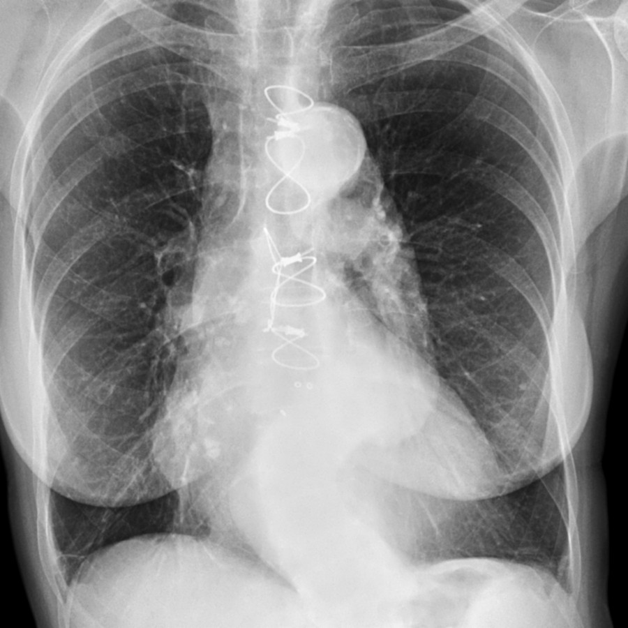
**Atherosclerotic**
*Frontal radiograph shows a dilated tortuous aorta with diffuse calcifications. Intimal disease further exacerbates medial degeneration by increasing wall stress and restricting blood flow.*
**Atherosclerotic**
*Frontal radiograph shows a dilated tortuous aorta with diffuse calcifications. Intimal disease further exacerbates medial degeneration by increasing wall stress and restricting blood flow.*
 and intimal calcifications
  . This patient had a diffusely dilated and tortuous aorta.](images/app.statdx.com_image_thumbnail_8505ecd7-1260-41f6-b29e-7cc482a87972_annotated_true_size_900_quality_90_2d77718fa1670e0b41e020eeaa77f8108c069f04.jpg)
**Atherosclerotic**
*Axial CECT shows intimal disease with mural thrombus and intimal calcifications . This patient had a diffusely dilated and tortuous aorta.*
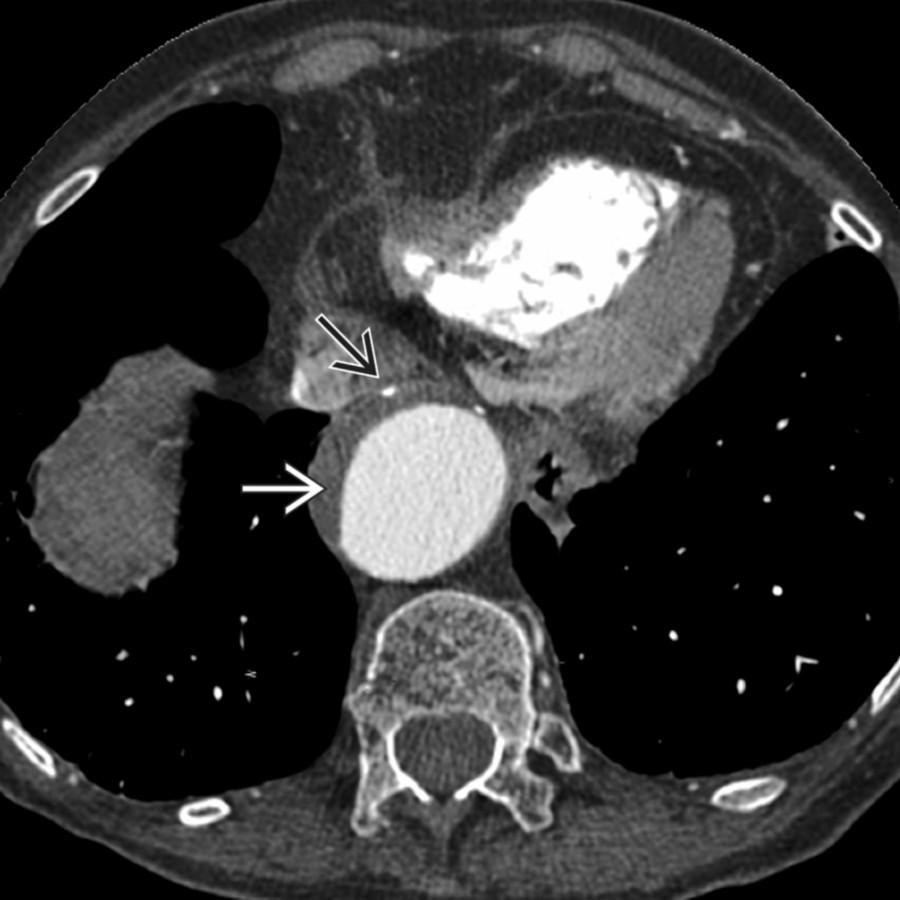
**Atherosclerotic**
*Axial CECT shows intimal disease with mural thrombus and intimal calcifications . This patient had a diffusely dilated and tortuous aorta.*
 , which can easily be detected on unenhanced CT.](images/app.statdx.com_image_thumbnail_36faa14f-20e2-4768-860d-c5732ebbad71_annotated_true_size_900_quality_90_7d3bf4488b5b41077857c1e2f202ec1d9002df4e.jpg)
**Atherosclerotic**
*Coronal CECT shows extravasation of contrast from a dilated abdominal aorta. Note extravasated blood , which can easily be detected on unenhanced CT.*
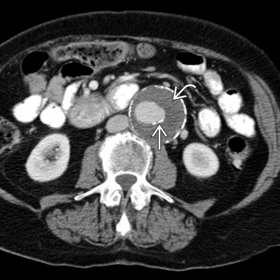
**Atherosclerotic**
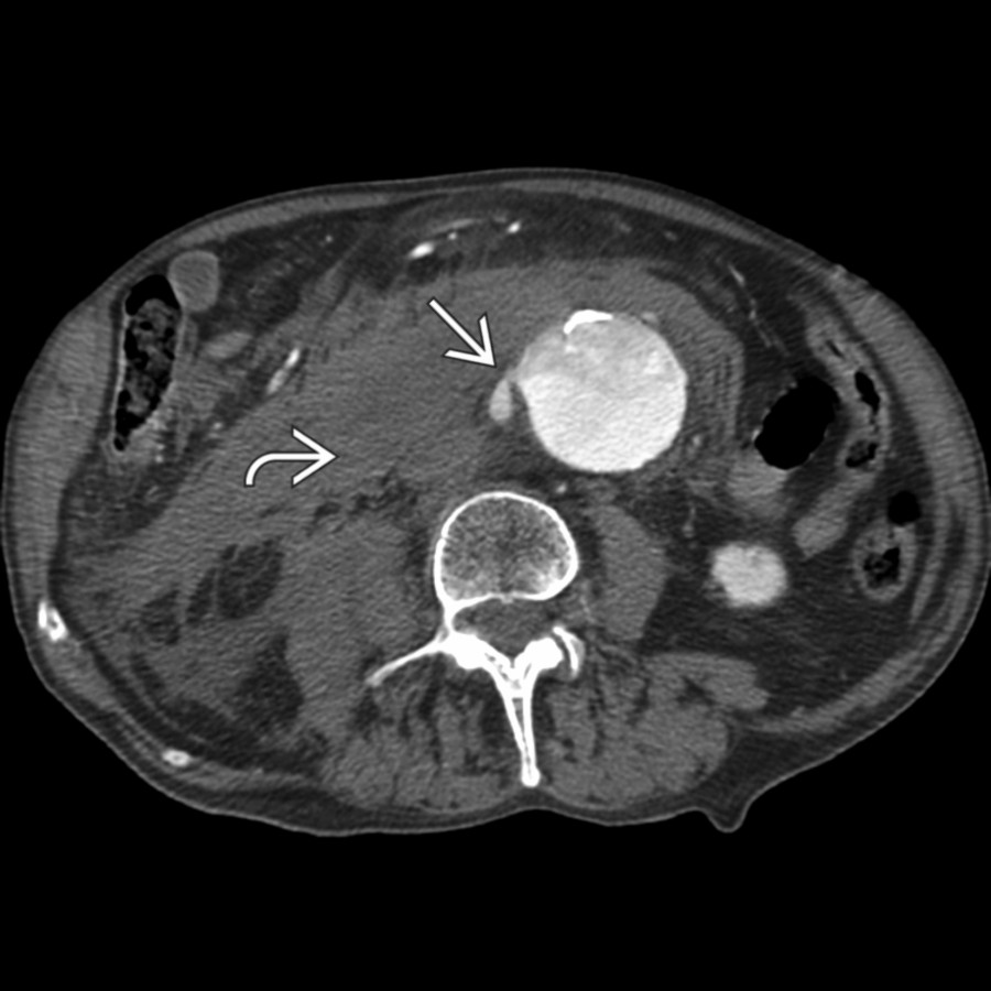
*Axial CECT shows dilated abdominal aorta with extensive mural thrombus . Calcifications occur when the thrombus is chronic and does not represent displaced intimal calcifications.*
**Degenerative**
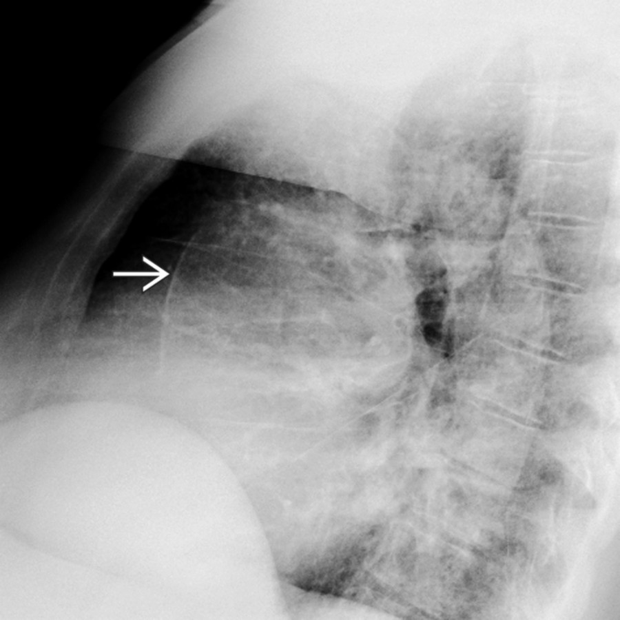
*Lateral radiograph shows diffuse aortic calcifications in a patient with longstanding hypertension and a dilated ascending aorta.*
**Aortic Stenosis**
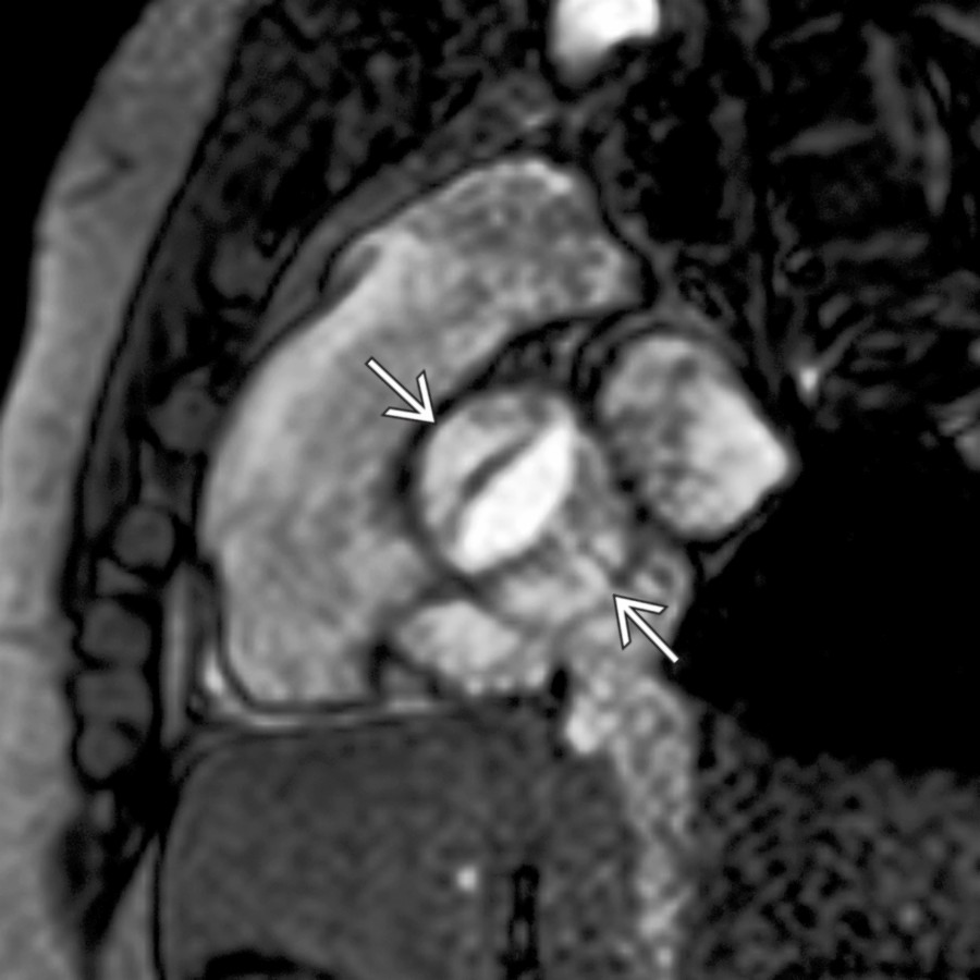
*Double oblique cine MR shows a bicuspid aortic valve in a young patient with a dilated ascending aorta. This image can be used to calculate valve area to quantify stenosis.*
**Aortic Stenosis**
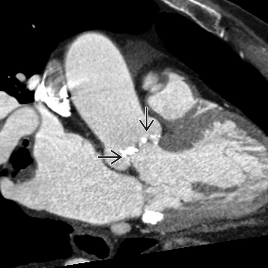
*Left ventricular outflow CECT shows calcifications on the aortic cusps in an older patient with an ascending aortic aneurysm.*
**Atherosclerotic**
*Coronal CECT shows extravasation of contrast from a dilated abdominal aorta. Note extravasated blood , which can easily be detected on unenhanced CT.*

**Atherosclerotic**
*Axial CECT shows dilated abdominal aorta with extensive mural thrombus . Calcifications occur when the thrombus is chronic and does not represent displaced intimal calcifications.*

**Degenerative**
*Lateral radiograph shows diffuse aortic calcifications in a patient with longstanding hypertension and a dilated ascending aorta.*

**Aortic Stenosis**
*Double oblique cine MR shows a bicuspid aortic valve in a young patient with a dilated ascending aorta. This image can be used to calculate valve area to quantify stenosis.*

**Aortic Stenosis**
*Left ventricular outflow CECT shows calcifications on the aortic cusps in an older patient with an ascending aortic aneurysm.*
 in acute dissection. Note the "bird beak" sign and "cob web" sign , which help identify the false lumen . This patient had a bicuspid valve and was treated with a modified Bentall procedure.](images/app.statdx.com_image_thumbnail_a7cd6f67-be8a-41fd-bce7-707554356e3c_annotated_true_size_900_quality_90_e6aec4e49575d93221be48bc8b7734e82c35b9ee.jpg)
**Aortic Dissection**
*Axial CECT shows ascending aortic false lumen dilation in acute dissection. Note the "bird beak" sign and "cob web" sign , which help identify the false lumen . This patient had a bicuspid valve and was treated with a modified Bentall procedure.*
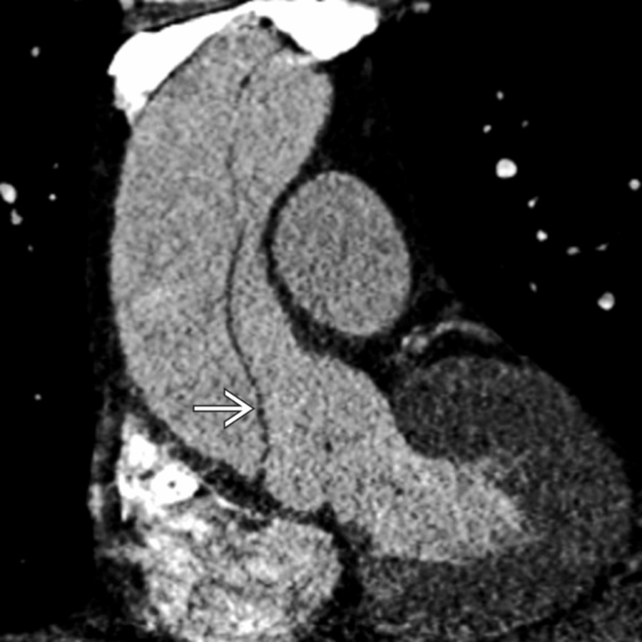
**Aortic Dissection**
*Double oblique CECT shows dilation of the ascending aorta in a hypertensive patient presenting with anterior chest pain. Note the intimal flap . This patient was treated with emergent surgery.*
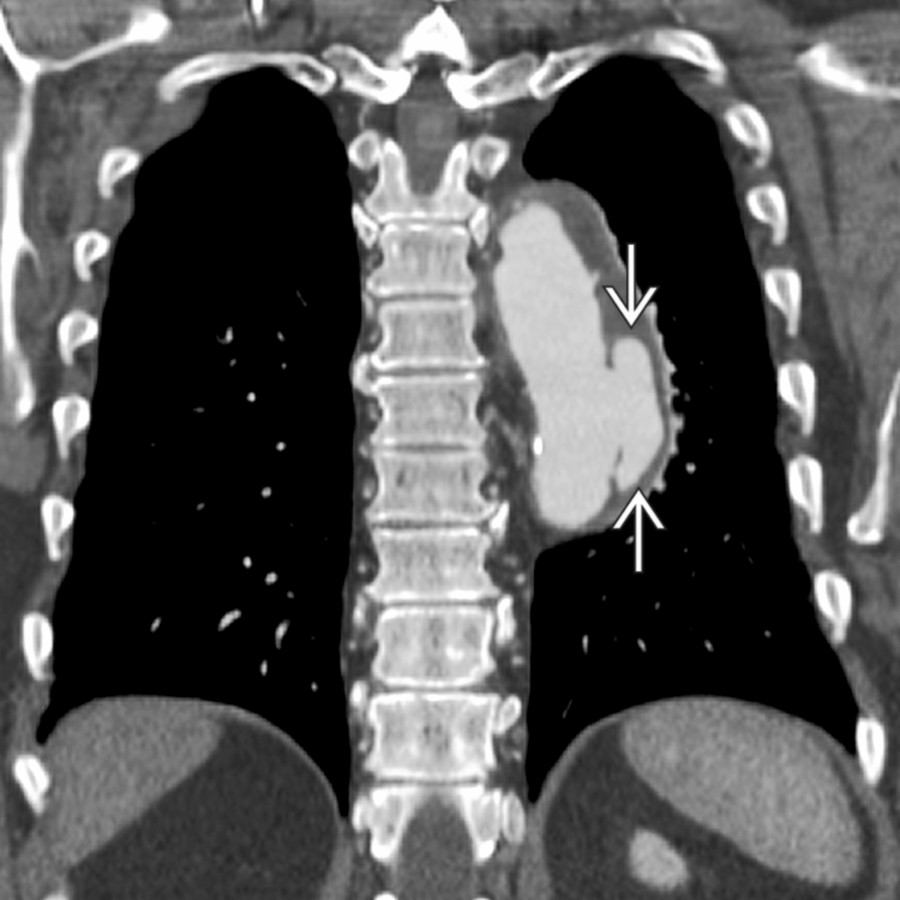
**Mycotic Aneurysm**
*Coronal CECT shows pseudoaneurysm in the mid descending aorta thought to be a mycotic aneurysm. Aside from this aneurysm, there was a paucity of disease throughout the remaining aorta.*

**Penetrating Atherosclerotic Ulcer**
*Axial black-blood MR shows an aortic wall defect that extends beyond the expected contour of the aortic lumen. High signal in this penetrating aortic ulcer is due to slow-flowing blood and not thrombosis.*

**Penetrating Atherosclerotic Ulcer**
*Coronal CECT shows a previously diagnosed penetrating atherosclerotic ulcer that progressed to frank rupture. Note the extravasated blood .*

**Posttraumatic Pseudoaneurysm**
*Volume-rendered image shows focal dilation of the aortic lumen at the level of the ligamentum arteriosum. This patient suffered a high-speed deceleration injury, presented with a traumatic pseudoaneurysm, and was treated with endovascular repair.*

**Connective Tissue Disease**
*Double oblique coronal left ventricular outflow view shows aortic root dilation and loss of sinotubular junction morphology in a patient with Marfan disease.*
### Additional Images

**Posttraumatic Pseudoaneurysm**
*Sagittal oblique conventional angiographic view shows a traumatic pseudoaneurysm prior to treatment with endovascular stenting. This patient suffered high-speed deceleration trauma.*
in acute dissection. Note the "bird beak" sign and "cob web" sign , which help identify the false lumen . This patient had a bicuspid valve and was treated with a modified Bentall procedure.](images/app.statdx.com_image_thumbnail_a7cd6f67-be8a-41fd-bce7-707554356e3c_annotated_true_size_900_quality_90_e6aec4e49575d93221be48bc8b7734e82c35b9ee.jpg)
**Aortic Dissection**
*Axial CECT shows ascending aortic false lumen dilation in acute dissection. Note the "bird beak" sign and "cob web" sign , which help identify the false lumen . This patient had a bicuspid valve and was treated with a modified Bentall procedure.*

**Aortic Dissection**
*Double oblique CECT shows dilation of the ascending aorta in a hypertensive patient presenting with anterior chest pain. Note the intimal flap . This patient was treated with emergent surgery.*

**Mycotic Aneurysm**
*Coronal CECT shows pseudoaneurysm in the mid descending aorta thought to be a mycotic aneurysm. Aside from this aneurysm, there was a paucity of disease throughout the remaining aorta.*

**Penetrating Atherosclerotic Ulcer**
*Axial black-blood MR shows an aortic wall defect that extends beyond the expected contour of the aortic lumen. High signal in this penetrating aortic ulcer is due to slow-flowing blood and not thrombosis.*

**Penetrating Atherosclerotic Ulcer**
*Coronal CECT shows a previously diagnosed penetrating atherosclerotic ulcer that progressed to frank rupture. Note the extravasated blood .*

**Posttraumatic Pseudoaneurysm**
*Volume-rendered image shows focal dilation of the aortic lumen at the level of the ligamentum arteriosum. This patient suffered a high-speed deceleration injury, presented with a traumatic pseudoaneurysm, and was treated with endovascular repair.*

**Connective Tissue Disease**
*Double oblique coronal left ventricular outflow view shows aortic root dilation and loss of sinotubular junction morphology in a patient with Marfan disease.*
### Additional Images

**Posttraumatic Pseudoaneurysm**
*Sagittal oblique conventional angiographic view shows a traumatic pseudoaneurysm prior to treatment with endovascular stenting. This patient suffered high-speed deceleration trauma.*