---
title: "Dolichoectasia"
docid: "31d50b93-b057-4da3-86b5-4cc8fb0bc806"
breadcrumbs:
- "Brain"
- "Differential Diagnosis"
- "Arteries"
- "Anatomically Based Differentials"
- "Fusiform Arterial Enlargement"
---
# ESSENTIAL INFORMATION
- ## Key Differential Diagnosis Issues
- Ectasia = elongated/tortuous artery
- Fusiform aneurysm
- Long-segment fusiform arterial dilatation
- Can be acute (dissecting) or chronic (atherosclerosis, nonatherosclerotic vasculopathy)
- ## Helpful Clues for Common Diagnoses
- **Dolichoectasia**
- Dilated/elongated arteries ± slow flow
- Vessel layers intact
- Older patients, chronic hypertension
- Vertebrobasilar > internal carotid artery (ICA)
- Ectasia often extends into branches
- **Atherosclerotic Fusiform Aneurysm**
- Thick wall ± organized thrombus
- Variable slow flow
- **Dissecting Aneurysm/Pseudoaneurysm**
- Focal arterial dilatation
- Trauma = most common etiology
- Next to hard/fixed structures (bone, dura)
- **Nonaneurysmal Dissection**
- Vertebral > basilar > ICA
- Lacks changes of atherosclerosis in other vessels
- Can be spontaneous or traumatic
- ## Helpful Clues for Less Common Diagnoses
- **CNS Vasculitis**
- Involves multiple vessels
- Alternating stenoses and fusiform dilatations
- **Inherited Connective Tissue Disorders**
- Abnormal connective tissue involves vessel walls
- Aneurysm and dissections
- Thoracic aorta most common location
- **HIV Infection**
- Vasculopathy may be present in children or adults
- Multifocal fusiform enlargement involving supraclinoid ICA and basilar artery
- No consistent infectious agent isolated
- ## Helpful Clues for Rare Diagnoses
- **Giant Serpentine Aneurysm**
- Large, partially thrombosed mass
- Distal branches arise from aneurysm dome
- Lacks definable neck
- ICA/middle cerebral artery > vertebrobasilar artery
- **Atypical Saccular Aneurysm**
- Arises from vessel bifurcations
- Long "aspect ratio" → fusiform appearance
- Often multilobulated, bizarre
## Images
### Selected Images
 in an octogenarian related to dolichoectasia. Fusiform dolichoectasia is a common finding in the vertebrobasilar arteries in older patients.](images/app.statdx.com_image_38e1497f-31e3-49db-8b46-4ab561bf1eac_fb437f8b_20251018T080401Z.jpg)
*Axial CT shows fusiform dilatation and tortuosity of the basilar artery in an octogenarian related to dolichoectasia. Fusiform dolichoectasia is a common finding in the vertebrobasilar arteries in older patients.*
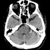
*Axial CT shows fusiform dilatation and tortuosity of the basilar artery in an octogenarian related to dolichoectasia. Fusiform dolichoectasia is a common finding in the vertebrobasilar arteries in older patients.*
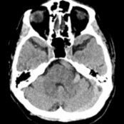
*Axial CT shows fusiform dilatation and tortuosity of the basilar artery in an octogenarian related to dolichoectasia. Fusiform dolichoectasia is a common finding in the vertebrobasilar arteries in older patients.*
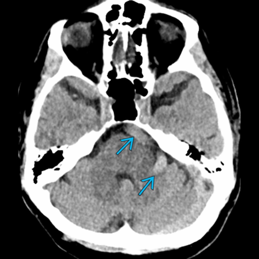
*Axial CT shows fusiform dilatation and tortuosity of the basilar artery in an octogenarian related to dolichoectasia. Fusiform dolichoectasia is a common finding in the vertebrobasilar arteries in older patients.*

*Axial CT shows fusiform dilatation and tortuosity of the basilar artery in an octogenarian related to dolichoectasia. Fusiform dolichoectasia is a common finding in the vertebrobasilar arteries in older patients.*

*Axial CT shows fusiform dilatation and tortuosity of the basilar artery in an octogenarian related to dolichoectasia. Fusiform dolichoectasia is a common finding in the vertebrobasilar arteries in older patients.*
 . No significant mural thrombus was noted in this fusiform aneurysm.](images/app.statdx.com_image_thumbnail_f63a9221-71c6-4320-af66-1ed581202eb9_size_168_quality_85_8a3ae196_20251018T080328Z.jpg)
*Coronal CTA shows fusiform dilatation of the right supraclinoid internal carotid artery (ICA). Irregularity from atherosclerotic disease can be seen of the M1 segment of the middle cerebral artery . No significant mural thrombus was noted in this fusiform aneurysm.*
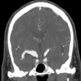
*Coronal CTA shows fusiform dilatation of the right supraclinoid internal carotid artery (ICA). Irregularity from atherosclerotic disease can be seen of the M1 segment of the middle cerebral artery . No significant mural thrombus was noted in this fusiform aneurysm.*
 , T2
, T2  , and T1 pre- and post DANTE VWI sequences shows peripheral enhancement, suggestive of instability.](images/app.statdx.com_image_thumbnail_68b41ae0-9f3a-4484-8b02-69d6772626db_size_168_quality_85_8d79e53e_20251018T080328Z.jpg)
*Dissecting pseudoaneurysm in the V4 segment of the right vertebral artery seen on 3D TOF MRA , T2 , and T1 pre- and post DANTE VWI sequences shows peripheral enhancement, suggestive of instability.*

*3D MIP MRA of the vertebrobasilar arteries in a teenage female with a history of type 4 Ehlers-Danlos shows fusiform dilatation of the vertebral artery . The affected gene is COL3A1, and this specific type of Ehlers-Danlos has a higher risk of aneurysm and vascular rupture.*
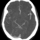
*Axial MIP from CT arteriography shows fusiform dilatation of the left middle cerebral artery bifurcation in this child with a history of Marfan syndrome.*

*Coronal MIP reformat from CT arteriography shows bilateral fusiform aneurysms of supraclinoid ICAs . This patient also had thoracic aortic aneurysm, which is consistent with familial thoracic aortic aneurysm and dissection and is associated with a mutation of ACTA2. This gene is responsible for a component of vascular smooth muscle.*
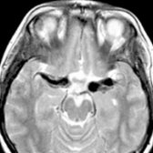
*Axial T2WI MR shows strikingly enlarged middle cerebral arteries in this child with congenital HIV/AIDS (an uncommon but well-recognized cause of pediatric fusiform arteriopathy). The stroke-like presentations of HIV infection may relate to vasculopathies, including large-vessel aneurysmal vasculopathy.*
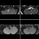
*Dissecting pseudoaneurysm in the V4 segment of the right vertebral artery seen on 3D TOF MRA , T2 , and T1 pre- and post DANTE VWI sequences shows peripheral enhancement, suggestive of instability.*

*3D MIP MRA of the vertebrobasilar arteries in a teenage female with a history of type 4 Ehlers-Danlos shows fusiform dilatation of the vertebral artery . The affected gene is COL3A1, and this specific type of Ehlers-Danlos has a higher risk of aneurysm and vascular rupture.*

*Axial MIP from CT arteriography shows fusiform dilatation of the left middle cerebral artery bifurcation in this child with a history of Marfan syndrome.*

*Coronal MIP reformat from CT arteriography shows bilateral fusiform aneurysms of supraclinoid ICAs . This patient also had thoracic aortic aneurysm, which is consistent with familial thoracic aortic aneurysm and dissection and is associated with a mutation of ACTA2. This gene is responsible for a component of vascular smooth muscle.*

*Axial T2WI MR shows strikingly enlarged middle cerebral arteries in this child with congenital HIV/AIDS (an uncommon but well-recognized cause of pediatric fusiform arteriopathy). The stroke-like presentations of HIV infection may relate to vasculopathies, including large-vessel aneurysmal vasculopathy.*
 with associated mural thrombus
  seen on sagittal CTA.](images/app.statdx.com_image_thumbnail_8fb2594c-2abc-43d9-84bc-b8b67664090e_size_168_quality_85_e273dd7e_20251018T080329Z.jpg)
*Axial NECT demonstrates a giant serpentine aneurysm in the basilar artery with associated mural thrombus seen on sagittal CTA.*
### Additional Images
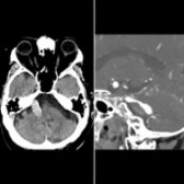
*Axial NECT demonstrates a giant serpentine aneurysm in the basilar artery with associated mural thrombus seen on sagittal CTA.*
### Additional Images
 .](images/app.statdx.com_image_thumbnail_5a50f257-ef7f-4680-94e0-670c586154aa_size_168_quality_85_75bc027c_20251018T080401Z.jpg)
*Sagittal T1WI MR shows an elongated basilar artery with a slow-flow, thickened wall . The apex of the tortuous basilar artery indents the hypothalamus, 3rd ventricle .*
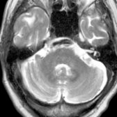
*Axial T2WI MR shows an elongated, tortuous basilar artery with a thickened arterial wall , typical for atherosclerosis-associated fusiform ectasia.*
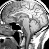
*Sagittal T1WI MR shows an elongated basilar artery with a slow-flow, thickened wall . The apex of the tortuous basilar artery indents the hypothalamus, 3rd ventricle .*

*Axial T2WI MR shows an elongated, tortuous basilar artery with a thickened arterial wall , typical for atherosclerosis-associated fusiform ectasia.*
 . This is an unusual example because of the location (ICA, middle cerebral artery).](5b2eae83-b875-4f2c-9903-07a167a394a0)
*Lateral angiography shows a large fusiform middle cerebral artery aneurysm that extends into smaller, more distal branches . This is an unusual example because of the location (ICA, middle cerebral artery).*
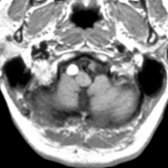
*Axial T1WI MR shows an enlarged right vertebral artery with high signal intensity as well as an absent flow void of the left vertebral artery .*
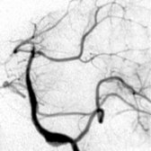
*Anteroposterior oblique view of the left vertebral angiogram shows focal elongations and widening of the basilar artery in a 6-year-old child with Ehlers-Danlos type 4.*

*Axial MRA submentovertex view shows an unusual nonatherosclerotic giant serpentine fusiform aneurysm. The patent channel lies within the clot in the partially thrombosed lumen.*
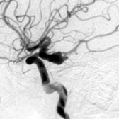
*Lateral angiography in 30-year-old man with a subarachnoid hemorrhage shows an elongated, bizarre-appearing, multilobulated aneurysm with long aspect ratio, tit-like projections.*
. This is an unusual example because of the location (ICA, middle cerebral artery).](5b2eae83-b875-4f2c-9903-07a167a394a0)
*Lateral angiography shows a large fusiform middle cerebral artery aneurysm that extends into smaller, more distal branches . This is an unusual example because of the location (ICA, middle cerebral artery).*

*Axial T1WI MR shows an enlarged right vertebral artery with high signal intensity as well as an absent flow void of the left vertebral artery .*

*Anteroposterior oblique view of the left vertebral angiogram shows focal elongations and widening of the basilar artery in a 6-year-old child with Ehlers-Danlos type 4.*

*Axial MRA submentovertex view shows an unusual nonatherosclerotic giant serpentine fusiform aneurysm. The patent channel lies within the clot in the partially thrombosed lumen.*

*Lateral angiography in 30-year-old man with a subarachnoid hemorrhage shows an elongated, bizarre-appearing, multilobulated aneurysm with long aspect ratio, tit-like projections.*