---
title: "Enhancing Suprasellar Mass"
docid: "528eaea9-095f-4fab-ad42-5ad84e215a2d"
authors:
- key: "318f80ab-6abb-4067-a809-2ebdaa5a30c9"
value: "Kalen Riley, MD, MBA"
- key: "5cff4116-3654-4b3a-bb75-5ebe0b8c9850"
value: "Anne G. Osborn, MD, FACR"
- key: "8d5254e9-8dda-478b-8f08-bdee97a32c79"
value: "Karen L. Salzman, MD, FACR"
breadcrumbs:
-
name: "Brain"
slug: "brain"
treeNodeId: "6d8829f1-14d7-45af-8675-255189aa526a"
-
name: "Differential Diagnosis"
slug: "differential-diagnosis"
treeNodeId: "a7fdd139-664e-4bb8-8d18-400e4733ff60"
-
name: "Sella/Juxtasellar, Pineal Region"
slug: "sellajuxtasellar-pineal-region"
treeNodeId: "5e38b9c1-3137-47e3-aa83-1fc82cb4099a"
-
name: "Anatomically Based Differentials"
slug: "anatomically-based-differentials"
treeNodeId: "7a51b2ca-8fee-4c16-aff3-b7189f68ea60"
-
name: "Enhancing Suprasellar Mass"
slug: "enhancing-suprasellar-mass"
treeNodeId: null
category: "Brain"
cmeTopicId: "01b1cc14-8442-4bbd-93ff-bd8895b90f87"
documentVersionId: "39bca65a-aa9f-4818-9b63-d727551a81f1"
imageCount: 19
lastUpdated: "01/26/23"
pageDescription: "Enhancing Suprasellar Mass"
pageKeywords: "Brain, Differential Diagnosis, Sella/Juxtasellar, Pineal Region, Anatomically Based Differentials, Enhancing Suprasellar Mass"
pageTitle: "Enhancing Suprasellar Mass | STATdx"
enhancedTitle: "Enhancing Suprasellar Mass"
type: "DDX"
references: true
breadcrumbs:
- "Brain"
- "Differential Diagnosis"
- "Sella/Juxtasellar, Pineal Region"
- "Anatomically Based Differentials"
- "Enhancing Suprasellar Mass"
---
# ESSENTIAL INFORMATION
- ## Key Differential Diagnosis Issues
- Effect of age on differential diagnosis important
- Common lesions ("big 5") account for > 75% of all suprasellar masses
- Most other lesions < 1-2% each
- Differential diagnosis narrows if mass confined to infundibulum
- ## Helpful Clues for Common Diagnoses
- **Pituitary Macroadenoma vs. Meningioma**
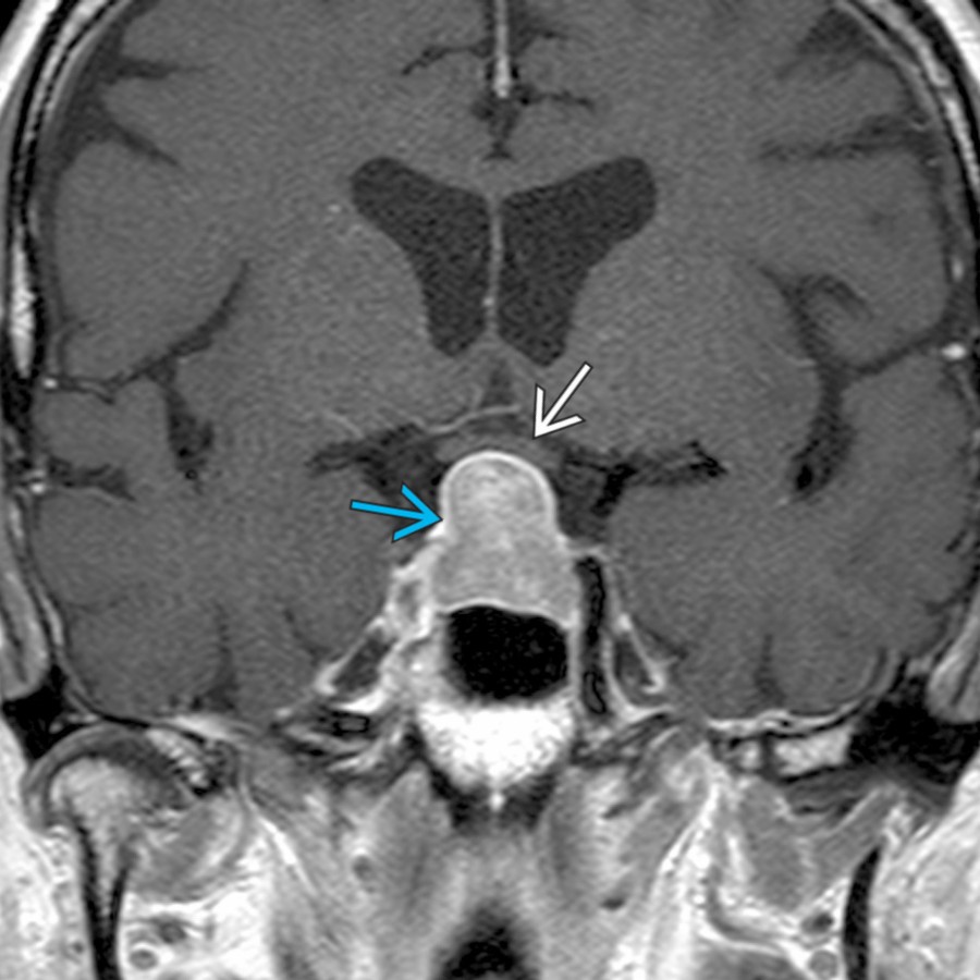
- Macroadenoma: Gland cannot be identified separate from mass
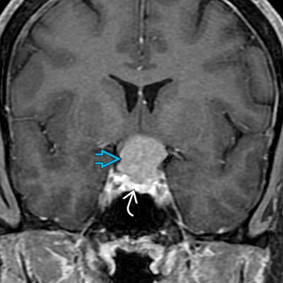
- Meningioma: Mass distinct from gland (diaphragma sellae separates mass above from pituitary below)
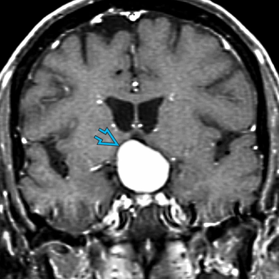
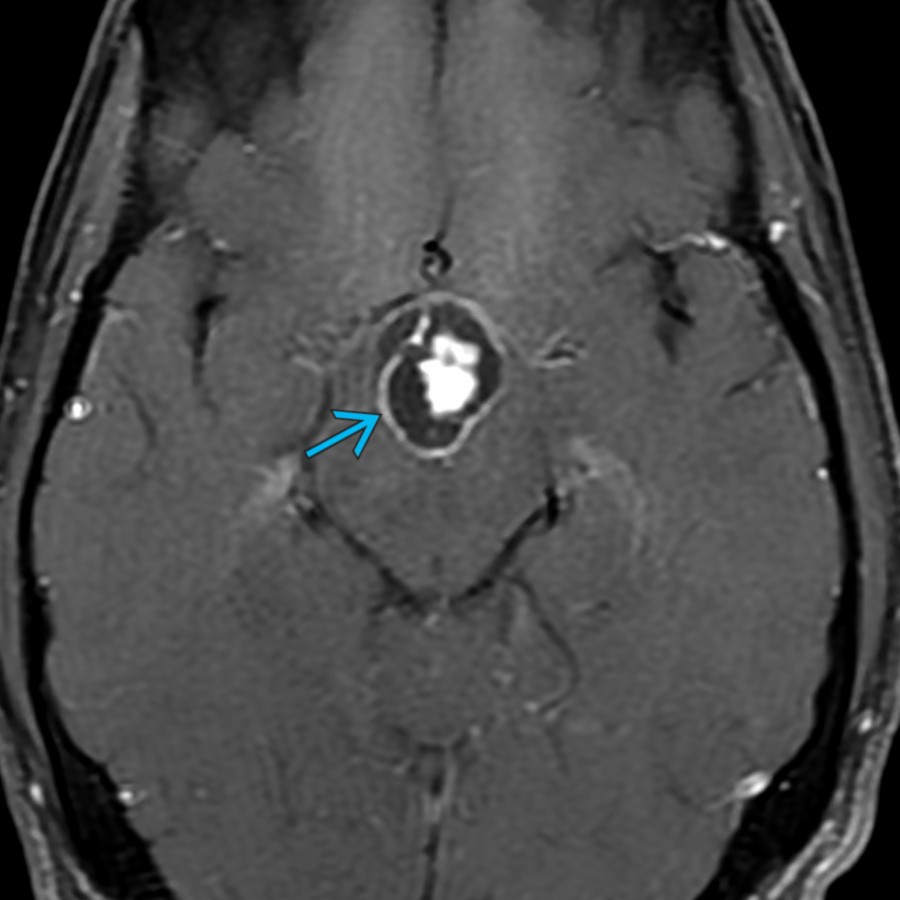
- **S****accular****Aneurysm**
- Coronal plane helps distinguish aneurysm from pituitary
- Look for phase artifact, flow void on MR
- **Craniopharyngioma**
- Adamantinomatous: 90% Ca⁺⁺, 90% cystic, 90% enhance
- Papillary may be solid, noncalcified, enhance strongly
- **Pilocytic Astrocytoma**
- More common in children than adults
- T2 hyperintense, variable enhancement
- Expands hypothalamus, optic chiasm
- May extend into optic nerves/tracts
- **Pilomyxoid Astrocytoma**
- Subtype of pilocytic astrocytoma, more aggressive
- Infant > child
- Large, bulky tumor with lateral extension to temporal lobe common
- Hemorrhage in 20-25%
- ## Helpful Clues for Less Common Diagnoses
- **Pituitary Hyperplasia**
- May be physiologic (pregnancy, lactation, puberty)
- Pathologic in end-organ failure (thyroid, ovarian, etc.)
- **Neurosarcoid**
- Older patients, enhancing mass
- **Langerhans Cell Histiocytosis**
- Children and young adults; enhancing mass
- May cause thickened infundibulum
- Absence of pituitary "bright spot"
- **Germinoma**
- Children > > adults; enhancing mass
- CT hyperdense
- May cause thickened infundibulum
- Absence of the pituitary "bright spot"
- **Lymphocytic Hypophysitis**
- Can mimic pituitary adenoma on imaging
- Most common in peripartum female
- May be autoimmune, inflammatory, granulomatous process, drug related
- May be secondary to checkpoint inhibitor therapy
- **Astrocytoma, IDH-Mutant**
- May affect hypothalamic and suprasellar regions
- T2 hyperintense; variable enhancement
- WHO grades 2-4
- ## Helpful Clues for Rare Diagnoses
- Solitary**metastasis** to gland &/or stalk rare
- **Lymphoma**, **leukemia** usually with systemic disease
- **Pituicytoma, g****ranular cell tumor**, and **spindle cell oncocytoma** are rare tumors, which may occur along infundibulum &/or posterior pituitary or may mimic macroadenoma
- ## Alternative Differential Approaches
- **Adult**: Consider adenoma, meningioma, and aneurysm
- **Child**: Craniopharyngioma, astrocytoma, Langerhans cell histiocytosis, and germinoma
## References
# Selected References
1. [Kinoshita Y et al: Natural course of Rathke's cleft cysts and risk factors for progression. J Neurosurg. 1-7, 2022](http://www.ncbi.nlm.nih.gov/pubmed/?term=36057119%5Bpmid%5D)
1. [Pascual JM et al: Duct-like recess in the infundibular portion of third ventricle craniopharyngiomas: an MRI sign identifying the papillary type. AJNR Am J Neuroradiol. 43(9):1333-40, 2022](http://www.ncbi.nlm.nih.gov/pubmed/?term=35953277%5Bpmid%5D)
1. [Poyuran R et al: Nonneoplastic and noninfective cysts of the central nervous system: a histopathological study. Neuropathology. ePub, 2022](http://www.ncbi.nlm.nih.gov/pubmed/?term=36210745%5Bpmid%5D)
1. [Jipa A et al: Imaging of the sellar and parasellar regions. Clin Imaging. 77:254-75, 2021](http://www.ncbi.nlm.nih.gov/pubmed/?term=34153590%5Bpmid%5D)
1. [Kurokawa R et al: MRI findings of immune checkpoint inhibitor-induced hypophysitis: possible association with fibrosis. AJNR Am J Neuroradiol. 41(9):1683-9, 2020](http://www.ncbi.nlm.nih.gov/pubmed/?term=32763900%5Bpmid%5D)
1. [Go JL et al: Imaging of the sella and parasellar region. Radiol Clin North Am. 55(1):83-101, 2017](http://www.ncbi.nlm.nih.gov/pubmed/?term=27890190%5Bpmid%5D)
1. [Seeburg DP et al: Imaging of the sella and parasellar region in the pediatric population. Neuroimaging Clin N Am. 27(1):99-121, 2017](http://www.ncbi.nlm.nih.gov/pubmed/?term=27889026%5Bpmid%5D)
1. [Zamora C et al: Sellar and parasellar imaging. Neurosurgery. 80(1):17-38, 2017](http://www.ncbi.nlm.nih.gov/pubmed/?term=28362892%5Bpmid%5D)
1. [Patel KS et al: Intraoperative magnetic resonance imaging assessment of non-functioning pituitary adenomas during transsphenoidal surgery. Pituitary. 19(2):222-31, 2016](http://www.ncbi.nlm.nih.gov/pubmed/?term=26323592%5Bpmid%5D)
1. [Petrakakis I et al: The sellar and suprasellar region: a "hideaway" of rare lesions. Clinical aspects, imaging findings, surgical outcome and comparative analysis. Clin Neurol Neurosurg. 149:154-65, 2016](http://www.ncbi.nlm.nih.gov/pubmed/?term=27540757%5Bpmid%5D)
1. [Wang J et al: The clinicopathological features of pituicytoma and the differential diagnosis of sellar glioma. Neuropathology. 36(5):432-440, 2016](http://www.ncbi.nlm.nih.gov/pubmed/?term=26919073%5Bpmid%5D)
1. [Wu AW et al: Chondroid chordoma of the sella turcica mimicking a pituitary adenoma. Ear Nose Throat J. 94(10-11):E47-9, 2015](http://www.ncbi.nlm.nih.gov/pubmed/?term=26535833%5Bpmid%5D)
1. DW Louis, et. al (Editors). WHO Classification of Tumours of the Central Nervous System. International Agency for Research on Cancer, Lyon, 2016. Chapter 17: Tumours of the sellar region, pp 323-334.
## Images
### Selected Images
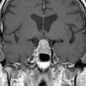
 . The pituitary gland cannot be separated from the mass and indeed is the mass. Note the superior displacement of the optic chiasm
  by the macroadenoma.](images/app.statdx.com_image_thumbnail_1cbcbdf8-0e55-4af0-b4ff-011551b12f25_annotated_true_size_900_quality_90_c8f5de23.jpg)
**Pituitary Macroadenoma**
*Coronal T1 C+ MR shows an enhancing intrasellar and suprasellar mass . The pituitary gland cannot be separated from the mass and indeed is the mass. Note the superior displacement of the optic chiasm by the macroadenoma.*
**Pituitary Macroadenoma**
*Coronal T1 C+ MR shows an enhancing intrasellar and suprasellar mass . The pituitary gland cannot be separated from the mass and indeed is the mass. Note the superior displacement of the optic chiasm by the macroadenoma.*
**Pituitary Macroadenoma**
*Coronal T1 C+ MR shows an enhancing intrasellar and suprasellar mass . The pituitary gland cannot be separated from the mass and indeed is the mass. Note the superior displacement of the optic chiasm by the macroadenoma.*

**Pituitary Macroadenoma**
*Coronal T1 C+ MR shows an enhancing intrasellar and suprasellar mass . The pituitary gland cannot be separated from the mass and indeed is the mass. Note the superior displacement of the optic chiasm by the macroadenoma.*
 separate from the normal pituitary gland below
  . Meningiomas often have associated hyperostosis of the adjacent bone. Additionally, these benign tumors enhance homogeneously and may have an associated dural tail.](images/app.statdx.com_image_thumbnail_e113dc69-0754-4224-952a-c02882a414df_annotated_true_size_900_quality_90_3104d9f2.jpg)
**Meningioma**
*Coronal T1 C+ FS MR shows a suprasellar enhancing mass separate from the normal pituitary gland below . Meningiomas often have associated hyperostosis of the adjacent bone. Additionally, these benign tumors enhance homogeneously and may have an associated dural tail.*
**Meningioma**
*Coronal T1 C+ FS MR shows a suprasellar enhancing mass separate from the normal pituitary gland below . Meningiomas often have associated hyperostosis of the adjacent bone. Additionally, these benign tumors enhance homogeneously and may have an associated dural tail.*
 caused by large basilar tip aneurysm with slow intraluminal flow. Aneurysms are the 3rd most common suprasellar mass in an adult.](images/app.statdx.com_image_thumbnail_67a712f3-90f1-4ab8-b4c4-c2c5a25c9e4a_annotated_true_size_900_quality_90_1ca9fa52.jpg)
**Saccular Aneurysm**
*Axial T1 C+ MR shows a suprasellar enhancing mass with prominent phase artifact caused by large basilar tip aneurysm with slow intraluminal flow. Aneurysms are the 3rd most common suprasellar mass in an adult.*
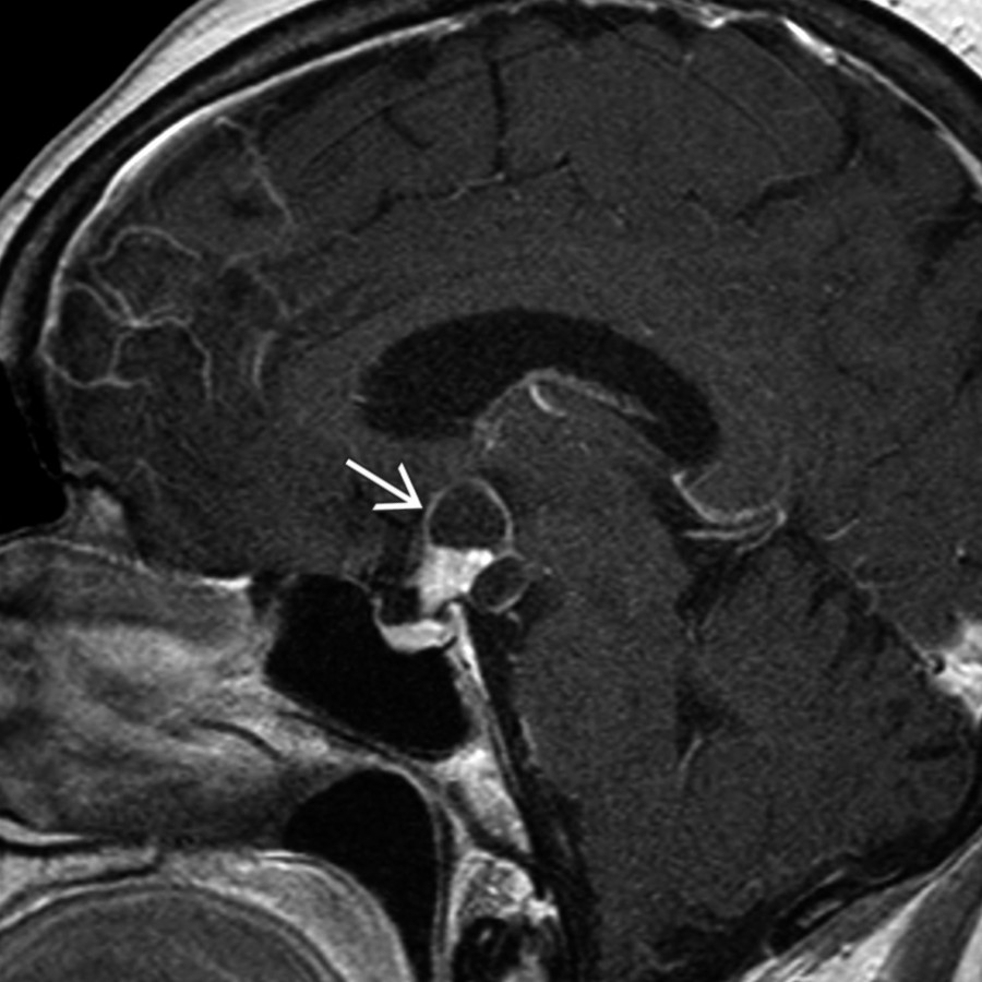
**Craniopharyngioma**
*Sagittal T1 C+ MR shows an enhancing, partially solid and cystic suprasellar mass related to an adamantinomatous craniopharyngioma. These WHO grade 1 tumors are the most common pediatric intracranial tumor of nonglial origin.*
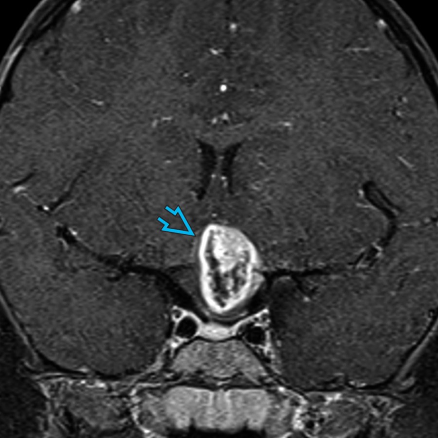
**Pilomyxoid Astrocytoma**
*Coronal T1 C+ FS MR shows a heterogeneously enhancing suprasellar mass in a child that was found to represent a pilomyxoid astrocytoma, WHO grade 1. Pilomyxoid astrocytoma is a subtype of pilocytic astrocytoma that is often more aggressive.*
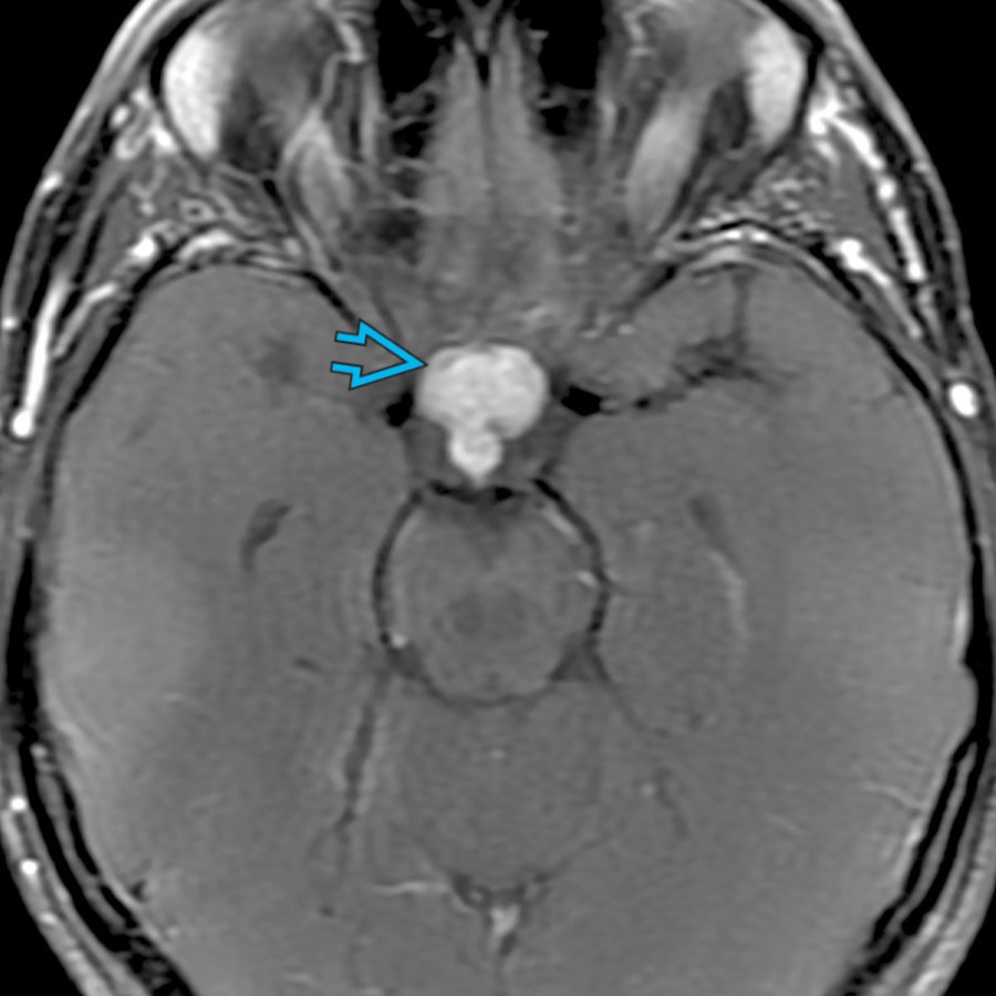
**Neurosarcoid**
*Axial T1 C+ FS MR shows a lobular, enhancing suprasellar mass related to neurosarcoid. Patients with neurosarcoid often have associated pulmonary hilar adenopathy.*
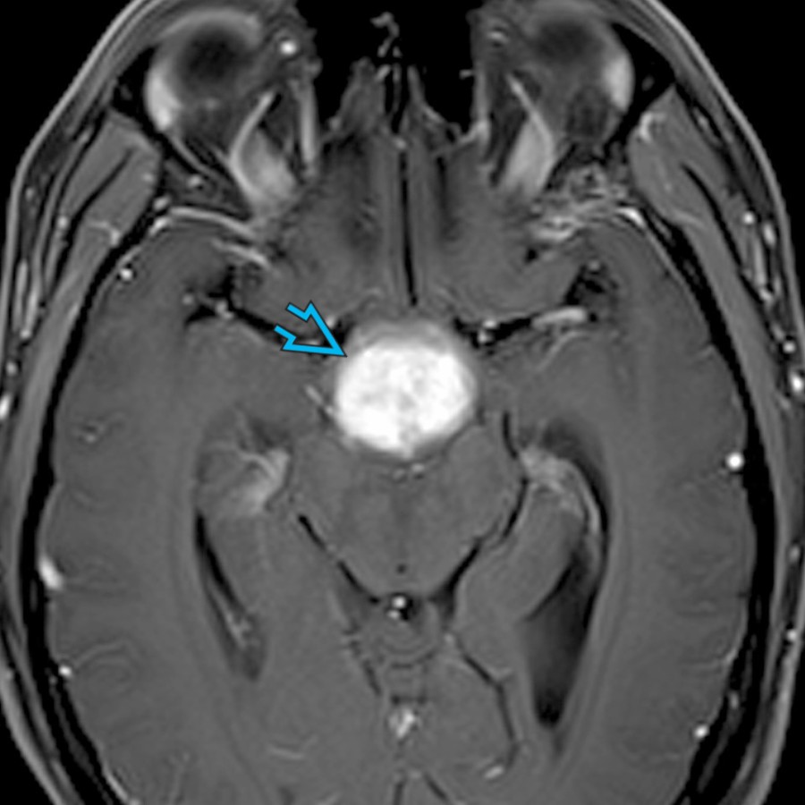
**Langerhans Cell Histiocytosis**
*Axial T1 C+ FS MR shows a diffusely enhancing suprasellar mass in a young adult who first presented with diabetes insipidus and a thick, enhancing infundibulum with absence of a posterior pituitary bright spot. Nine months later, patient returned with a much larger suprasellar mass.*
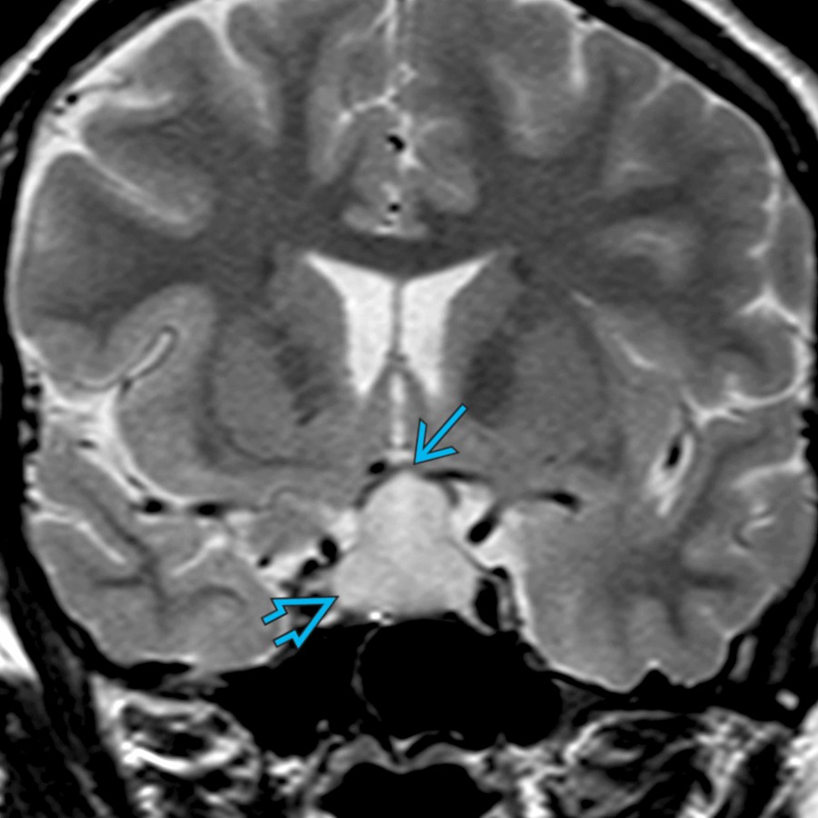
**Lymphocytic Hypophysitis**
*Coronal T2 MR in a pregnant patient with acute vision problems shows a sellar and suprasellar mass with superior displacement of the optic chiasm . Imaging of lymphocytic hypophysitis may mimic a pituitary macroadenoma.*
### Additional Images
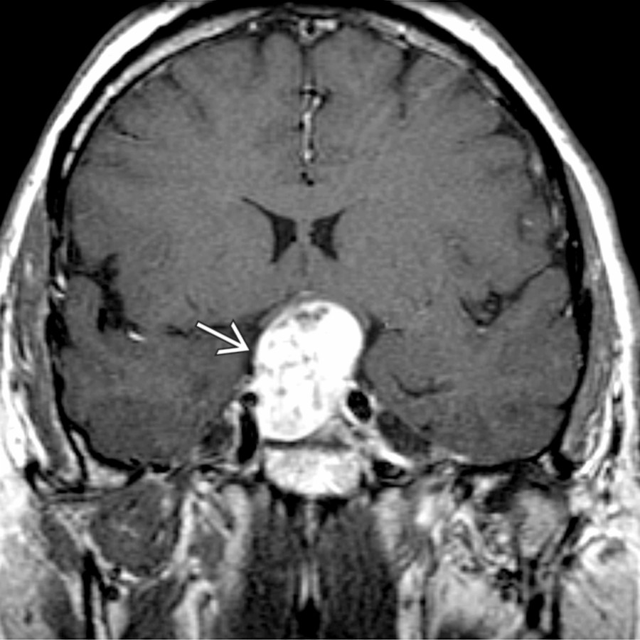
**Pituitary Macroadenoma**
*Coronal T1 C+ MR shows an inhomogeneously enhancing intra- and suprasellar mass ; the pituitary gland cannot be found separate from it and indeed is the mass.*
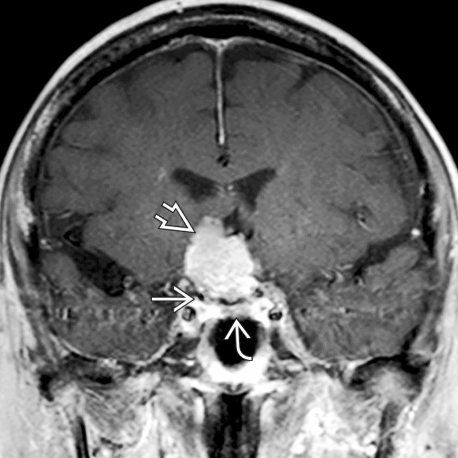
**Meningioma**
*Coronal T1 C+ FS MR shows a suprasellar enhancing mass separated from the normal pituitary gland below by a thin black line of diaphragma sellae .*
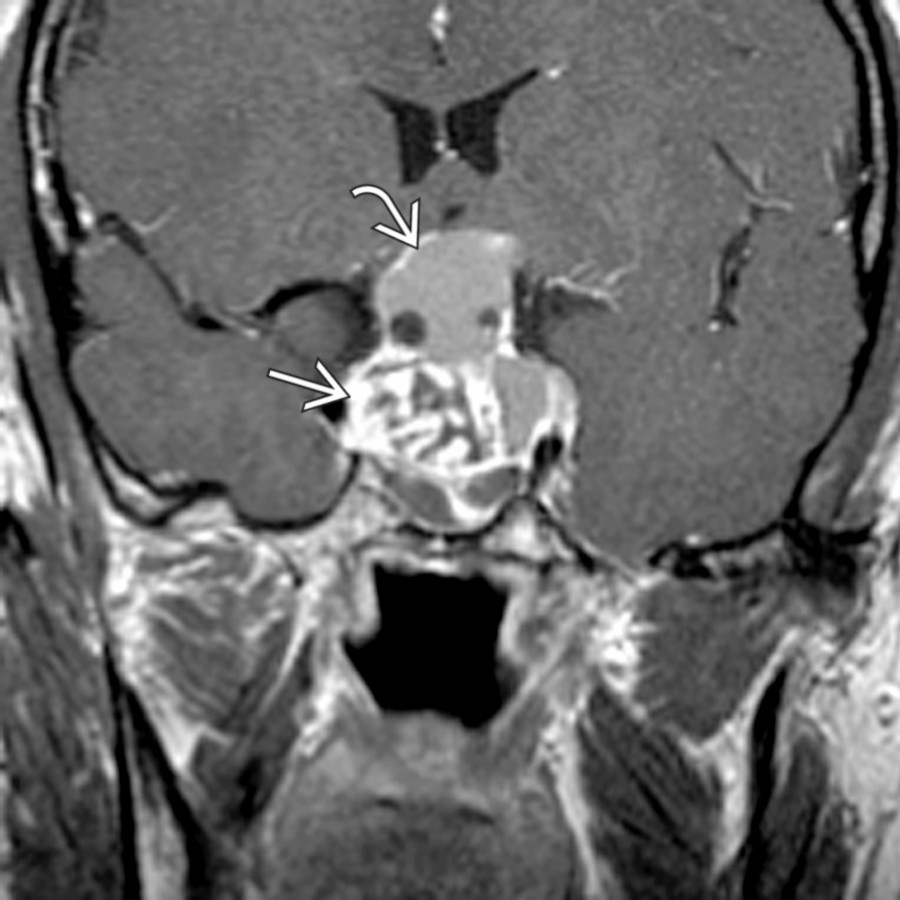
**Craniopharyngioma**
*Coronal T1 C+ MR in a child shows a intra-/suprasellar mass. An apical nonenhancing portion is surrounded by a thin enhancing rim . The solid portion enhances strongly but heterogeneously .*
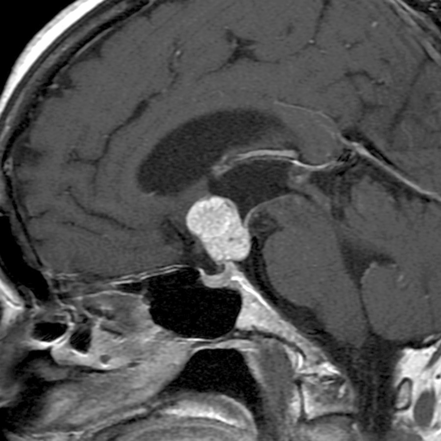
**Craniopharyngioma**
*Sagittal T1 C+ FS MR shows a papillary craniopharyngioma in an adult man. Papillary craniopharyngiomas are most often solid masses.*
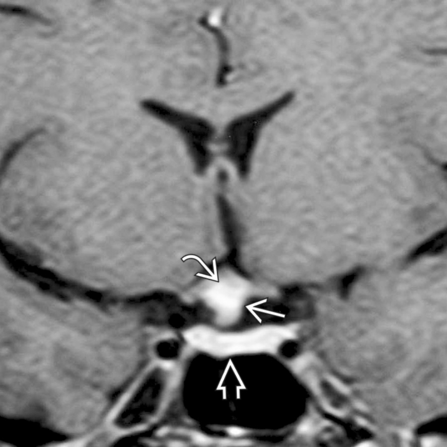
**Astrocytoma, IDH-Mutant**
*Coronal T1 C+ MR shows an enhancing suprasellar mass separate from the pituitary gland below . Note involvement of the optic chiasm .*
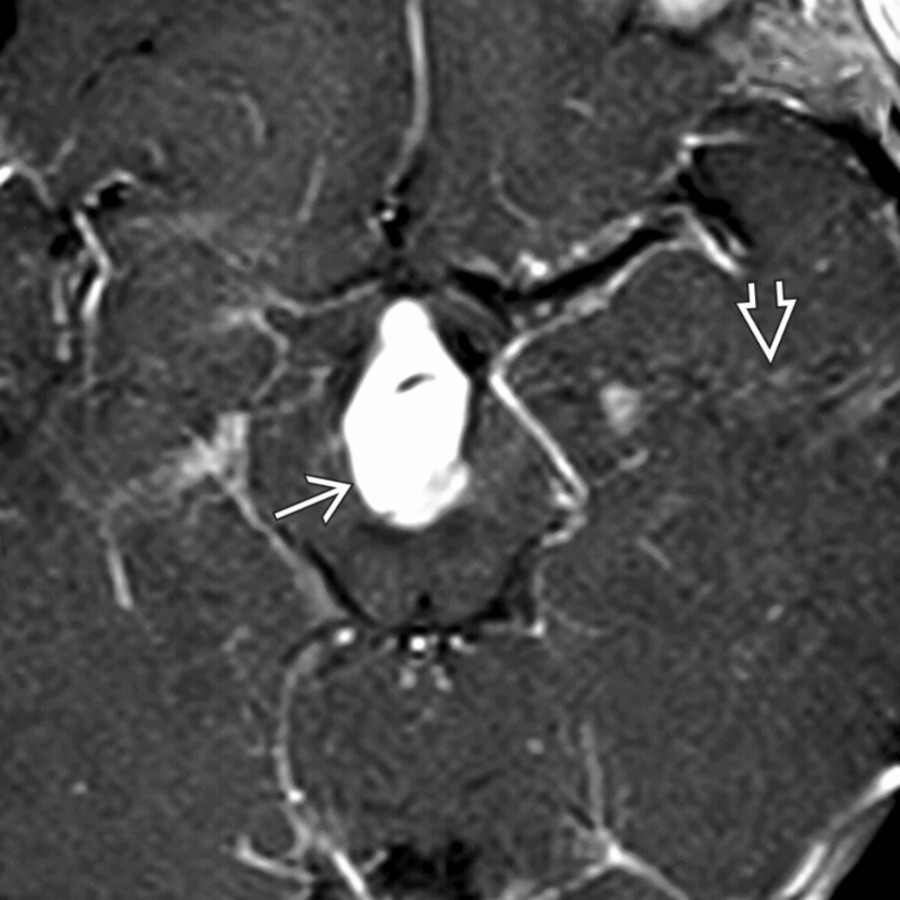
**Saccular Aneurysm**
*Axial T1 C+ MR shows a suprasellar enhancing mass with prominent phase artifact caused by large basilar tip aneurysm with slow intraluminal flow. Aneurysms are the 3rd most common suprasellar mass in an adult.*

**Craniopharyngioma**
*Sagittal T1 C+ MR shows an enhancing, partially solid and cystic suprasellar mass related to an adamantinomatous craniopharyngioma. These WHO grade 1 tumors are the most common pediatric intracranial tumor of nonglial origin.*

**Pilomyxoid Astrocytoma**
*Coronal T1 C+ FS MR shows a heterogeneously enhancing suprasellar mass in a child that was found to represent a pilomyxoid astrocytoma, WHO grade 1. Pilomyxoid astrocytoma is a subtype of pilocytic astrocytoma that is often more aggressive.*

**Neurosarcoid**
*Axial T1 C+ FS MR shows a lobular, enhancing suprasellar mass related to neurosarcoid. Patients with neurosarcoid often have associated pulmonary hilar adenopathy.*

**Langerhans Cell Histiocytosis**
*Axial T1 C+ FS MR shows a diffusely enhancing suprasellar mass in a young adult who first presented with diabetes insipidus and a thick, enhancing infundibulum with absence of a posterior pituitary bright spot. Nine months later, patient returned with a much larger suprasellar mass.*

**Lymphocytic Hypophysitis**
*Coronal T2 MR in a pregnant patient with acute vision problems shows a sellar and suprasellar mass with superior displacement of the optic chiasm . Imaging of lymphocytic hypophysitis may mimic a pituitary macroadenoma.*
### Additional Images

**Pituitary Macroadenoma**
*Coronal T1 C+ MR shows an inhomogeneously enhancing intra- and suprasellar mass ; the pituitary gland cannot be found separate from it and indeed is the mass.*

**Meningioma**
*Coronal T1 C+ FS MR shows a suprasellar enhancing mass separated from the normal pituitary gland below by a thin black line of diaphragma sellae .*

**Craniopharyngioma**
*Coronal T1 C+ MR in a child shows a intra-/suprasellar mass. An apical nonenhancing portion is surrounded by a thin enhancing rim . The solid portion enhances strongly but heterogeneously .*

**Craniopharyngioma**
*Sagittal T1 C+ FS MR shows a papillary craniopharyngioma in an adult man. Papillary craniopharyngiomas are most often solid masses.*

**Astrocytoma, IDH-Mutant**
*Coronal T1 C+ MR shows an enhancing suprasellar mass separate from the pituitary gland below . Note involvement of the optic chiasm .*
 .](images/app.statdx.com_image_thumbnail_ff3cfeac-da02-4993-b553-b3c4956a9a56_annotated_true_size_900_quality_90_da128db3.jpg)
**Pilocytic Astrocytoma**
*Sagittal T1 C+ MR in a 3-year-old boy with rotary nystagmus and vomiting shows a large suprasellar mass clearly separate from the pituitary gland below .*
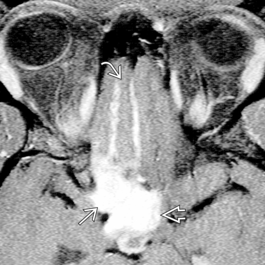
**Neurosarcoid**
*Axial T1 C+ FS MR shows an enhancing suprasellar mass involving the optic chiasm and hypothalamus with pial enhancement along the inferior frontal lobe . The lesion also involved the optic nerve.*
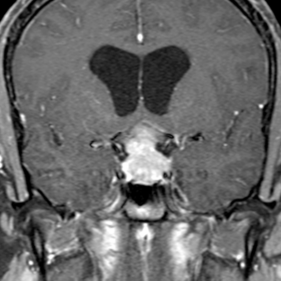
**Germinoma**
*Coronal T1 C+ FS MR in 22-year-old man with diabetes insipidus shows hydrocephalus and an enhancing sellar/suprasellar mass. Germinoma commonly presents with diabetes insipidus. A macroadenoma often has visual defects.*
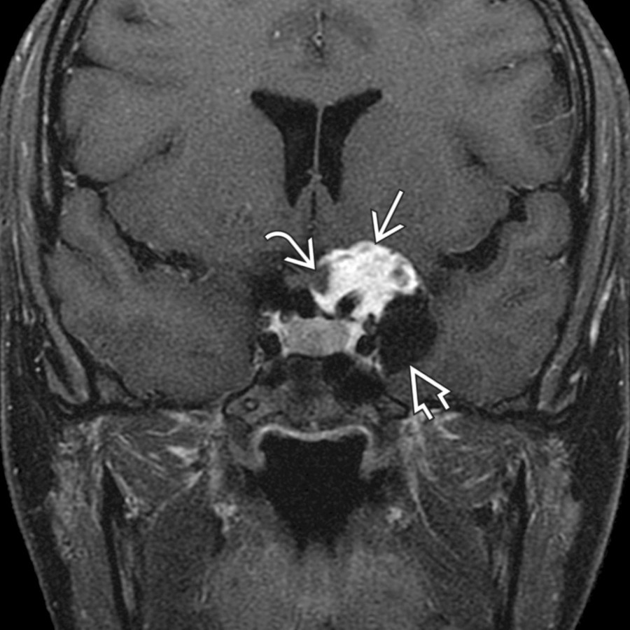
**Rare Sellar Region Tumor (Pituicytoma, Granular Cell Tumor, Spindle Cell Oncocytoma)**
*Coronal T1 C+ MR shows a diffusely enhancing suprasellar mass that involves the infundibulum. A pituicytoma was diagnosed at resection. These rare tumors are WHO grade 1 and usually arise along the infundibulum or neurohypophysis. (Courtesy A.V. Hasso, MD.)*
**Craniopharyngioma**
*Axial T1 C+ FS MR shows a cystic and solid suprasellar enhancing mass . In an adult, craniopharyngiomas may be adamantinomatous or papillary types. Adamantinomatous craniopharyngiomas enhance, calcify, and have cysts. Papillary craniopharyngiomas are commonly solid and enhancing. These are WHO grade 1 tumors.*
**Pilocytic Astrocytoma**
*Coronal T1 C+ FS MR in 25-year-old woman with visual complaints shows an enhancing suprasellar mass that partially encases the optic chiasm . There is also a parasellar cystic portion of the mass. Pilocytic astrocytomas often present as a solid and cystic cerebellar mass in a child or young adult. These WHO grade 1 tumors are typically T2 hyperintense with variable enhancement.*
.](images/app.statdx.com_image_thumbnail_ff3cfeac-da02-4993-b553-b3c4956a9a56_annotated_true_size_900_quality_90_da128db3.jpg)
**Pilocytic Astrocytoma**
*Sagittal T1 C+ MR in a 3-year-old boy with rotary nystagmus and vomiting shows a large suprasellar mass clearly separate from the pituitary gland below .*

**Neurosarcoid**
*Axial T1 C+ FS MR shows an enhancing suprasellar mass involving the optic chiasm and hypothalamus with pial enhancement along the inferior frontal lobe . The lesion also involved the optic nerve.*

**Germinoma**
*Coronal T1 C+ FS MR in 22-year-old man with diabetes insipidus shows hydrocephalus and an enhancing sellar/suprasellar mass. Germinoma commonly presents with diabetes insipidus. A macroadenoma often has visual defects.*

**Rare Sellar Region Tumor (Pituicytoma, Granular Cell Tumor, Spindle Cell Oncocytoma)**
*Coronal T1 C+ MR shows a diffusely enhancing suprasellar mass that involves the infundibulum. A pituicytoma was diagnosed at resection. These rare tumors are WHO grade 1 and usually arise along the infundibulum or neurohypophysis. (Courtesy A.V. Hasso, MD.)*

**Craniopharyngioma**
*Axial T1 C+ FS MR shows a cystic and solid suprasellar enhancing mass . In an adult, craniopharyngiomas may be adamantinomatous or papillary types. Adamantinomatous craniopharyngiomas enhance, calcify, and have cysts. Papillary craniopharyngiomas are commonly solid and enhancing. These are WHO grade 1 tumors.*

**Pilocytic Astrocytoma**
*Coronal T1 C+ FS MR in 25-year-old woman with visual complaints shows an enhancing suprasellar mass that partially encases the optic chiasm . There is also a parasellar cystic portion of the mass. Pilocytic astrocytomas often present as a solid and cystic cerebellar mass in a child or young adult. These WHO grade 1 tumors are typically T2 hyperintense with variable enhancement.*