---
title: "Enlarged Cardiac Silhouette"
docid: "13d59c6a-0f7f-4389-8d8c-f19ca039e446"
authors:
- key: "df804626-c042-4296-96e3-836a6da50fd6"
value: "Gregory Kicska, MD, PhD"
breadcrumbs:
-
name: "Cardiac"
slug: "cardiac"
treeNodeId: "fa90100b-619c-430e-8074-b5b9789bab39"
-
name: "Differential Diagnosis"
slug: "differential-diagnosis"
treeNodeId: "952326a0-b3ea-4a21-aa7a-d796cc9325ed"
-
name: "Enlarged Cardiac Silhouette"
slug: "enlarged-cardiac-silhouette"
treeNodeId: null
category: "Cardiac"
documentVersionId: "c4c4d705-71ba-4631-be39-f4b35c596710"
imageCount: 5
lastUpdated: "03/17/22"
pageDescription: "Enlarged Cardiac Silhouette"
pageKeywords: "Cardiac, Differential Diagnosis, Enlarged Cardiac Silhouette"
pageTitle: "Enlarged Cardiac Silhouette | STATdx"
enhancedTitle: "Enlarged Cardiac Silhouette"
type: "DDX"
breadcrumbs:
- "Cardiac"
- "Differential Diagnosis"
- "Enlarged Cardiac Silhouette"
---
# ESSENTIAL INFORMATION
- ## Key Differential Diagnosis Issues
- Pericardial space fluid: Globular enlargement
- Cardiac chamber enlargement: Characteristic contour abnormality, such as filling of retrosternal clear space in right ventricle enlargement
- Pericardial mass: Focal contour irregularity
- ## Helpful Clues for Common Diagnoses
- **Ischemic Cardiomyopathy**
- Subendocardial fat or calcium, left ventricle (LV) wall thinning in coronary distribution, dense coronary calcifications
- MR shows subendocardial or transmural delayed enhancement in coronary artery distribution
- **Valvular Disease**
- Valvular calcifications most common
- MR cine or phase contrast shows flow jets
- **Heart Failure Exacerbation**
- Coexistent signs of pulmonary edema
- **Pericardial****Effusion**
- New globular heart enlargement on radiograph, fluid-density pericardial fluid on CT
- Hemopericardium suggested by high-density pericardial fluid or neoplasm history (lung, breast, melanoma)
- ## Helpful Clues for Less Common Diagnoses
- **Nonischemic Dilated Cardiomyopathy**
- Dilated LV, thin wall, LVEF < 40%
- Either no delayed enhancement present or enhancement is not subendocardial
- **Pericardial Mass**
- Pericardial cyst: Circumscribed fluid density at right more than left cardiophrenic angle
- Pericardial fat pad: Fat density most commonly at right cardiophrenic angle
- ## Helpful Clues for Rare Diagnoses
- **Left Ventricle Aneurysm**
- True aneurysm
- Post infarct wall thinning, dilatation, and associated thrombus
- Most often present along apical anterior or lateral wall
- False aneurysm
- Ruptured myocardium contained by pericardial adhesions
- Most commonly seen at infero-lateral or inferior basal LV wall segments
- Neck narrower than internal diameter
## Images
### Selected Images
 in the septal and anterior wall at the base. The patient had hypokinesis and wall thinning at this location.](images/app.statdx.com_image_thumbnail_cbe4ba3d-6512-494a-8a88-92e1e0c7ddd0_annotated_true_size_900_quality_90_631757b3c8bf7dd62d33b84ca04f40c67c117996.jpg)
**Ischemic Cardiomyopathy**
*Short-axis delayed contrast-enhanced image shows subendocardial enhancement in the septal and anterior wall at the base. The patient had hypokinesis and wall thinning at this location.*
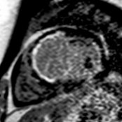
**Ischemic Cardiomyopathy**
*Short-axis delayed contrast-enhanced image shows subendocardial enhancement in the septal and anterior wall at the base. The patient had hypokinesis and wall thinning at this location.*
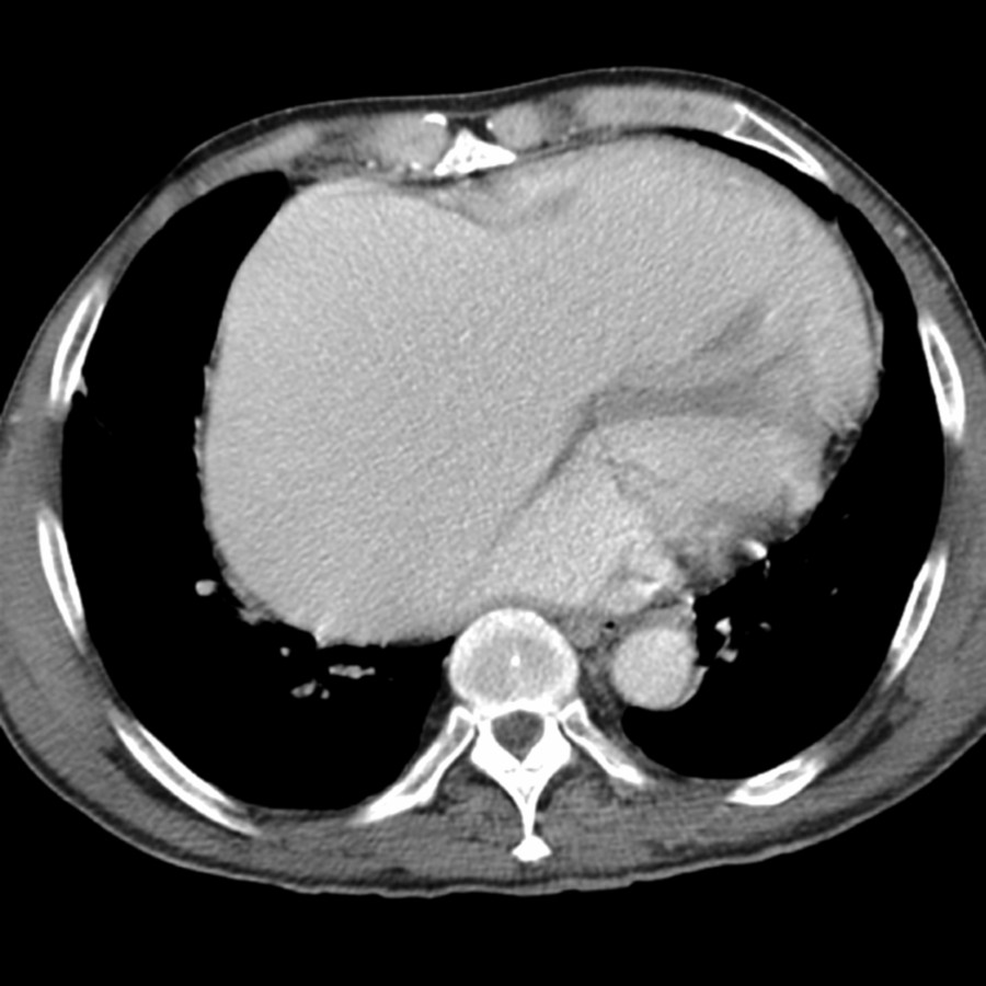
**Valvular Disease**
*Axial CECT shows an enlarged right atrium in a patient with severe tricuspid regurgitation. Radiograph showed rightward deviation of the right heart border. Regurgitant jet was seen on MR.*
### Additional Images
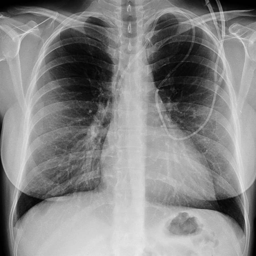
**Heart Failure Exacerbation**
*Frontal radiograph shows a normal cardiac silhouette in a patient with ischemic cardiomyopathy and a 40% ejection fraction. Compare with the next image, which was acquired 1 month later.*
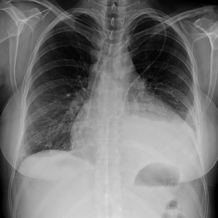
**Heart Failure Exacerbation**
*Frontal radiograph shows marked enlargement of the cardiac silhouette. The patient presented with increasing shortness of breath. Large pericardial effusion was found with presumed etiology of heart failure exacerbation.*
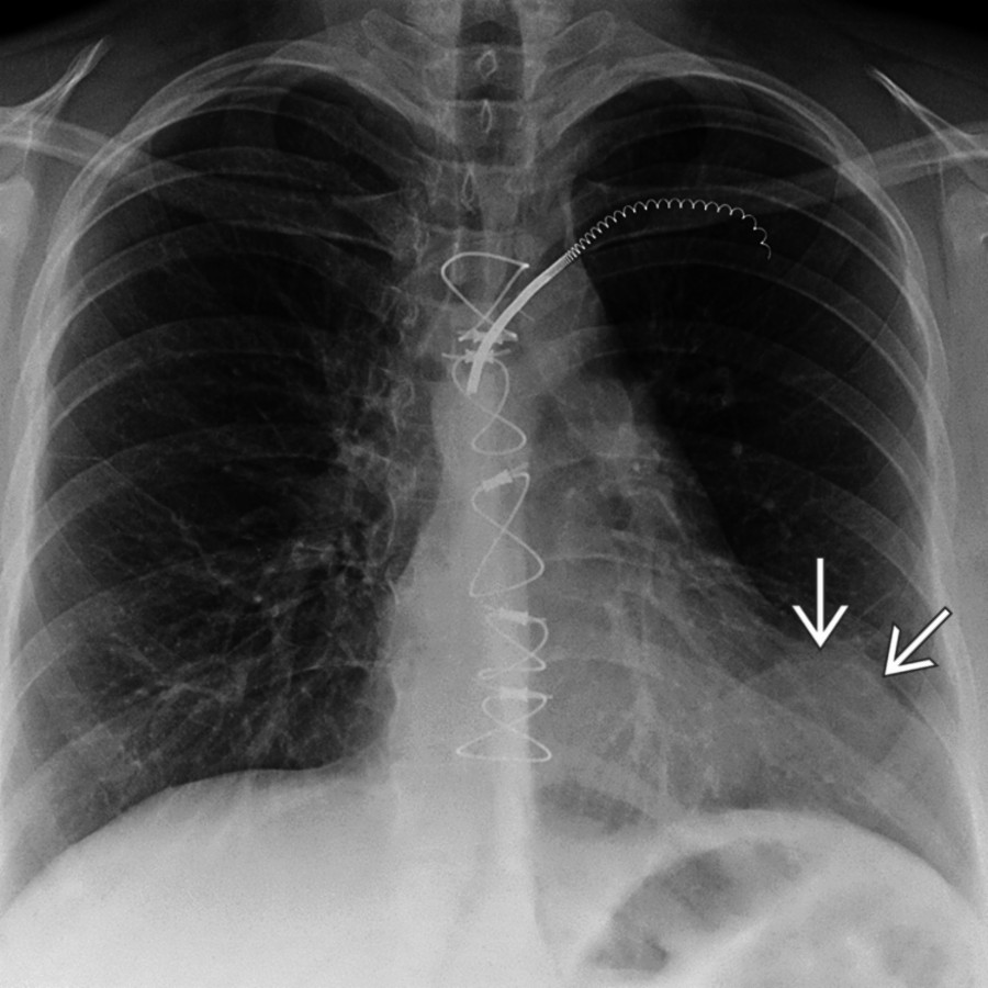
**Left Ventricle Aneurysm**
*Frontal radiograph shows left cardiac border contour deformity . CT showed abdominal fat, which had herniated into the left cardiophrenic space.*
in the septal and anterior wall at the base. The patient had hypokinesis and wall thinning at this location.](images/app.statdx.com_image_thumbnail_cbe4ba3d-6512-494a-8a88-92e1e0c7ddd0_annotated_true_size_900_quality_90_631757b3c8bf7dd62d33b84ca04f40c67c117996.jpg)
**Ischemic Cardiomyopathy**
*Short-axis delayed contrast-enhanced image shows subendocardial enhancement in the septal and anterior wall at the base. The patient had hypokinesis and wall thinning at this location.*

**Ischemic Cardiomyopathy**
*Short-axis delayed contrast-enhanced image shows subendocardial enhancement in the septal and anterior wall at the base. The patient had hypokinesis and wall thinning at this location.*

**Valvular Disease**
*Axial CECT shows an enlarged right atrium in a patient with severe tricuspid regurgitation. Radiograph showed rightward deviation of the right heart border. Regurgitant jet was seen on MR.*
### Additional Images

**Heart Failure Exacerbation**
*Frontal radiograph shows a normal cardiac silhouette in a patient with ischemic cardiomyopathy and a 40% ejection fraction. Compare with the next image, which was acquired 1 month later.*

**Heart Failure Exacerbation**
*Frontal radiograph shows marked enlargement of the cardiac silhouette. The patient presented with increasing shortness of breath. Large pericardial effusion was found with presumed etiology of heart failure exacerbation.*

**Left Ventricle Aneurysm**
*Frontal radiograph shows left cardiac border contour deformity . CT showed abdominal fat, which had herniated into the left cardiophrenic space.*