---
title: "Finger-in-Glove Sign"
docid: "81c5db2f-b8f6-4092-bcd2-ffb8aa3ab18a"
authors:
- key: "fa682303-6426-4b97-8c60-783051febb88"
value: "Jorge Alberto Carrillo-Bayona, MD"
breadcrumbs:
-
name: "Chest"
slug: "chest"
treeNodeId: "23b17a2b-c629-4f3b-b960-0bfdc5d138ca"
-
name: "Differential Diagnosis"
slug: "differential-diagnosis"
treeNodeId: "41e2b114-bb5b-4430-86a8-4c667ef0f50a"
-
name: "Airways"
slug: "airways"
treeNodeId: "dbd08dc8-069b-455c-af2f-73376c667e07"
-
name: "General Imaging Patterns"
slug: "general-imaging-patterns"
treeNodeId: "569ccd87-4cb8-4a9e-83f4-c8ac16b26c5e"
-
name: "Finger-in-Glove Sign"
slug: "finger-in-glove-sign"
treeNodeId: null
category: "Chest"
documentVersionId: "b09d26e0-dd26-493f-a120-ba11c7e04745"
imageCount: 8
lastUpdated: "02/10/20"
pageDescription: "Finger-in-Glove Sign"
pageKeywords: "Chest, Differential Diagnosis, Airways, General Imaging Patterns, Finger-in-Glove Sign"
pageTitle: "Finger-in-Glove Sign | STATdx"
enhancedTitle: "Finger-in-Glove Sign"
type: "DDX"
breadcrumbs:
- "Chest"
- "Differential Diagnosis"
- "Airways"
- "General Imaging Patterns"
- "Finger-in-Glove Sign"
---
# ESSENTIAL INFORMATION
- ## Key Differential Diagnosis Issues
- Finger-in-glove sign: Mucoid impaction with resultant dilated or impacted bronchi
- Bronchial obstruction: Atresia, foreign body, neoplasm
- Nonobstructive entities: Allergic bronchopulmonary aspergillosis, cystic fibrosis
- Radiography/CT: Branching tubular opacities that typically radiate from hilum toward lung periphery
- Differential diagnosis of bronchial branching tubular opacities
- Arteriovenous malformation
- Exhibits contrast enhancement
- Demonstrates afferent and efferent vessels: Feeding artery(ies) and draining vein(s)
- ## Helpful Clues for Common Diagnoses
- **Allergic Bronchopulmonary Aspergillosis**
- Immunologic disorder caused by hypersensitivity to*Aspergillus fumigatus*
- Predisposing conditions: Asthma, cystic fibrosis
- Laboratory findings: Positive aspergillus skin test, elevated IgE levels against *Aspergillus fumigatus*
- Total IgE levels > 1,000 IU/mL
- IgG antibodies against*A. fumigatus* in serum
- Total eosinophil count > 500 cells/mL
- Imaging: Branching tubular opacities, high attenuation on CT due to intrinsic calcium oxalate
- **Congenital Bronchial Atresia**
- Focal atresia of segmental bronchus
- Typically affects left upper lobe apicoposterior segment
- Recurrent infection in 20% of patients
- CT: Tubular or branching opacity surrounded by hyperlucent lung due to air-trapping and oligemia
- ## Helpful Clues for Less Common Diagnoses
- **Bronchiectasis**
- Cystic fibrosis
- Autosomal recessive disorder that affects regulation of chloride transport
- Recurrent infection, progressive bronchial wall injury
- Diffuse bronchiectasis (upper lobe predominant); central and peripheral airways affected
- Primary ciliary dyskinesia
- Structural abnormality that affects ciliary motion
- Bronchiectasis, sinusitis, infertility
- Basilar predominant airway involvement
- **Benign Airway Neoplasm**
- Hamartoma
- Endobronchial (1.4% of cases)
- Fat &/or calcification in endobronchial lesion supports diagnosis
- Lipoma
- Endobronchial lipoma (0.1-0.5% of lung tumors)
- Nonenhancing homogeneous fat attenuation lesion
- **Malignant Airway Neoplasm**
- Carcinoid tumor
- Low-grade neuroendocrine neoplasm
- Intrinsic calcification; may be eccentric
- Marked contrast enhancement
- Lung cancer
- Centrally obstructing mass
- Mucoid impaction uncommon
- **Foreign Body**
- Patients < 15 years of age (70%)
- Most frequent location: Right lower lobe bronchus and bronchus intermedius
- Adults: Chronic clinical course, recurrent pneumonia
## Images
### Selected Images
 corresponding to mucoid impaction in dilated airways.](images/app.statdx.com_image_thumbnail_827b9b74-083a-4a9c-abb6-c4b077a6a9d3_size_168_quality_85_cc4c584a_20251018T064244Z.jpg)
**Allergic Bronchopulmonary Aspergillosis**
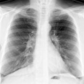
*PA chest radiograph of a patient with asthma and allergic bronchopulmonary aspergillosis shows right basilar tubular opacities corresponding to mucoid impaction in dilated airways.*
**Allergic Bronchopulmonary Aspergillosis**
*PA chest radiograph of a patient with asthma and allergic bronchopulmonary aspergillosis shows right basilar tubular opacities corresponding to mucoid impaction in dilated airways.*

**Allergic Bronchopulmonary Aspergillosis**
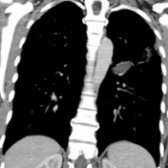
*Coronal NECT of the same patient shows right basilar branching tubular opacities that correspond to right lower lobe bronchiectasis with intrinsic mucus plugs . Impacted mucus may exhibit high attenuation. Allergic bronchopulmonary aspergillosis is due to hypersensitivity to fungal antigens.*
**Congenital Bronchial Atresia**
*Coronal CECT of a 27-year-old man with bronchial atresia shows a nonenhancing left upper lobe tubular opacity that corresponds to an atretic bronchus impacted with mucus .*

**Congenital Bronchial Atresia**
*Coronal CECT of the same patient shows focal air-trapping in the left upper lobe distal to the impacted atretic bronchus and mucocele. Bronchial atresia most frequently affects the left upper lobe apicoposterior bronchus. Affected patients are typically asymptomatic.*
**Allergic Bronchopulmonary Aspergillosis**
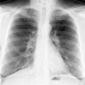
*PA chest radiograph of a patient with asthma and allergic bronchopulmonary aspergillosis shows right basilar tubular opacities corresponding to mucoid impaction in dilated airways.*

**Allergic Bronchopulmonary Aspergillosis**
*PA chest radiograph of a patient with asthma and allergic bronchopulmonary aspergillosis shows right basilar tubular opacities corresponding to mucoid impaction in dilated airways.*

**Allergic Bronchopulmonary Aspergillosis**
*Coronal NECT of the same patient shows right basilar branching tubular opacities that correspond to right lower lobe bronchiectasis with intrinsic mucus plugs . Impacted mucus may exhibit high attenuation. Allergic bronchopulmonary aspergillosis is due to hypersensitivity to fungal antigens.*

**Congenital Bronchial Atresia**
*Coronal CECT of a 27-year-old man with bronchial atresia shows a nonenhancing left upper lobe tubular opacity that corresponds to an atretic bronchus impacted with mucus .*

**Congenital Bronchial Atresia**
*Coronal CECT of the same patient shows focal air-trapping in the left upper lobe distal to the impacted atretic bronchus and mucocele. Bronchial atresia most frequently affects the left upper lobe apicoposterior bronchus. Affected patients are typically asymptomatic.*
 within dilated peripheral airways.](images/app.statdx.com_image_thumbnail_9da07da7-411f-4382-9f5d-2f7fa7d05601_size_168_quality_85_365199d9_20251018T064244Z.jpg)
**Bronchiectasis**
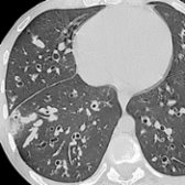
*Axial NECT of a 20-year-old man with cystic fibrosis shows multifocal bronchiectasis , bronchial wall thickening, and soft tissue tubular opacities that correspond to impacted mucus within dilated peripheral airways.*
**Bronchiectasis**
*Axial NECT of a 20-year-old man with cystic fibrosis shows multifocal bronchiectasis , bronchial wall thickening, and soft tissue tubular opacities that correspond to impacted mucus within dilated peripheral airways.*
 .](4b74c2f7-f68a-485b-9d38-1715615bdbde)
**Bronchiectasis**
*Axial NECT of a 34-year-old-man with primary ciliary dyskinesia and Kartagener syndrome shows bilateral upper lobe branching tubular opacities that correspond to impacted mucus within bronchiectatic airways. Note coexistent situs inversus and right aortic arch .*
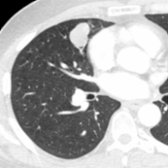
**Malignant Airway Neoplasm**
*Axial CECT of a patient with a history of chronic cough and blood-tinged sputum shows a polylobular middle lobe soft tissue mass that exhibits an endobronchial component and obstructs a small airway in the middle lobe. Carcinoid tumor was diagnosed at surgery.*

**Malignant Airway Neoplasm**
*Sagittal CECT of the same patient shows the centrally obstructing carcinoid tumor and peripheral mucoid impaction . Carcinoid tumor is a low-grade malignant neoplasm with frequent endobronchial involvement.*
.](4b74c2f7-f68a-485b-9d38-1715615bdbde)
**Bronchiectasis**
*Axial NECT of a 34-year-old-man with primary ciliary dyskinesia and Kartagener syndrome shows bilateral upper lobe branching tubular opacities that correspond to impacted mucus within bronchiectatic airways. Note coexistent situs inversus and right aortic arch .*

**Malignant Airway Neoplasm**
*Axial CECT of a patient with a history of chronic cough and blood-tinged sputum shows a polylobular middle lobe soft tissue mass that exhibits an endobronchial component and obstructs a small airway in the middle lobe. Carcinoid tumor was diagnosed at surgery.*

**Malignant Airway Neoplasm**
*Sagittal CECT of the same patient shows the centrally obstructing carcinoid tumor and peripheral mucoid impaction . Carcinoid tumor is a low-grade malignant neoplasm with frequent endobronchial involvement.*