---
title: "Frontotemporal Lobar Degeneration"
docid: "49510d0e-acf7-45cb-9eb1-53f8193b0b6d"
authors:
- key: "1fa14dfd-71ea-4960-908e-e720313bc63a"
value: "Santhosh Gaddikeri, MD"
- key: "a25c450b-3d34-4f64-bba3-cc0834813df6"
value: "Miral D. Jhaveri, MD, MBA"
breadcrumbs:
-
name: "Brain"
slug: "brain"
treeNodeId: "6d8829f1-14d7-45af-8675-255189aa526a"
-
name: "Diagnosis"
slug: "diagnosis"
treeNodeId: "51c00394-446e-4a38-94af-d3b1d14d34e8"
-
name: "Pathology-Based Diagnoses"
slug: "pathology-based-diagnoses"
treeNodeId: "d9d3a8ed-f21b-4831-8c77-591a3500ef77"
-
name: "Acquired Toxic/Metabolic/Degenerative Disorders"
slug: "acquired-toxicmetabolicdegenerativ-"
treeNodeId: "ba3cfeaf-64d9-4117-91e8-d2ce58783fc5"
-
name: "Dementias and Degenerative Disorders"
slug: "dementias-and-degenerative-disorde-"
treeNodeId: "6381104d-7a4c-4be5-bb19-3cd90837d547"
-
name: "Frontotemporal Lobar Degeneration"
slug: "frontotemporal-lobar-degeneration"
treeNodeId: null
category: "Brain"
cmeTopicId: "144a695e-95a4-4ec7-8df6-1c4db98375f6"
documentVersionId: "857bab94-d633-4e44-a081-50f182d9ed83"
imageCount: 30
lastUpdated: "09/30/20"
pageDescription: "Frontotemporal Lobar Degeneration"
pageKeywords: "Brain, Diagnosis, Pathology-Based Diagnoses, Acquired Toxic/Metabolic/Degenerative Disorders, Dementias and Degenerative Disorders, Frontotemporal Lobar Degeneration"
pageTitle: "Frontotemporal Lobar Degeneration | STATdx"
enhancedTitle: "Frontotemporal Lobar Degeneration"
type: "DX"
references: true
tables: 1
breadcrumbs:
- "Brain"
- "Diagnosis"
- "Pathology-Based Diagnoses"
- "Acquired Toxic/Metabolic/Degenerative Disorders"
- "Dementias and Degenerative Disorders"
- "Frontotemporal Lobar Degeneration"
---
# KEY FACTS
- ## Terminology
- Clinical subtypes
- Behavioral variant frontotemporal dementia **(bvFTD)**
- Primary progressive aphasia syndromes **(PPA)**
- Semantic variant **(sv-PPA)**: Previously known as semantic dementia
- Nonfluent/agrammatic variant **(nfv-PPA)**: Previously known as progressive nonfluent aphasia
- Logopenic variant **(lv-PPA)**
- Frontotemporal dementia with motor symptoms
- ## Imaging
- Early
- PET shows frontotemporal ↓ glucose metabolism
- Late: Frontotemporal atrophy with knife-like gyri on MR
- Subtypes have characteristic cortical atrophy patterns
- ## Top Differential Diagnoses
- Alzheimer dementia (AD)
- Vascular dementia
- Corticobasal ganglionic degeneration (CBD)
- Dementia with Lewy bodies (DLB)
- ## Clinical Issues
- Clinical syndromes (some overlap)
- **bvFTD**: Disinhibition, apathy & loss of empathy, hyperorality, & compulsive behavior
- **sv-PPA**: Impaired single-word comprehension & object naming with preserved fluency, repetition, & grammar
- **nfv-PPA**: Effortful speech production of phonemes (linguistic units of sound) & orofacial apraxia
- **lv-PPA**: Impaired word finding & repetition with errors in speech & naming
- Younger age group than AD
- FTLD most common cause of early-onset (< 65 years) dementia
- Median survival: 6-11 years from symptom onset & 3-4 years from diagnosis
# TERMINOLOGY
- ## Abbreviations
- Frontotemporal lobar degeneration (FTLD)
- Clinical subtypes
- Behavioral variant frontotemporal dementia **(bvFTD)**
- Primary progressive aphasia syndromes **(PPA)**
- Semantic variant **(sv-PPA)**: Previously known as semantic dementia
- Nonfluent/agrammatic variant **(nfv-PPA)**: Previously known as progressive nonfluent aphasia
- Logopenic variant**(lv-PPA)**: Has Alzheimer pathology & is not included as 1 of 3 clinical FTD syndromes
- Frontotemporal dementia (FTD) with motor symptoms
- Corticobasal degeneration (CBD)
- Progressive supranuclear palsy (PSP)
- FTD with motor neuron disease
- FTD with amyotrophic lateral sclerosis (ALS)
- ## Synonyms
- Pick disease no longer used
- Referred to pathologic variant with Pick bodies
- ## Definitions
- Heterogeneous family of neurodegenerative disorders characterized by focal lobar degeneration of frontal &/or temporal lobes
# IMAGING
- ## General Features
- ### Best diagnostic clue
- Structural & functional imaging are supportive but not diagnostic of FTD
- PET showing frontotemporal ↓ glucose metabolism
- Anterior frontotemporal atrophy with knife-like gyri
- ### Location
- Anterior temporal/frontal lobes, orbitofrontal cortex, medial temporal region
- Relative sparing of parietooccipital lobes
- ### Morphology
- Knife blade appearance of atrophic gyri
- ± marked asymmetry
- May have worst atrophy in dominant hemisphere
- ## CT Findings
- ### NECT
- Frontal lobe atrophy often most prominent feature
- ↑ size of frontal horns (larger than rest of lateral ventricles)
- ## MR Findings
- ### T1WI
- Atrophy of frontal & temporal lobes, often asymmetric
- Knife-like gyri with normal signal
- Dilated frontal sulci reflecting atrophy
- Relative sparing of parietooccipital lobes
- ### T2WI
- ± hyperintensity in frontotemporal white matter (WM)
- ### FLAIR
- ± hyperintensity in frontotemporal WM
- ### MRS
- ↓ NAA glutamate + glutamine (neuronal loss), ↑ myoinositol (↑ glial content) in frontal lobes
- ↓ NAA in posterior cingulate gyri
- Reflects ↓ neuronal population, viability
- ± lactate peak in frontal lobes
- MR voxel-based morphometry
- Subtypes have characteristic cortical atrophy patterns
- Frontal vs. temporal, left vs. right help discriminate
- **bvFTD**: Atrophy of frontal & temporal lobes
- Anterior insula, anterior cingulate, orbitofrontal cortex, & amygdala (early changes occur in right hemisphere)
- **sv-PPA**: Typically anterior temporal lobe atrophy (asymmetric to left)
- Entire temporal lobe can be involved
- Ventromedial & superior frontal lobes
- Right temporal atrophy as disease progresses
- **nfv-PPA**: Selective left posterior frontoinsular region
- **lv-PPA**: Predominant atrophy of left posterior temporal cortex & parietal lobe
- DTI
- Widespread damage to WM tracts reported
- **bvFTD**: Uncinate fasciculus, inferior longitudinal fasciculus, & anterior commissural fibers
- **sv-PPA**: Inferior longitudinal & uncinate fasciculi
- **nfv-FTD**: Left superior longitudinal fasciculus
- **lv-PPA**: Widespread dorsal & ventral WM tracts
- ## Nuclear Medicine Findings
- ### PET
- Functional imaging more sensitive than MR in early-stage disease
- FDG PET: ↓ metabolic activity in frontotemporal cortex
- Amyloid PET helps differentiate FTLD from Alzheimer disease (AD)
- HMPAO-SPECT
- Sensitive technique for early detection of FTD
- Occurs before atrophy is evident
- **bvFTD**: ↓ perfusion frontal & anterior temporal lobes
- Asymmetric, left or right dominant
- **sv-PPA**: Prominent anterior temporal hypoperfusion, left > right
- **nfv-PPA**: Asymmetric frontal hypoperfusion often involving insular cortex
- **lv-PPA**: ↓ perfusion in left parietal inferior lobule & posterolateral temporal lobe
- SPECT perfusion deficits predominantly in frontal & anterior temporal lobes with preserved perfusion posteriorly
- Helps distinguish FTD from AD
- Reduced frontal perfusion is not specific to FTD but also occurs in some cases of schizophrenia, depression, HIV encephalopathy, Creutzfeldt-Jakob disease, AD
- ## Imaging Recommendations
- ### Best imaging tool
- PET/SPECT; MR voxel-based morphometry
- ### Protocol advice
- Routine T1WI, T2WI, coronal T2WI MR
# DIFFERENTIAL DIAGNOSIS
- [Alzheimer Disease](/document/alzheimer-disease/f71f5cf5-b1af-4c6d-b145-b4c10eec7b58)
- Parietal & temporal cortical atrophy with disproportionate hippocampal volume loss
- Increased rate of atrophy in FTD compared to AD
- Often coexisting microvascular disease, WM hyperintensities, microhemorrhages
- Amyloid imaging (11C-labeled Pittsburgh Compound-B) helps to differentiate AD from other dementias
- [Vascular Dementia](/document/vascular-dementia/f59dab57-c511-4369-8fcc-592421a4b8d1)
- 2nd most common dementia (15-30%)
- WM & deep gray lacunae
- Hyperintense lesions on T2WI & focal atrophy is suggestive of chronic infarcts
- [Corticobasal Degeneration](/document/corticobasal-degeneration/23f97d4e-8724-4229-b9f8-08f63906ebd8)
- Prominent extrapyramidal, cortical symptoms
- Severe frontoparietal atrophy contralateral to more severely affected clinically
- Atrophy of paracentral structures
- [Dementia With Lewy Bodies](/document/dementia-with-lewy-bodies/e8e46d1d-46d2-4e5a-880f-f025a84c5871)
- Hypometabolism of entire brain, especially visual cortex
- Visual & auditory hallucinations, paranoid delusions
# PATHOLOGY
- ## General Features
- ### Etiology
- Tau protein (hyperphosphorylated microtubular protein) or TDP-43 (TAR DNA-binding protein-43)
- Rare cases change on fused-in-sarcoma (FUS) protein
- ### Genetics
- FTD is highly heritable without clear inheritance pattern
- 25-40% of FTD is familial, > 50% of bvFTD is autosomal dominant
- Mutations in following 3 genes together constitute 15% of FTD cases
- Most common: Hexanucleotide expansion in chromosome 9 open reading frame 72 (*C9orf72*) gene
- Microtubule-associated protein tau (*MAPT*) gene
- Granulin precursor (*GRN*) gene
- ## Staging, Grading, & Classification
- Histopathologic classification of FTLD based on abnormal inclusions
- FTLD-tau: Tau inclusion (hyperphosphorylated tau protein)/Pick bodies
- FTLD-TDP: Tau-negative & TDP-43-positive inclusions (subtypes: Type A, B, C, & D)
- FTLD-FUS: Tau-/TDP-negative & FUS-positive inclusions
- FTLD-ALS/dipeptide repeats (DPR): TDP-negative DPR protein aggregates
- FTLD-ni: No inclusions
- FTD clinical syndromes correlate with brain atrophy patterns & not with pathologic subtypes
- ## Gross Pathologic & Surgical Features
- Gross atrophy of frontal &/or anterior temporal lobes
- Firm cortical gray matter (gliosis) &/or basal ganglia atrophy
- Soft, retracted subcortical WM
- ## Microscopic Features
- Loss of pyramidal neurons & microvacuolar degeneration in layer II & III of frontal & temporal cortex
- Subjacent WM shows axonal & myelin loss
- FTLD-related tauopathies
- Pick disease: Prototypical tauopathy of FTLD
- Characterized by Pick bodies: Solitary, round or oval, argyrophilic inclusions in cytoplasm of neurons
- Commonly found in dentate gyrus of hippocampus, amygdala, frontal & temporal neocortex
# CLINICAL ISSUES
- ## Presentation
- ### Most common signs/symptoms
- Personality, behavior, & language changes
- Memory loss, confusion, cognitive & speech dysfunction, apathy, & abulia
- ### Clinical profile
- **bvFTD**: Disinhibition, apathy & loss of empathy, hyperorality, & compulsive behavior
- 15-20% may develop concomitant motor neuron disease (MND)
- **sv-PPA**: Impaired single-word comprehension & object naming with preserved fluency, repetition, & grammar
- **nfv-PPA**: Effortful speech production of phonemes (linguistic units of sound) & orofacial apraxia
- **lv-PPA**: Impaired word finding & repetition with errors in speech & naming
- ## Demographics
- ### Age
- More common cause of early-onset (midlife) dementia
- Mean age of onset is 58 years; rare < 40 & > 75 years
- Peak incidence 45-65 years
- ### Sex
- bvFTD & sv-FTD: Male preponderance
- nfv-PPA: Female predominance
- ### Ethnicity
- Familial forms of Pick complex dementias particularly common in people of Scandinavian origin
- ### Epidemiology
- FTLD more common cause of early-onset (< 65 years) dementia
- Age > 65 years account for 20-25% of cases of FTLD
- Prevalence: 3.5-15/100,000 person-years
- FTLD accounts for ~ 5% of all pathologic diagnoses in patients with dementia
- ## Natural History & Prognosis
- Insidious onset of behavioral & cognitive dysfunction
- Speech & language disturbance are often more profound than memory disorder
- Median survival 6-11 years from symptom onset & 3-4 years from diagnosis
- Currently no FDA-approved disease-modifying drugs available for treatment of FTD
- Some patients develop artistic talents during course of dementia (disinhibition of "creative" brain areas)
# DIAGNOSTIC CHECKLIST
- ## Consider
- Other common forms of dementia (AD, dementia with Lewy bodies)
- ## Image Interpretation Pearls
- Bilateral frontal lobe atrophy should make one consider diagnosis of FTD
- Bilateral asymmetric anterior temporal lobe atrophy: sv-PPA
- ## Reporting Tips
- Report pattern of cortical volume loss
52f7139c-657b-4ff0-8be5-0f3d7523130e
## References
# Selected References
1. [Ishii K: Diagnostic imaging of dementia with Lewy bodies, frontotemporal lobar degeneration, and normal pressure hydrocephalus. Jpn J Radiol. 38(1):64-76, 2020](http://www.ncbi.nlm.nih.gov/pubmed/?term=31549279%5Bpmid%5D)
1. [Raji CA et al: Overview of MR imaging volumetric quantification in neurocognitive disorders. Top Magn Reson Imaging. 28(6):311-5, 2019](http://www.ncbi.nlm.nih.gov/pubmed/?term=31794503%5Bpmid%5D)
1. [Risacher SL et al: Neuroimaging in aging and neurologic diseases. Handb Clin Neurol. 167:191-227, 2019](http://www.ncbi.nlm.nih.gov/pubmed/?term=31753134%5Bpmid%5D)
1. [Shepherd TM et al: Clinical use of integrated positron emission tomography-magnetic resonance imaging for dementia patients. Top Magn Reson Imaging. 28(6):299-310, 2019](http://www.ncbi.nlm.nih.gov/pubmed/?term=31794502%5Bpmid%5D)
1. [Zukotynski K et al: PET/CT of dementia. AJR Am J Roentgenol. 211(2):246-59, 2018](http://www.ncbi.nlm.nih.gov/pubmed/?term=29949415%5Bpmid%5D)
1. [Mann DM et al: Frontotemporal lobar degeneration: pathogenesis, pathology and pathways to phenotype. Brain Pathol. 27(6):723-36, 2017](http://www.ncbi.nlm.nih.gov/pubmed/?term=28100023%5Bpmid%5D)
1. [Bang J et al: Frontotemporal dementia. Lancet. 386(10004):1672-82, 2015](http://www.ncbi.nlm.nih.gov/pubmed/?term=26595641%5Bpmid%5D)
1. [Shivamurthy VK et al: Brain FDG PET and the diagnosis of dementia. AJR Am J Roentgenol. 204(1):W76-85, 2015](http://www.ncbi.nlm.nih.gov/pubmed/?term=25539279%5Bpmid%5D)
1. [Bott NT et al: Frontotemporal dementia: diagnosis, deficits and management. Neurodegener Dis Manag. 4(6):439-54, 2014](http://www.ncbi.nlm.nih.gov/pubmed/?term=25531687%5Bpmid%5D)
1. [Chare L et al: New criteria for frontotemporal dementia syndromes: clinical and pathological diagnostic implications. J Neurol Neurosurg Psychiatry. 85(8):865-70, 2014](http://www.ncbi.nlm.nih.gov/pubmed/?term=24421286%5Bpmid%5D)
1. [Diehl-Schmid J et al: Imaging frontotemporal lobar degeneration. Curr Neurol Neurosci Rep. 14(10):489, 2014](http://www.ncbi.nlm.nih.gov/pubmed/?term=25171901%5Bpmid%5D)
1. [Bhogal P et al: The common dementias: a pictorial review. Eur Radiol. 23(12):3405-17, 2013](http://www.ncbi.nlm.nih.gov/pubmed/?term=24081643%5Bpmid%5D)
1. [Risacher SL et al: Neuroimaging biomarkers of neurodegenerative diseases and dementia. Semin Neurol. 33(4):386-416, 2013](http://www.ncbi.nlm.nih.gov/pubmed/?term=24234359%5Bpmid%5D)
1. [Gorno-Tempini ML et al: Classification of primary progressive aphasia and its variants. Neurology. 76(11):1006-14, 2011](http://www.ncbi.nlm.nih.gov/pubmed/?term=21325651%5Bpmid%5D)
1. [Rascovsky K et al: Sensitivity of revised diagnostic criteria for the behavioural variant of frontotemporal dementia. Brain. 134(Pt 9):2456-77, 2011](http://www.ncbi.nlm.nih.gov/pubmed/?term=21810890%5Bpmid%5D)
1. [Krueger CE et al: Longitudinal rates of lobar atrophy in frontotemporal dementia, semantic dementia, and Alzheimer's disease. Alzheimer Dis Assoc Disord. 24(1):43-8, 2010](http://www.ncbi.nlm.nih.gov/pubmed/?term=19571735%5Bpmid%5D)
1. [Mackenzie IR et al: Nomenclature and nosology for neuropathologic subtypes of frontotemporal lobar degeneration: an update. Acta Neuropathol. 119(1):1-4, 2010](http://www.ncbi.nlm.nih.gov/pubmed/?term=19924424%5Bpmid%5D)
1. [King RD et al: Characterization of atrophic changes in the cerebral cortex using fractal dimensional analysis. Brain Imaging Behav. 3(2):154-66, 2009](http://www.ncbi.nlm.nih.gov/pubmed/?term=20740072%5Bpmid%5D)
1. [Lindberg O et al: Cortical morphometric subclassification of frontotemporal lobar degeneration. AJNR Am J Neuroradiol. 30(6):1233-9, 2009](http://www.ncbi.nlm.nih.gov/pubmed/?term=19346314%5Bpmid%5D)
1. [Moon WJ et al: Atrophy measurement of the anterior commissure and substantia innominata with 3T high-resolution MR imaging: does the measurement differ for patients with frontotemporal lobar degeneration and Alzheimer disease and for healthy subjects? AJNR Am J Neuroradiol. 29(7):1308-13, 2008](http://www.ncbi.nlm.nih.gov/pubmed/?term=18436612%5Bpmid%5D)
1. [Mihara M et al: Magnetic resonance spectroscopic study of Alzheimer's disease and frontotemporal dementia/Pick complex. Neuroreport. 17(4):413-6, 2006](http://www.ncbi.nlm.nih.gov/pubmed/?term=16514368%5Bpmid%5D)
1. [Whitwell JL et al: Magnetic resonance imaging signatures of tissue pathology in frontotemporal dementia. Arch Neurol. 62(9):1402-8, 2005](http://www.ncbi.nlm.nih.gov/pubmed/?term=16157747%5Bpmid%5D)
## Tables
# Imaging Features for Various Clinical Subtypes of Frontotemporal Dementia
| Clinical Subtypes | Imaging Features |
| --- | --- |
| bvFTD | MR: Atrophy of frontal & temporal lobes; asymmetric right frontal &/or temporal lobe atrophy may occur NM: Decreased perfusion & metabolism in frontal &/or temporal lobes, usually asymmetric to right side |
| sv-PPA | MR: Typically atrophy of left anterior & inferior temporal lobe; right temporal lobe atrophies as disease progresses NM: Decreased perfusion & metabolism in anterior temporal lobes (L > > R) |
| nfv-PPA | MR: Selective left perisylvian & frontal atrophy NM: Asymmetric decreased perfusion & metabolism in frontal lobes (L > > R) often involving insular cortex |
| lv-PPA | MR: Prominent atrophy of left angular & middle temporal gyri NM: Decreased perfusion & metabolism in left parietal inferior lobule & posterolateral temporal lobe |
## Images
### Selected Images
 . Parietooccipital lobes are spared. Gyri around the central sulcus are normal.](images/app.statdx.com_image_thumbnail_ccf3ab42-d18d-44f4-9b3f-cc9fd9510d11_size_168_quality_85_4a79edbc_20251014T204712Z.jpg)
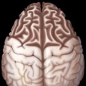
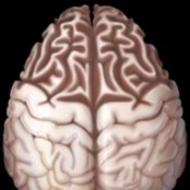
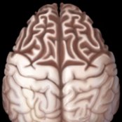
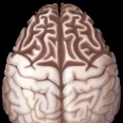
*Graphic depicts the classic disproportionate frontal lobe atrophy of late-stage frontotemporal dementia (FTD). The sulci are widened & gyri are knife-like . Parietooccipital lobes are spared. Gyri around the central sulcus are normal.*
*Graphic depicts the classic disproportionate frontal lobe atrophy of late-stage frontotemporal dementia (FTD). The sulci are widened & gyri are knife-like . Parietooccipital lobes are spared. Gyri around the central sulcus are normal.*
*Graphic depicts the classic disproportionate frontal lobe atrophy of late-stage frontotemporal dementia (FTD). The sulci are widened & gyri are knife-like . Parietooccipital lobes are spared. Gyri around the central sulcus are normal.*
*Graphic depicts the classic disproportionate frontal lobe atrophy of late-stage frontotemporal dementia (FTD). The sulci are widened & gyri are knife-like . Parietooccipital lobes are spared. Gyri around the central sulcus are normal.*
*Graphic depicts the classic disproportionate frontal lobe atrophy of late-stage frontotemporal dementia (FTD). The sulci are widened & gyri are knife-like . Parietooccipital lobes are spared. Gyri around the central sulcus are normal.*
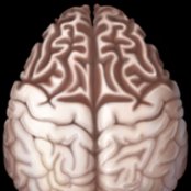
*Graphic depicts the classic disproportionate frontal lobe atrophy of late-stage frontotemporal dementia (FTD). The sulci are widened & gyri are knife-like . Parietooccipital lobes are spared. Gyri around the central sulcus are normal.*
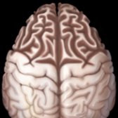
*Graphic depicts the classic disproportionate frontal lobe atrophy of late-stage frontotemporal dementia (FTD). The sulci are widened & gyri are knife-like . Parietooccipital lobes are spared. Gyri around the central sulcus are normal.*

*Graphic depicts the classic disproportionate frontal lobe atrophy of late-stage frontotemporal dementia (FTD). The sulci are widened & gyri are knife-like . Parietooccipital lobes are spared. Gyri around the central sulcus are normal.*

*Graphic depicts the classic disproportionate frontal lobe atrophy of late-stage frontotemporal dementia (FTD). The sulci are widened & gyri are knife-like . Parietooccipital lobes are spared. Gyri around the central sulcus are normal.*

*Graphic depicts the classic disproportionate frontal lobe atrophy of late-stage frontotemporal dementia (FTD). The sulci are widened & gyri are knife-like . Parietooccipital lobes are spared. Gyri around the central sulcus are normal.*

*Graphic depicts the classic disproportionate frontal lobe atrophy of late-stage frontotemporal dementia (FTD). The sulci are widened & gyri are knife-like . Parietooccipital lobes are spared. Gyri around the central sulcus are normal.*

*Graphic depicts the classic disproportionate frontal lobe atrophy of late-stage frontotemporal dementia (FTD). The sulci are widened & gyri are knife-like . Parietooccipital lobes are spared. Gyri around the central sulcus are normal.*
 .](images/app.statdx.com_image_thumbnail_8cab5d42-f1b2-44ba-ba3b-b0c17fdb3217_size_168_quality_85_547ec7bb_20251014T204712Z.jpg)
*Coronal NECT MPR through the frontal lobes of a 62-year-old man with behavioral variant frontotemporal dementia (bvFTD) shows asymmetric atrophy of right frontal lobe with relatively preserved left frontal lobe volume .*
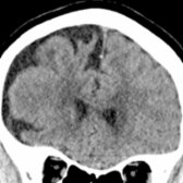
*Coronal NECT MPR through the frontal lobes of a 62-year-old man with behavioral variant frontotemporal dementia (bvFTD) shows asymmetric atrophy of right frontal lobe with relatively preserved left frontal lobe volume .*
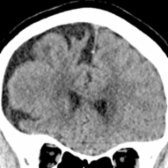
*Coronal NECT MPR through the frontal lobes of a 62-year-old man with behavioral variant frontotemporal dementia (bvFTD) shows asymmetric atrophy of right frontal lobe with relatively preserved left frontal lobe volume .*

*Coronal NECT MPR through the frontal lobes of a 62-year-old man with behavioral variant frontotemporal dementia (bvFTD) shows asymmetric atrophy of right frontal lobe with relatively preserved left frontal lobe volume .*
 and frontal operculum .](images/app.statdx.com_image_thumbnail_04291565-16df-407f-8345-c6415c6edd9a_size_168_quality_85_4aced880_20251014T204712Z.jpg)
*Coronal T2WI MR of a 55-year-old woman with nfv-PPA shows diffuse brain parenchymal atrophy, which is more pronounced in the left perisylvian region due to atrophy of adjacent inferior & lateral temporal convexity and frontal operculum .*
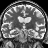
*Coronal T2WI MR of a 55-year-old woman with nfv-PPA shows diffuse brain parenchymal atrophy, which is more pronounced in the left perisylvian region due to atrophy of adjacent inferior & lateral temporal convexity and frontal operculum .*
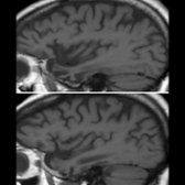
*Parasagittal T1WI MR through left (top) & right (bottom) temporal lobes of a 55-year-old man with sv-PPA shows asymmetric temporal lobe atrophy on left vs. right . Also note relatively preserved frontal lobe volume.*
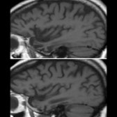
*Parasagittal T1WI MR through left (top) & right (bottom) temporal lobes of a 55-year-old man with sv-PPA shows asymmetric temporal lobe atrophy on left vs. right . Also note relatively preserved frontal lobe volume.*
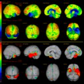
*FDG PET raw data & 3D-SSP images of a 59-year-old man with sv-PPA demonstrate decreased metabolic activity in the anterior & medial left temporal lobe & to a lesser degree in anterior & medial temporal lobe . (Courtesy M. Matesan, MD.)*
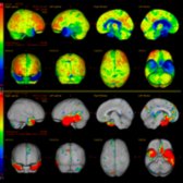
*FDG PET raw data & 3D-SSP images of a 59-year-old man with sv-PPA demonstrate decreased metabolic activity in the anterior & medial left temporal lobe & to a lesser degree in anterior & medial temporal lobe . (Courtesy M. Matesan, MD.)*
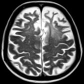
*Axial T2WI MR in a patient with classic FTD shows predominantly frontal lobar atrophy with knife-like gyri . In this case, an associated region of hyperintense white matter is present.*
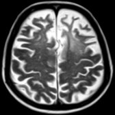
*Axial T2WI MR in a patient with classic FTD shows predominantly frontal lobar atrophy with knife-like gyri . In this case, an associated region of hyperintense white matter is present.*
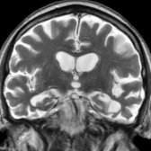
*Coronal T2WI MR of a 55-year-old woman with nfv-PPA shows diffuse brain parenchymal atrophy, which is more pronounced in the left perisylvian region due to atrophy of adjacent inferior & lateral temporal convexity and frontal operculum .*

*Coronal T2WI MR of a 55-year-old woman with nfv-PPA shows diffuse brain parenchymal atrophy, which is more pronounced in the left perisylvian region due to atrophy of adjacent inferior & lateral temporal convexity and frontal operculum .*

*Parasagittal T1WI MR through left (top) & right (bottom) temporal lobes of a 55-year-old man with sv-PPA shows asymmetric temporal lobe atrophy on left vs. right . Also note relatively preserved frontal lobe volume.*

*Parasagittal T1WI MR through left (top) & right (bottom) temporal lobes of a 55-year-old man with sv-PPA shows asymmetric temporal lobe atrophy on left vs. right . Also note relatively preserved frontal lobe volume.*

*FDG PET raw data & 3D-SSP images of a 59-year-old man with sv-PPA demonstrate decreased metabolic activity in the anterior & medial left temporal lobe & to a lesser degree in anterior & medial temporal lobe . (Courtesy M. Matesan, MD.)*

*FDG PET raw data & 3D-SSP images of a 59-year-old man with sv-PPA demonstrate decreased metabolic activity in the anterior & medial left temporal lobe & to a lesser degree in anterior & medial temporal lobe . (Courtesy M. Matesan, MD.)*

*Axial T2WI MR in a patient with classic FTD shows predominantly frontal lobar atrophy with knife-like gyri . In this case, an associated region of hyperintense white matter is present.*

*Axial T2WI MR in a patient with classic FTD shows predominantly frontal lobar atrophy with knife-like gyri . In this case, an associated region of hyperintense white matter is present.*
 .](images/app.statdx.com_image_thumbnail_c6cdd9e8-f0d6-4aca-831a-511ed76ddc55_size_168_quality_85_8fd2489d_20251014T204712Z.jpg)
*Axial FDG PET of a 55-year-old woman with sv-PPA (clinical subtype of FTD) shows decreased metabolic activity in the left temporal lobe & to a lesser degree in the right temporal lobe . Note relative sparing of parietal & occipital lobes .*
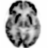
*Axial FDG PET of a 55-year-old woman with sv-PPA (clinical subtype of FTD) shows decreased metabolic activity in the left temporal lobe & to a lesser degree in the right temporal lobe . Note relative sparing of parietal & occipital lobes .*
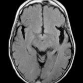
*Axial FLAIR MR in the same patient shows asymmetric atrophy of bilateral temporal lobes (left > right) with knife-like gyri . (Courtesy M. Matesan, MD.)*
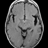
*Axial FLAIR MR in the same patient shows asymmetric atrophy of bilateral temporal lobes (left > right) with knife-like gyri . (Courtesy M. Matesan, MD.)*
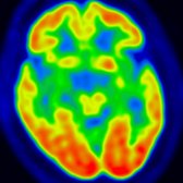
*Axial color-coded FDG PET in a 65-year-old man with apathy & slowly progressive behavioral changes with clinical diagnosis of bvFTD shows decreased metabolic activity in bilateral anterior & medial temporal lobes with relative sparing of bilateral parietooccipital lobes .*
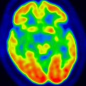
*Axial color-coded FDG PET in a 65-year-old man with apathy & slowly progressive behavioral changes with clinical diagnosis of bvFTD shows decreased metabolic activity in bilateral anterior & medial temporal lobes with relative sparing of bilateral parietooccipital lobes .*
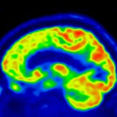
*Sagittal reformat of color-coded FDG PET in the same patient shows decreased metabolic activity in the frontal lobe . (Courtesy J. Singh, MD.)*
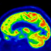
*Sagittal reformat of color-coded FDG PET in the same patient shows decreased metabolic activity in the frontal lobe . (Courtesy J. Singh, MD.)*
### Additional Images
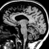
*Sagittal T1WI MR demonstrates marked atrophy of the frontal lobe in a patient with FTD.*
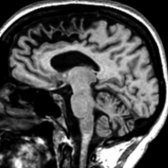
*Sagittal T1WI MR demonstrates marked atrophy of the frontal lobe in a patient with FTD.*
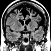
*Coronal T1WI MR in a patient with FTD shows prominent atrophy of both frontal lobes, which appears more pronounced on the left side.*
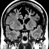
*Coronal T1WI MR in a patient with FTD shows prominent atrophy of both frontal lobes, which appears more pronounced on the left side.*
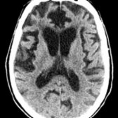
*Axial NECT demonstrates predominantly frontal lobe volume loss from FTD.*
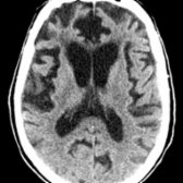
*Axial NECT demonstrates predominantly frontal lobe volume loss from FTD.*
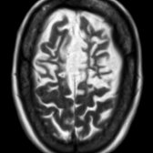
*Axial T2WI MR in a patient with FTD shows marked frontal lobe atrophy.*

*Axial T2WI MR in a patient with FTD shows marked frontal lobe atrophy.*

*Axial FLAIR MR shows frontal lobe volume loss as well as associated hyperintense signal in white matter.*
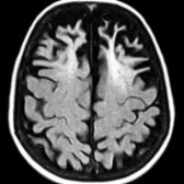
*Axial FLAIR MR shows frontal lobe volume loss as well as associated hyperintense signal in white matter.*
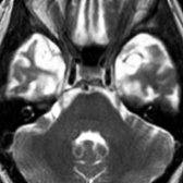
*Axial T2WI MR in a patient with FTD demonstrates marked volume loss as well as associated hyperintense signal within bilateral temporal lobes.*
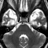
*Axial T2WI MR in a patient with FTD demonstrates marked volume loss as well as associated hyperintense signal within bilateral temporal lobes.*
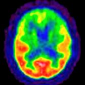
*FDG PET in a patient with FTD & dementia depicts glucose hypometabolism (green regions in the cortex) in the frontal lobes. (Courtesy N. Foster, MD & the University of Michigan PET Center.)*
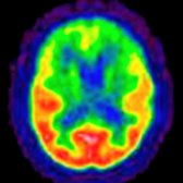
*FDG PET in a patient with FTD & dementia depicts glucose hypometabolism (green regions in the cortex) in the frontal lobes. (Courtesy N. Foster, MD & the University of Michigan PET Center.)*

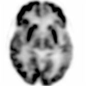
*FDG PET in the same patient shows a decreased rate of glucose metabolism in the frontal & temporal lobes, consistent with FTD. (Courtesy N. Foster, MD & the University of Michigan PET Center.)*

*FDG PET in the same patient shows a decreased rate of glucose metabolism in the frontal & temporal lobes, consistent with FTD. (Courtesy N. Foster, MD & the University of Michigan PET Center.)*
*Axial FDG PET of a 55-year-old woman with sv-PPA (clinical subtype of FTD) shows decreased metabolic activity in the left temporal lobe & to a lesser degree in the right temporal lobe . Note relative sparing of parietal & occipital lobes .*

*Axial FDG PET of a 55-year-old woman with sv-PPA (clinical subtype of FTD) shows decreased metabolic activity in the left temporal lobe & to a lesser degree in the right temporal lobe . Note relative sparing of parietal & occipital lobes .*

*Axial FLAIR MR in the same patient shows asymmetric atrophy of bilateral temporal lobes (left > right) with knife-like gyri . (Courtesy M. Matesan, MD.)*

*Axial FLAIR MR in the same patient shows asymmetric atrophy of bilateral temporal lobes (left > right) with knife-like gyri . (Courtesy M. Matesan, MD.)*

*Axial color-coded FDG PET in a 65-year-old man with apathy & slowly progressive behavioral changes with clinical diagnosis of bvFTD shows decreased metabolic activity in bilateral anterior & medial temporal lobes with relative sparing of bilateral parietooccipital lobes .*

*Axial color-coded FDG PET in a 65-year-old man with apathy & slowly progressive behavioral changes with clinical diagnosis of bvFTD shows decreased metabolic activity in bilateral anterior & medial temporal lobes with relative sparing of bilateral parietooccipital lobes .*

*Sagittal reformat of color-coded FDG PET in the same patient shows decreased metabolic activity in the frontal lobe . (Courtesy J. Singh, MD.)*

*Sagittal reformat of color-coded FDG PET in the same patient shows decreased metabolic activity in the frontal lobe . (Courtesy J. Singh, MD.)*
### Additional Images

*Sagittal T1WI MR demonstrates marked atrophy of the frontal lobe in a patient with FTD.*

*Sagittal T1WI MR demonstrates marked atrophy of the frontal lobe in a patient with FTD.*

*Coronal T1WI MR in a patient with FTD shows prominent atrophy of both frontal lobes, which appears more pronounced on the left side.*

*Coronal T1WI MR in a patient with FTD shows prominent atrophy of both frontal lobes, which appears more pronounced on the left side.*

*Axial NECT demonstrates predominantly frontal lobe volume loss from FTD.*

*Axial NECT demonstrates predominantly frontal lobe volume loss from FTD.*

*Axial T2WI MR in a patient with FTD shows marked frontal lobe atrophy.*

*Axial T2WI MR in a patient with FTD shows marked frontal lobe atrophy.*

*Axial FLAIR MR shows frontal lobe volume loss as well as associated hyperintense signal in white matter.*

*Axial FLAIR MR shows frontal lobe volume loss as well as associated hyperintense signal in white matter.*

*Axial T2WI MR in a patient with FTD demonstrates marked volume loss as well as associated hyperintense signal within bilateral temporal lobes.*

*Axial T2WI MR in a patient with FTD demonstrates marked volume loss as well as associated hyperintense signal within bilateral temporal lobes.*

*FDG PET in a patient with FTD & dementia depicts glucose hypometabolism (green regions in the cortex) in the frontal lobes. (Courtesy N. Foster, MD & the University of Michigan PET Center.)*

*FDG PET in a patient with FTD & dementia depicts glucose hypometabolism (green regions in the cortex) in the frontal lobes. (Courtesy N. Foster, MD & the University of Michigan PET Center.)*

*FDG PET in the same patient shows a decreased rate of glucose metabolism in the frontal & temporal lobes, consistent with FTD. (Courtesy N. Foster, MD & the University of Michigan PET Center.)*

*FDG PET in the same patient shows a decreased rate of glucose metabolism in the frontal & temporal lobes, consistent with FTD. (Courtesy N. Foster, MD & the University of Michigan PET Center.)*
 . The parietal & occipital lobes are relatively well preserved.](images/app.statdx.com_image_thumbnail_4f435ef8-d0f0-4cbc-9a09-5b92318e8482_size_168_quality_85_2f2854c4_20251014T204712Z.jpg)
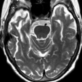
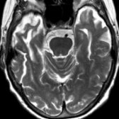
*Axial T2WI MR in a 57-year-old woman with probable FTD shows temporal lobe atrophy with knife-like gyri . The parietal & occipital lobes are relatively well preserved.*
*Axial T2WI MR in a 57-year-old woman with probable FTD shows temporal lobe atrophy with knife-like gyri . The parietal & occipital lobes are relatively well preserved.*
*Axial T2WI MR in a 57-year-old woman with probable FTD shows temporal lobe atrophy with knife-like gyri . The parietal & occipital lobes are relatively well preserved.*

*Axial T2WI MR in a 57-year-old woman with probable FTD shows temporal lobe atrophy with knife-like gyri . The parietal & occipital lobes are relatively well preserved.*
 lobes. There is relative preservation of glucose metabolism in the occipital & parietal lobes
  .](images/app.statdx.com_image_thumbnail_96ca8a9c-4048-495b-8d92-22a54ed7e9de_size_168_quality_85_ae34a1a3_20251014T193509Z.jpg)
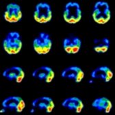
*FDG PET in a patient with FTD shows marked hypometabolism in the frontal & temporal lobes. There is relative preservation of glucose metabolism in the occipital & parietal lobes .*
*FDG PET in a patient with FTD shows marked hypometabolism in the frontal & temporal lobes. There is relative preservation of glucose metabolism in the occipital & parietal lobes .*
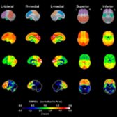
*Sagittal SSP FDG PET scan in a 72-year-old man with FTD shows a normal elderly control map (2nd row), the patient's glucose metabolism (3rd row), & Z-score statistical map (bottom row). The frontal lobes are strikingly hypometabolic. The temporal lobes are somewhat less severely affected.*
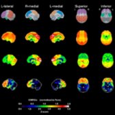
*Sagittal SSP FDG PET scan in a 72-year-old man with FTD shows a normal elderly control map (2nd row), the patient's glucose metabolism (3rd row), & Z-score statistical map (bottom row). The frontal lobes are strikingly hypometabolic. The temporal lobes are somewhat less severely affected.*
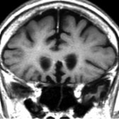
*Coronal T1WI MR in the same patient shows focal atrophy of the olfactory gyri . This finding was initially overlooked & the patient was given the imaging diagnosis of Alzheimer disease. Review & subsequent clinical evaluation confirmed FTD.*
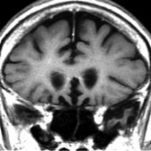
*Coronal T1WI MR in the same patient shows focal atrophy of the olfactory gyri . This finding was initially overlooked & the patient was given the imaging diagnosis of Alzheimer disease. Review & subsequent clinical evaluation confirmed FTD.*
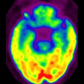
*Axial FDG PET in a patient with FTD shows decreased glucose metabolism in frontal & temporal lobes. The parietal & occipital lobes are spared.*
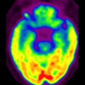
*Axial FDG PET in a patient with FTD shows decreased glucose metabolism in frontal & temporal lobes. The parietal & occipital lobes are spared.*
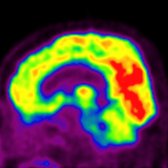
*Sagittal FDG PET in the same patient with FTD depicts glucose hypometabolism in the frontal lobes . The occipital lobes are normal.*
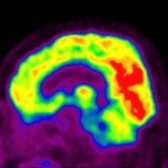
*Sagittal FDG PET in the same patient with FTD depicts glucose hypometabolism in the frontal lobes . The occipital lobes are normal.*
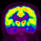
*Coronal FDG PET in a patient with FTD exhibits glucose hypometabolism in frontal and temporal lobes .*
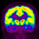
*Coronal FDG PET in a patient with FTD exhibits glucose hypometabolism in frontal and temporal lobes .*
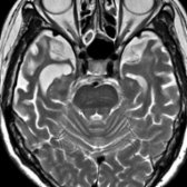
*Axial T2WI MR in a patient with semantic dementia subtype of FTLD shows marked atrophy of anterolateral temporal lobes . Note the relative preservation of the hippocampi .*
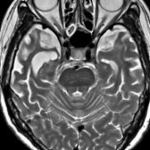
*Axial T2WI MR in a patient with semantic dementia subtype of FTLD shows marked atrophy of anterolateral temporal lobes . Note the relative preservation of the hippocampi .*
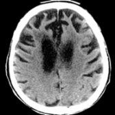
*Axial NECT in a 78-year-old patient diagnosed with late-stage FTD through the upper lateral ventricles shows striking frontal lobar atrophy with classic knife-like gyri characteristic of FTD. In contrast, the parietal & occipital lobes appear relatively spared.*
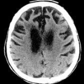
*Axial NECT in a 78-year-old patient diagnosed with late-stage FTD through the upper lateral ventricles shows striking frontal lobar atrophy with classic knife-like gyri characteristic of FTD. In contrast, the parietal & occipital lobes appear relatively spared.*
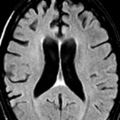
*Axial FLAIR MR in a patient with early findings of FTD shows relatively mild frontal lobe volume loss compared to the normal appearance of the parietal lobes.*
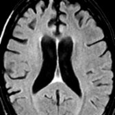
*Axial FLAIR MR in a patient with early findings of FTD shows relatively mild frontal lobe volume loss compared to the normal appearance of the parietal lobes.*
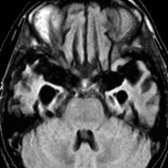
*Axial FLAIR MR shows atrophy of both the frontal & temporal lobes. Note compensatory enlargement of the temporal horns due to temporal lobe volume loss.*
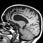
*Sagittal T1WI MR shows predominantly frontal & temporal lobe atrophy. Some gyri demonstrate a classic knife-like appearance () reflecting severe atrophy of a gyrus.*
.](images/app.statdx.com_image_thumbnail_96ca8a9c-4048-495b-8d92-22a54ed7e9de_size_168_quality_85_ae34a1a3_20251014T193509Z.jpg)
*FDG PET in a patient with FTD shows marked hypometabolism in the frontal & temporal lobes. There is relative preservation of glucose metabolism in the occipital & parietal lobes .*

*FDG PET in a patient with FTD shows marked hypometabolism in the frontal & temporal lobes. There is relative preservation of glucose metabolism in the occipital & parietal lobes .*

*Sagittal SSP FDG PET scan in a 72-year-old man with FTD shows a normal elderly control map (2nd row), the patient's glucose metabolism (3rd row), & Z-score statistical map (bottom row). The frontal lobes are strikingly hypometabolic. The temporal lobes are somewhat less severely affected.*

*Sagittal SSP FDG PET scan in a 72-year-old man with FTD shows a normal elderly control map (2nd row), the patient's glucose metabolism (3rd row), & Z-score statistical map (bottom row). The frontal lobes are strikingly hypometabolic. The temporal lobes are somewhat less severely affected.*

*Coronal T1WI MR in the same patient shows focal atrophy of the olfactory gyri . This finding was initially overlooked & the patient was given the imaging diagnosis of Alzheimer disease. Review & subsequent clinical evaluation confirmed FTD.*

*Coronal T1WI MR in the same patient shows focal atrophy of the olfactory gyri . This finding was initially overlooked & the patient was given the imaging diagnosis of Alzheimer disease. Review & subsequent clinical evaluation confirmed FTD.*

*Axial FDG PET in a patient with FTD shows decreased glucose metabolism in frontal & temporal lobes. The parietal & occipital lobes are spared.*

*Axial FDG PET in a patient with FTD shows decreased glucose metabolism in frontal & temporal lobes. The parietal & occipital lobes are spared.*

*Sagittal FDG PET in the same patient with FTD depicts glucose hypometabolism in the frontal lobes . The occipital lobes are normal.*

*Sagittal FDG PET in the same patient with FTD depicts glucose hypometabolism in the frontal lobes . The occipital lobes are normal.*

*Coronal FDG PET in a patient with FTD exhibits glucose hypometabolism in frontal and temporal lobes .*

*Coronal FDG PET in a patient with FTD exhibits glucose hypometabolism in frontal and temporal lobes .*

*Axial T2WI MR in a patient with semantic dementia subtype of FTLD shows marked atrophy of anterolateral temporal lobes . Note the relative preservation of the hippocampi .*

*Axial T2WI MR in a patient with semantic dementia subtype of FTLD shows marked atrophy of anterolateral temporal lobes . Note the relative preservation of the hippocampi .*

*Axial NECT in a 78-year-old patient diagnosed with late-stage FTD through the upper lateral ventricles shows striking frontal lobar atrophy with classic knife-like gyri characteristic of FTD. In contrast, the parietal & occipital lobes appear relatively spared.*

*Axial NECT in a 78-year-old patient diagnosed with late-stage FTD through the upper lateral ventricles shows striking frontal lobar atrophy with classic knife-like gyri characteristic of FTD. In contrast, the parietal & occipital lobes appear relatively spared.*

*Axial FLAIR MR in a patient with early findings of FTD shows relatively mild frontal lobe volume loss compared to the normal appearance of the parietal lobes.*

*Axial FLAIR MR in a patient with early findings of FTD shows relatively mild frontal lobe volume loss compared to the normal appearance of the parietal lobes.*

*Axial FLAIR MR shows atrophy of both the frontal & temporal lobes. Note compensatory enlargement of the temporal horns due to temporal lobe volume loss.*

*Sagittal T1WI MR shows predominantly frontal & temporal lobe atrophy. Some gyri demonstrate a classic knife-like appearance () reflecting severe atrophy of a gyrus.*