---
title: "Giant Cell Arteritis"
docid: "208eca17-81b8-448c-b8be-80e274dccc42"
authors:
- key: "ee6ece9d-ad74-458c-a8df-11628ae7f879"
value: "Arzu Canan, MD"
- key: "3d1e4c57-c1cf-4c89-b0f0-5d82b29a31e1"
value: "Suhny Abbara, MD, FACR, MSCCT, FNASCI"
- key: "10bb95ac-a27a-4ebe-833b-e59fea07734b"
value: "Santiago Mart\u00ednez-Jim\u00e9nez, MD, FACR"
breadcrumbs:
-
name: "Cardiac"
slug: "cardiac"
treeNodeId: "fa90100b-619c-430e-8074-b5b9789bab39"
-
name: "Diagnosis"
slug: "diagnosis"
treeNodeId: "5c92cf4f-e9d5-4059-9c13-22255c51c121"
-
name: "Aorta"
slug: "aorta"
treeNodeId: "4d206a6b-1a82-467c-9199-0df25ab749d7"
-
name: "Giant Cell Arteritis"
slug: "giant-cell-arteritis"
treeNodeId: null
category: "Cardiac"
documentVersionId: "9aabd711-33a6-4598-b5e2-495eff0adf14"
imageCount: 16
lastUpdated: "11/14/24"
pageDescription: "Giant Cell Arteritis"
pageKeywords: "Cardiac, Diagnosis, Aorta, Giant Cell Arteritis"
pageTitle: "Giant Cell Arteritis | STATdx"
enhancedTitle: "Giant Cell Arteritis"
type: "DX"
references: true
breadcrumbs:
- "Cardiac"
- "Diagnosis"
- "Aorta"
- "Giant Cell Arteritis"
---
# KEY FACTS
- ## Terminology
- Chronic, systemic, large or medium-sized, often granulomatous vasculitis
- Often involves thoracic aorta and major branches
- Often involves temporal artery
- ## Imaging
- CTA
- Concentric aortic thickening (> 2 mm)
- Aortic aneurysm; classically ascending aorta
- Aortic dissection: Intimomedial flap
- MR
- Assessment of active inflammation
- Delayed enhancement after gadolinium
- Ultrasonography
- High specificity and sensitivity; operator dependent
- Hypoechoic halo temporal &/or axillary arteries
- PET
- Active inflammation demonstrates ↑ FDG uptake
- ## Top Differential Diagnoses
- Takayasu arteritis
- May be identical to GCA
- Extremely rare in patients > 50 years
- Atherosclerotic disease
- May be difficult to differentiate radiographically, though clinical symptoms often facilitate process
- Similar age group
- ## Clinical Issues
- Headache, visual disturbances, jaw claudication
- Polymyalgia rheumatica
- Serologic markers
- ↑ sedimentation rate
- ↑ C-reactive protein
- Thrombocytosis
- Treatment
- Corticosteroids
# TERMINOLOGY
- ## Abbreviations
- Giant cell arteritis (GCA)
- ## Synonyms
- Temporal arteritis
- Cranial GCA (C-GCA) often referred to as temporal arteritis; terminology not longer recommended, as sparing of temporal artery is not uncommon and because disease may involve large vessels
- Horton disease
- ## Definitions
- Granulomatous autoimmune vasculitis affecting larger arteries and aorta
- C-GCA: Often involves temporal artery and other head/neck vessels, but may also involve aorta and major branches
- Large-vessel GCA (LV-GCA): Often involves thoracic aorta and major branches
- Frequently associated with polymyalgia rheumatica (PMR)
- Aching and morning stiffness in shoulders, hip girdle, and neck
# IMAGING
- ## General Features
- ### Location
- Temporal artery
- Aorta and aortic branches
- ## CT Findings
- ### NECT
- Typically, GCA involving aorta is not as apparent or dense as intramural hematoma; however, there can be hyperdensity if associated with hemorrhage or calcification
- Transmural calcification is often similar to calcified atherosclerotic plaques (common)
- ### CTA
- Concentric aortic thickening (> 2 mm)
- Aortic stenosis
- Aortic aneurysm; classically ascending aorta
- Aortic dissection: Intimomedial flap
- Limited role in C-GCA
- ## MR Findings
- Equally accurate as CT for morphologic assessment on several sequences (e.g., T1WI, T2WI, HASTE, SSFP, etc.)
- Contrast-enhanced MRA is more accurate to assess areas of stenosis and aneurysm
- Assessment of active inflammation
- Contrast-enhanced sequences: Delayed enhancement (i.e., ↑ signal) of vessel wall after gadolinium
- Fat-saturated STIR sequence: High signal of thickened vessel wall
- Cranial (temporal artery) involvement
- High sensitivity and specificity
- Mural thickening (> 0.5 mm)
- Mural high T2 signal and contrast enhancement
- ## Ultrasonographic Findings
- ### Grayscale ultrasound
- C-GCA
- High specificity and sensitivity; operator dependent
- Hypoechoic halo (i.e., **halo sign**) in temporal &/or axillary arteries
- **Compression sign**: Persistence of halo during compression of vessel lumen by ultrasound probe
- ### Color Doppler
- Always in conjunction with grayscale ultrasound
- Helpful to localize temporal artery
- ## Angiographic Findings
- Stenosis (often long, regular, and smooth-walled)
- Occlusion
- Aneurysm
- Limited in diagnosis of early vasculitis
- ## Nuclear Medicine Findings
- ### PET
- LV-GCA: Active inflammation demonstrates ↑ FDG uptake
- Subclinical inflammation of large vessels in 80% with GCA and ~ 30% PMR
- Response to treatment correlates with ↓ FDG uptake
- Limited role in C-GCA, not recommended
- ## Imaging Recommendations
- ### Best imaging tool
- MR
- STIR: Thickening and high signal of aortic wall
- Contrast-enhanced MR: Thickening and enhancement of aortic wall
- MRA is helpful to detect areas of stenosis and aneurysm
- ### Protocol advice
- Consider concomitant NECT to differentiate from intramural hematoma
- Caveat: GCA can occasionally be hyperdense
- PET
- Recognized role in patient with fever &/or inflammation of unknown origin
- Unclear role in follow-up, especially asymptomatic patients without elevated inflammatory markers
# DIFFERENTIAL DIAGNOSIS
- [Takayasu Arteritis](/document/takayasu-arteritis/3b589c7b-d975-4f2c-a5b1-ff83dd856ee7)
- May have similar imaging appearance to GCA
- Rare in patients > 50 years old
- [Other Systemic Vasculitides](/document/polyarteritis-nodosa/5c1ed46f-9132-4903-830d-1907a0774c7d)
- e.g., polyarteritis nodosa, syphilitic aortitis
- Occurs most often in small and medium-sized arteries
- Biopsy and pattern of distribution often help differentiation
- ## Fibromuscular Dysplasia
- Most often affects renal arteries
- Can also involve carotid arteries
- Results in stenoses; occasional spontaneous dissection
- [Atherosclerotic Disease](/document/atherosclerosis/41278e3c-2240-4122-b555-8776d0918082)
- May be difficult to differentiate radiographically, though clinical symptoms often facilitate process
- Similar age group
# PATHOLOGY
- ## General Features
- ### Etiology
- Unknown
- Most accepted hypothesis: Antigen-driven disease mediated by T cells and macrophages that reach aortic wall via vasa vasorum
- ## Staging, Grading, & Classification
- Temporal artery biopsy remains diagnostic gold standard for C-GCA
- Predictors of positive temporal artery biopsy
- Jaw claudication
- Neck pain
- C-reactive protein > 2.45 mg/dL
- Sedimentation rate > 47 mm/h
- Thrombocytosis
- Pallid optic disc edema
- Temporal artery abnormalities
- Temporal artery biopsy can be negative (10-15%)
- ## Gross Pathologic & Surgical Features
- Involvement of aorta (65.0%)
- Involvement of main aortic tributaries (57.5%)
- Brachiocephalic trunk (47.5%)
- Subclavian arteries (42.5%)
- Carotid arteries (35.0%)
- Femoral arteries (30.0%)
- Splanchnic arteries (22.5%)
- Axillary arteries (17.5%)
- Iliac arteries (15.0%)
- Renal arteries (7.5%)
- ## Microscopic Features
- Focal chronic inflammatory cell infiltrates
- Granulomas in vessel wall formed by CD4(+) T cells and macrophages
- Focal areas of intimal hyperplasia
- Proliferation of smooth muscle cells, which leads to narrowing of arterial lumen and eventually ischemia
- Focal areas of fragmentation of inner elastic lamina
- Focal concentric scars around inner elastic lamina
# CLINICAL ISSUES
- ## Presentation
- ### Most common signs/symptoms
- Headache
- Visual disturbances
- Jaw claudication
- ### Other signs/symptoms
- PMR
- Present in 50% of patients at diagnosis of GCA
- 20% of PMR will develop GCA
- Clinical manifestations
- Morning stiffness
- Pain (shoulder > hip or neck)
- Synovitis and bursitis
- Swelling and tenosynovitis
- ↓ range of motion
- Muscle tenderness
- Subjective weakness
- Systemic signs and symptoms (e.g., malaise, fatigue, depression, anorexia, weight loss, fever)
- ### Clinical profile
- Clinical phenotypes
- C-GCA (temporal arteritis with headache and visual disturbance)
- LV-GCA (arm/limb claudication, chest pain)
- PMR
- Phenotypes can overlap
- Serologic markers
- ↑ erythrocyte sedimentation rate
- ↑ C-reactive protein
- Thrombocytosis
- Association with HLA-DRB1*04
- LV-GCA linked to other systematic diseases, such as Behçet disease or hyper-IgG4 syndrome
- Factors for aneurysm formation
- Aortic insufficiency
- Murmur at time of diagnosis
- Hyperlipemia
- ↑ eritrosedimentation in combination with polymyalgia symptoms
- ↑ levels of IL-2
- ## Demographics
- ### Age
- Patients > 50 years old
- Incidence ↑ steadily with age
- ### Sex
- Women > men
- ### Ethnicity
- More common in people of Northern European and Scandinavian descent
- ### Epidemiology
- Prevalence in USA: 1 in 160,000
- Lifetime risk of developing GCA in USA: 1% in women and 0.5% in men
- ## Natural History & Prognosis
- Prognosis for visual recovery is poor
- ↑ risk aortic aneurysm formation and dissection: 17-fold and 2.5x higher risk of thoracic and abdominal aortic aneurysms
- ↓ survival rate
- Involvement of coronary arteries may result in myocardial infarction or congestive heart failure
- Bowel necrosis (uncommon)
- 15-30% of PMR cases eventually develop GCA
- ## Treatment
- GCA and PMR: Corticosteroids
- Aspirin
- Other (2nd-line therapy)
- Methotrexate
- Azathioprine
- Tocilizumab (IL-6 receptor alpha inhibitor)
# DIAGNOSTIC CHECKLIST
- ## Consider
- Annual surveillance to assess for aneurysm and dissection
- Alternatives for follow-up
- Chest radiograph + echocardiogram + abdominal Doppler ultrasound
- Contrast-enhanced CT of chest and abdomen
a728420c-dc69-475b-9a5a-54a5aee00e78
## References
# Selected References
1. [Pepper K: Giant cell arteritis. Postgrad Med. 135(sup1):22-32, 2023](http://www.ncbi.nlm.nih.gov/pubmed/?term=37021621%5Bpmid%5D)
1. [Braun J et al: The role of 18F-FDG positron emission tomography for the diagnosis of vasculitides. Clin Exp Rheumatol. 36 Suppl 114(5):108-14, 2018](http://www.ncbi.nlm.nih.gov/pubmed/?term=30296989%5Bpmid%5D)
1. [Dejaco C et al: The spectrum of giant cell arteritis and polymyalgia rheumatica: revisiting the concept of the disease. Rheumatology (Oxford). 56(4):506-15, 2017](http://www.ncbi.nlm.nih.gov/pubmed/?term=27481272%5Bpmid%5D)
1. [Gomułka K et al: Horton's disease: still an important medical problem in elderly patients: a review and case report. Postepy Dermatol Alergol. 34(5):510-3, 2017](http://www.ncbi.nlm.nih.gov/pubmed/?term=29507571%5Bpmid%5D)
1. [Buttgereit F et al: Polymyalgia rheumatica and giant cell arteritis: a systematic review. JAMA. 315(22):2442-58, 2016](http://www.ncbi.nlm.nih.gov/pubmed/?term=27299619%5Bpmid%5D)
1. [Aschwanden M et al: The ultrasound compression sign to diagnose temporal giant cell arteritis shows an excellent interobserver agreement. Clin Exp Rheumatol. 33(2 Suppl 89):S-113-5, 2015](http://www.ncbi.nlm.nih.gov/pubmed/?term=26016760%5Bpmid%5D)
1. [Khan A et al: Imaging in giant cell arteritis. Curr Rheumatol Rep. 17(8):527, 2015](http://www.ncbi.nlm.nih.gov/pubmed/?term=26113013%5Bpmid%5D)
1. [Hartlage GR et al: Multimodality imaging of aortitis. JACC Cardiovasc Imaging. 7(6):605-19, 2014](http://www.ncbi.nlm.nih.gov/pubmed/?term=24925329%5Bpmid%5D)
1. [Schmidt WA: Ultrasound in vasculitis. Clin Exp Rheumatol. 32(1 Suppl 80):S71-7, 2014](http://www.ncbi.nlm.nih.gov/pubmed/?term=24529335%5Bpmid%5D)
1. [Jennette JC et al: 2012 revised International Chapel Hill Consensus Conference Nomenclature of Vasculitides. Arthritis Rheum. 65(1):1-11, 2013](http://www.ncbi.nlm.nih.gov/pubmed/?term=23045170%5Bpmid%5D)
1. [Blockmans D: Diagnosis and extension of giant cell arteritis. Contribution of imaging techniques. Presse Med. 41(10):948-54, 2012](http://www.ncbi.nlm.nih.gov/pubmed/?term=22795837%5Bpmid%5D)
1. [Castañer E et al: Imaging findings in pulmonary vasculitis. Semin Ultrasound CT MR. 33(6):567-79, 2012](http://www.ncbi.nlm.nih.gov/pubmed/?term=23168065%5Bpmid%5D)
1. [Bossert M et al: Aortic involvement in giant cell arteritis: current data. Joint Bone Spine. 78(3):246-51, 2011](http://www.ncbi.nlm.nih.gov/pubmed/?term=21030278%5Bpmid%5D)
1. [Falardeau J: Giant cell arteritis. Neurol Clin. 28(3):581-91, 2010](http://www.ncbi.nlm.nih.gov/pubmed/?term=20637990%5Bpmid%5D)
1. [Bley TA et al: Diagnostic value of high-resolution MR imaging in giant cell arteritis. AJNR Am J Neuroradiol. 28(9):1722-7, 2007](http://www.ncbi.nlm.nih.gov/pubmed/?term=17885247%5Bpmid%5D)
## Images
### Selected Images
 density material surrounding the great vessels. (Courtesy C. S. Restrepo, MD.)](images/app.statdx.com_image_thumbnail_71136eb1-0d2b-49ff-b87d-6ce0d9841503_annotated_true_size_900_quality_90_ad0952eae49e4419ba1dd3c7298eb51db4e06285.jpg)
*Axial CTA of the chest in a patient with giant cell arteritis (GCA) shows soft tissue density material surrounding the great vessels. (Courtesy C. S. Restrepo, MD.)*
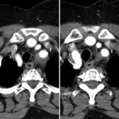
*Axial CTA of the chest in a patient with giant cell arteritis (GCA) shows soft tissue density material surrounding the great vessels. (Courtesy C. S. Restrepo, MD.)*
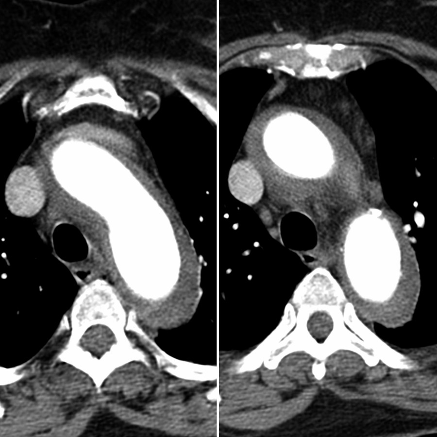
*Axial chest CTA in the same patient shows concentric thickening of the thoracic aorta, which is a common finding in patients with GCA but is indistinguishable from Takayasu arteritis. GCA is more common in patients > 50 years old. Concomitant NECT is recommended to help differentiate from intramural hematoma. (Courtesy C. S. Restrepo, MD.)*
*Axial CTA of the chest in a patient with giant cell arteritis (GCA) shows soft tissue density material surrounding the great vessels. (Courtesy C. S. Restrepo, MD.)*

*Axial CTA of the chest in a patient with giant cell arteritis (GCA) shows soft tissue density material surrounding the great vessels. (Courtesy C. S. Restrepo, MD.)*

*Axial chest CTA in the same patient shows concentric thickening of the thoracic aorta, which is a common finding in patients with GCA but is indistinguishable from Takayasu arteritis. GCA is more common in patients > 50 years old. Concomitant NECT is recommended to help differentiate from intramural hematoma. (Courtesy C. S. Restrepo, MD.)*
 . FDG PET has excellent sensitivity and specificity for the diagnosis of GCA and may be used when clinical or serological discrepancies arise during or after treatment of this condition.](images/app.statdx.com_image_thumbnail_d5540713-06cc-400b-ae45-79d96db42f48_annotated_true_size_900_quality_90_85ac1bb47663898ddb59c325c1ce42be3554a508.jpg)
*Coronal FDG PET/CT in the same patient shows marked uptake of FDG along the ascending aortic wall . FDG PET has excellent sensitivity and specificity for the diagnosis of GCA and may be used when clinical or serological discrepancies arise during or after treatment of this condition.*
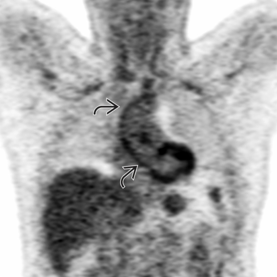
*Coronal FDG PET/CT in the same patient shows marked uptake of FDG along the ascending aortic wall . FDG PET has excellent sensitivity and specificity for the diagnosis of GCA and may be used when clinical or serological discrepancies arise during or after treatment of this condition.*
 .](images/app.statdx.com_image_thumbnail_b721097c-0fa2-4822-bf72-2e9b0f1c6aea_annotated_true_size_900_quality_90_d5fb0b72a59c6d4706daf9f2925186d5ccc3a505.jpg)
*Coronal FDG PET/CT in a patient with GCA shows diffuse uptake along the ascending aortic wall as well as along the subclavian and axillary arteries bilaterally .*
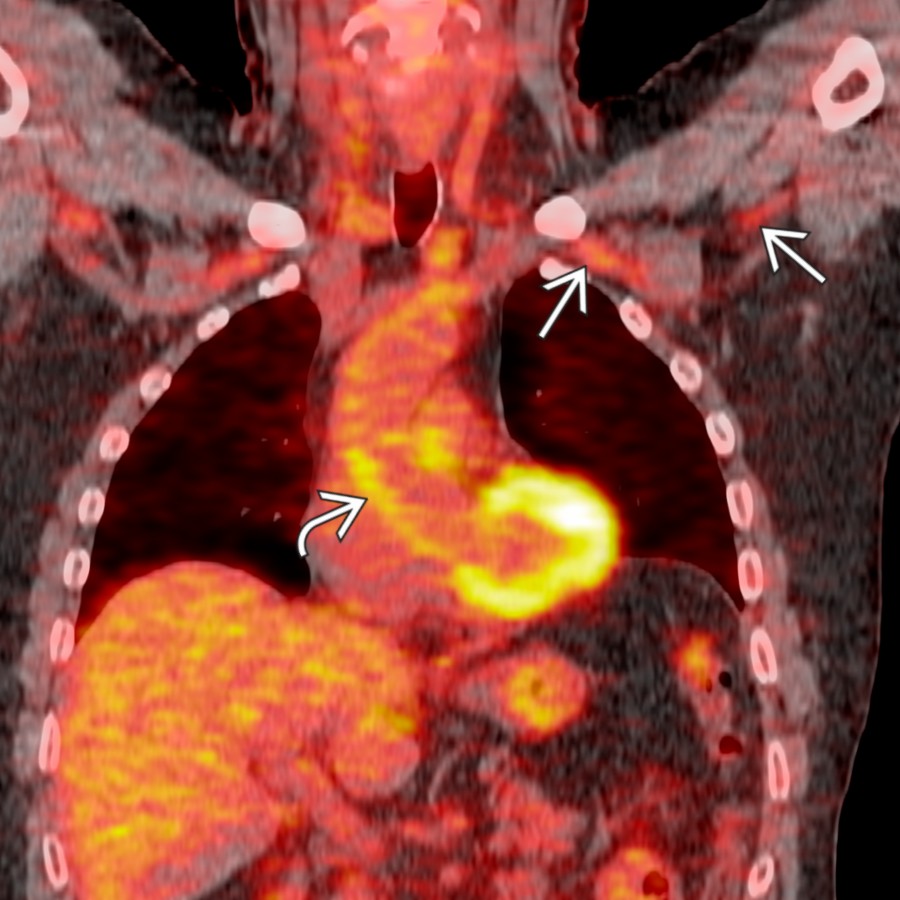
*Coronal FDG PET/CT in a patient with GCA shows diffuse uptake along the ascending aortic wall as well as along the subclavian and axillary arteries bilaterally .*
 and stranding of the periaortic fat. Note the reactive left pleural effusion
  .](images/app.statdx.com_image_thumbnail_387818b3-096b-4e25-925d-791c3ea4c545_annotated_true_size_900_quality_90_1dac0e9a51481ca48964e3fc914f406c9872013e.jpg)
*Axial CTA in a patient with GCA shows diffuse arterial wall thickening and stranding of the periaortic fat. Note the reactive left pleural effusion .*
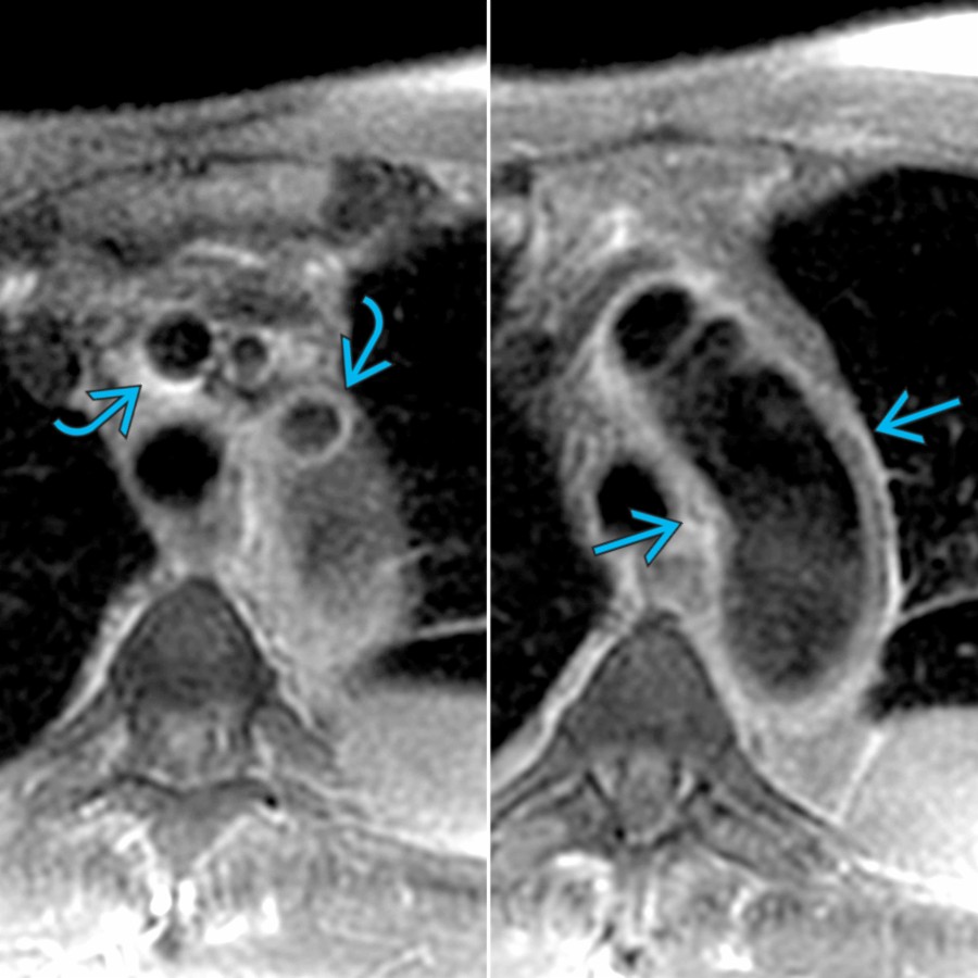
*Axial double inversion recovery FS MR in the same patient at different levels shows diffuse high signal of the aortic wall as well as head and neck vessels . MR is the preferred method to assess for active inflammation also seen in the form of vessel parietal enhancement after intravenous gadolinium.*
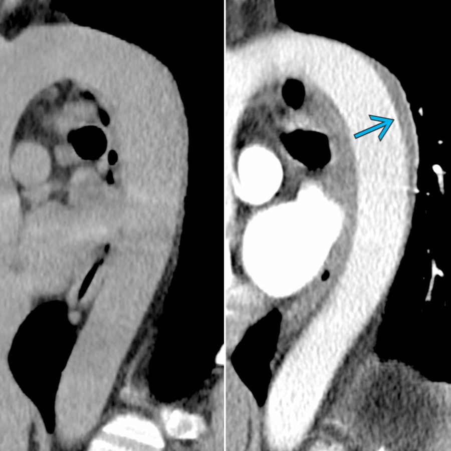
*Sagittal reformat CECT in a patient with GCA before and after contrast shows focal parietal thickening along the posterior descending thoracic aorta, only evident on CECT . Typically, vasculitis is not hyperdense on NECT as opposed to intramural hematoma.*
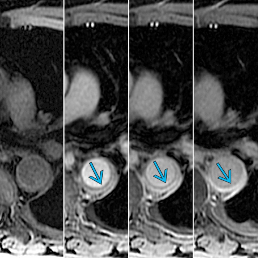
*3D GRE MR (unenhanced and post contrast) at the same level shows progressive enhancement of the aortic wall after administration of intravenous contrast .*
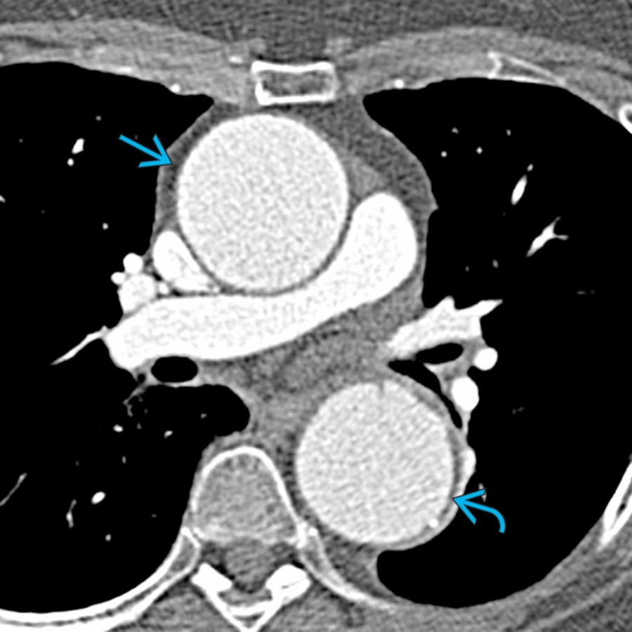
*Axial CTA in a patient with unsuspected GCA who underwent reconstruction of the ascending aorta due to aneurysm is shown. Note the aneurysmal ascending and descending aorta .*
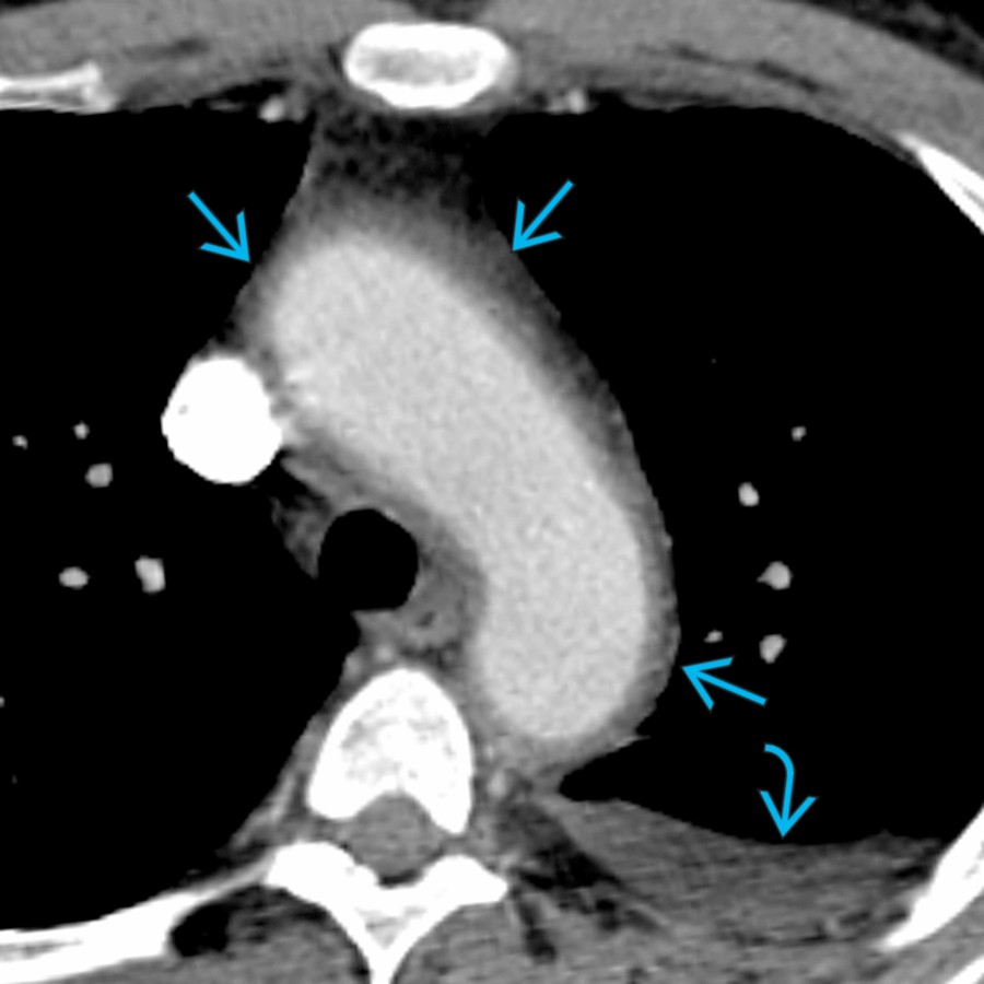
*Axial CTA in a patient with GCA shows diffuse arterial wall thickening and stranding of the periaortic fat. Note the reactive left pleural effusion .*

*Axial double inversion recovery FS MR in the same patient at different levels shows diffuse high signal of the aortic wall as well as head and neck vessels . MR is the preferred method to assess for active inflammation also seen in the form of vessel parietal enhancement after intravenous gadolinium.*

*Sagittal reformat CECT in a patient with GCA before and after contrast shows focal parietal thickening along the posterior descending thoracic aorta, only evident on CECT . Typically, vasculitis is not hyperdense on NECT as opposed to intramural hematoma.*

*3D GRE MR (unenhanced and post contrast) at the same level shows progressive enhancement of the aortic wall after administration of intravenous contrast .*

*Axial CTA in a patient with unsuspected GCA who underwent reconstruction of the ascending aorta due to aneurysm is shown. Note the aneurysmal ascending and descending aorta .*
 . Aneurysm is a very common complication of undiagnosed and untreated GCA only evident after resection.](images/app.statdx.com_image_thumbnail_0cb0e2d2-44cf-4f74-8fc7-64d634b973a7_annotated_true_size_900_quality_90_490afc268f5273006b244f2cb42ea010ec178f1a.jpg)
*Sagittal CECT MIP in the same patient shows diffuse aneurysmal thoracic aorta . Note also the aneurysmal right brachiocephalic trunk . Aneurysm is a very common complication of undiagnosed and untreated GCA only evident after resection.*
### Additional Images
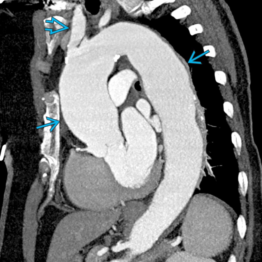
*Sagittal CECT MIP in the same patient shows diffuse aneurysmal thoracic aorta . Note also the aneurysmal right brachiocephalic trunk . Aneurysm is a very common complication of undiagnosed and untreated GCA only evident after resection.*
### Additional Images
 .](7f26d900-a43d-45d4-9957-141b82c7fb6c)
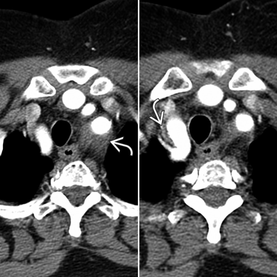
*Axial CTA in a young patient shows mural thickening of the supraaortic great vessels and stranding of the adjacent perivascular fat due to a vasculitis. Note the stenosis of the left common carotid artery .*

*Axial GRE MR following gadolinium administration shows marked circumferential mural thickening and enhancement of the descending thoracic aorta , consistent with active arteritis.*

*Axial GRE MR following gadolinium administration in the same patient confirms the presence of mural thickening and enhancement of the supraaortic arteries , consistent with active GCA.*

*Coronal contrast-enhanced MRA MIP in the same patient confirms multiple stenoses of the proximal pulmonary arteries without intraluminal thrombus. These are nonspecific features that are consistent with a pulmonary vasculitis, including GCA.*

*Axial CTA shows circumferential soft tissue thickening of the aortic arch in a patient with GCA. This represents an inflammatory reaction resulting in aortic mural thickening .*

*Axial CTA shows irregular mural thickening of the descending thoracic aorta and pulmonary arteries . Mural thickening represents a common sequela of inflammatory arteritis.*
.](7f26d900-a43d-45d4-9957-141b82c7fb6c)
*Axial CTA in a young patient shows mural thickening of the supraaortic great vessels and stranding of the adjacent perivascular fat due to a vasculitis. Note the stenosis of the left common carotid artery .*

*Axial GRE MR following gadolinium administration shows marked circumferential mural thickening and enhancement of the descending thoracic aorta , consistent with active arteritis.*

*Axial GRE MR following gadolinium administration in the same patient confirms the presence of mural thickening and enhancement of the supraaortic arteries , consistent with active GCA.*

*Coronal contrast-enhanced MRA MIP in the same patient confirms multiple stenoses of the proximal pulmonary arteries without intraluminal thrombus. These are nonspecific features that are consistent with a pulmonary vasculitis, including GCA.*

*Axial CTA shows circumferential soft tissue thickening of the aortic arch in a patient with GCA. This represents an inflammatory reaction resulting in aortic mural thickening .*

*Axial CTA shows irregular mural thickening of the descending thoracic aorta and pulmonary arteries . Mural thickening represents a common sequela of inflammatory arteritis.*