---
title: "Guillain-Barr\u00e9 Spectrum Disorders"
docid: "c1f52a65-920e-4e28-8a75-07dfa208f290"
authors:
- key: "b2e6dabb-ee1c-42a4-a332-9f0814c1c607"
value: "Surjith Vattoth, MD, FRCR"
breadcrumbs:
-
name: "Brain"
slug: "brain"
treeNodeId: "6d8829f1-14d7-45af-8675-255189aa526a"
-
name: "Diagnosis"
slug: "diagnosis"
treeNodeId: "51c00394-446e-4a38-94af-d3b1d14d34e8"
-
name: "Pathology-Based Diagnoses"
slug: "pathology-based-diagnoses"
treeNodeId: "d9d3a8ed-f21b-4831-8c77-591a3500ef77"
-
name: "Infectious, Inflammatory, and Demyelinating Disease"
slug: "infectious-inflammatory-and-demyel-"
treeNodeId: "7210f860-fe5f-4a2d-81cc-4fe06c769607"
-
name: "Inflammatory and Demyelinating Disease"
slug: "inflammatory-and-demyelinating-dis-"
treeNodeId: "62ab4dc3-dbf6-45a9-8532-f0e962aa62dc"
-
name: "Guillain-Barr\u00e9 Spectrum Disorders"
slug: "guillain-barr-spectrum-disorders"
treeNodeId: null
category: "Brain"
documentVersionId: "52016b28-7710-43a4-8cca-e659ab8227cf"
imageCount: 5
lastUpdated: "06/12/20"
pageDescription: "Guillain-Barr\u00e9 Spectrum Disorders"
pageKeywords: "Brain, Diagnosis, Pathology-Based Diagnoses, Infectious, Inflammatory, and Demyelinating Disease, Inflammatory and Demyelinating Disease, Guillain-Barr\u00e9 Spectrum Disorders"
pageTitle: "Guillain-Barr\u00e9 Spectrum Disorders | STATdx"
enhancedTitle: "Guillain-Barr\u00e9 Spectrum Disorders"
type: "DX"
references: true
breadcrumbs:
- "Brain"
- "Diagnosis"
- "Pathology-Based Diagnoses"
- "Infectious, Inflammatory, and Demyelinating Disease"
- "Inflammatory and Demyelinating Disease"
- "Guillain-Barr\u00e9 Spectrum Disorders"
---
# KEY FACTS
- ## Terminology
- Guillain-Barré syndrome (GBS), Miller Fisher syndrome (MFS), Bickerstaff brainstem encephalitis (BBE)
- **Classic GBS**: Acute-onset, rapidly progressive, ascending sensorimotor neuropathy; absent or ↓ tendon reflexes
- **Clinical variants of GBS**: Pure motor, paraparetic, bilateral facial palsy with paraesthesias, pharyngeal-cervical-brachial (PCB) variants and MFS
- **Other disorders often included in GBS spectrum** (controversial): Pure sensory variant, pure sensory ataxia, BBE
- ## Imaging
- Cauda equina (CE) nerve root enhancement in > 80%
- Isolated ventral CE in 17%, both ventral and dorsal in 65%
- Enhancement intensity of ventral CE ≥ dorsal CE
- Pial surface of distal cord/conus variable enhancement
- Pediatric GBS and MFS: CN enhancement in > 80%
- CNIII, CNVI, CNVII most common; others like CNII (and optic chiasm), CNV, CNXI, CNXII
- BBE: Brainstem T2 hyperintensities, may involve cerebellar peduncles and cerebellum
- ## Top Differential Diagnoses
- Subacute or chronic demyelinating polyneuropathies (SIDP/CIDP)
- CSF metastatic seeding, infections, arachnoiditis
- ## Pathology
- 6 temporally associated pathogens: *Campylobacter jejuni*, Cytomegalovirus (CMV), Hepatitis E virus, *Mycoplasma pneumoniae*, Epstein Barr virus (EBV), and Zika virus
- ## Clinical Issues
- Motor weakness and sensory signs in legs progressing to upper limbs and cranial muscles
- GBS spectrum/clinical variants with various findings
- Serum anti-GQ1b antibodies in up to 90% of MFS, 70% of BBE, and 8% of classic GBS
- Anti-GM1 antibodies more characteristic of classic GBS
- Intravenous immunoglobulins and plasma exchange therapy
- ## Diagnostic Checklist
- Consider GBS, variants or overlap syndromes in MR with ventral > dorsal cauda equina nerve root enhancement
- Look for cranial nerve root enhancement in MFS
- Look for brainstem signal abnormalities in BBE
# TERMINOLOGY
- ## Abbreviations
- Guillain-Barré syndrome (GBS), Miller Fisher syndrome (MFS), Bickerstaff brainstem encephalitis (BBE)
- Acute inflammatory demyelinating polyradiculoneuropathy (AIDP), acute motor axonal neuropathy (AMAN), acute motor sensory axonal neuropathy (AMSAN)
- ## Definitions
- Immune-mediated peripheral nerves and nerve roots disorder, usually triggered by infections
- **Classic GBS**: Acute-onset, rapidly progressive, ascending sensorimotor neuropathy; absent or ↓ tendon reflexes
- Initially in legs, progressing to arms and cranial muscles
- Usually reach maximum disability within 2 weeks
- **Electrophysiological subtypes**: **AIDP, AMAN, AMSAN**
- **Clinical variants of GBS**: Do not characteristically progress to classic GBS pattern of sensory loss and weakness
- Usually not "pure"; often partial overlap with classic GBS or other variants
- **Pure motor variant**: Motor weakness; no sensory signs
- **Paraparetic variant**: Paresis in lower limbs only
- **Bilateral facial palsy with paraesthesias variant**: Weakness limited to cranial nerves; ↓ reflexes
- **Pharyngeal-cervical-brachial (PCB) variant**: PCB muscle weakness, no lower limb weakness
- **MFS**: Ophthalmoplegia, areflexia, ataxia
- 15% overlap classic GBS
- Incomplete forms: **Acute ataxic neuropathy** (isolated ataxia); **acute ophthalmoplegia**
- **Other disorders often included in GBS spectrum**:****Due to similar clinical or pathophysiological features
- Inclusion in GBS spectrum debatable as these do not fulfil GBS diagnostic criteria
- **Pure sensory variant**: Overlapping clinical features of classic GBS, except motor signs and symptoms
- **Pure sensory ataxia**: Overlap with MFS
- **BBE**: Initially ophthalmoplegia, ataxia, areflexia (like MFS), later brainstem dysfunction, e.g., ↓ consciousness and pyramidal tract signs; often overlap classic GBS
# IMAGING
- ## General Features
- ### Best diagnostic clue
- Cauda equina (CE) nerve root enhancement > 80%
- Isolated ventral CE 17%, both ventral and dorsal 65%
- Enhancement intensity of ventral CE ≥ dorsal CE
- Pial surface of distal cord/conus variable enhancement
- Pediatric GBS and MFS: Cranial nerve enhancement > 80%
- CNIII, CNVI, CNVII most common; others like CNII (and optic chiasm), CNV, CNXI, CNXII
- Entire spinal cord T2-hyperintense posterior column described as delayed-onset finding in MFS case
- BBE: Brainstem T2 hyperintensities, may involve cerebellar peduncles and cerebellum
- Enhance only rarely; extremely rare, patchy spinal cord involvement described
- ## MR Findings
- ### T2WI
- May see slight nerve root thickening
- Brainstem hyperintensities in BBE
- ### T1WI C+
- Ventral > dorsal nerve root enhancement
- Cranial nerve enhancement
- ## Ultrasonographic Findings
- Enlarged nerves/roots during first 3 weeks, improve later
- ## Imaging Recommendations
- ### Best imaging tool
- MR of lumbar spine with contrast
- MR of brain with contrast if MFS or BBE suspected
# DIFFERENTIAL DIAGNOSIS
- ## Infections
- Bacterial or granulomatous meningitis
- Nerve root enhancement in minority of enterovirus D68 acute flaccid myelitis
- ## CSF Metastatic Seeding
- Nodular nerve root thickening and enhancement
- ## Subacute or Chronic Demyelinating Polyneuropathies
- Slower onset, protracted course
- CIDP: Nadir 2 months; SIDP: 4-8 weeks
- ## Arachnoiditis
- Secondary to hemorrhage, surgery, chemicals, chemotherapy (like vincristine), radiation
- ## Vasculitic Neuropathy
- Polyarteritis nodosa, Churg-Strauss syndrome
# PATHOLOGY
- ## General Features
- Infection in 6 weeks prior to onset in 2/3; triggering immune response causing GBS
- 6 temporally associated pathogens: *Campylobacter jejuni*, Cytomegalovirus (CMV), Hepatitis E virus, *Mycoplasma pneumoniae*, Epstein Barr virus (EBV), and Zika virus
- Absence of preceding illness does not exclude GBS
- Infections and other immunological stimuli (immunobiologicals like tumour necrosis factor antagonists, immune checkpoint inhibitors or type I interferons, surgery, amlgnancy) may be subclinical
- Serum antibodies against gangliosides in axolemma and other peripheral nerve components
- ## Gross Pathologic & Surgical Features
- Peripheral nerve and nerve root complement activation, macrophage infiltration, and edema
# CLINICAL ISSUES
- ## Presentation
- ### Most common signs/symptoms
- Motor weakness and sensory signs in legs progressing to upper limbs and cranial muscles
- Low back pain common symptom
- GBS spectrum/clinical variants with various findings
- Respiratory failure and mechanical ventilation in 20%
- Autonomic nervous system involvement with cardiac arrhythmias and unstable blood pressure
- ### Other signs/symptoms
- Serum **anti-GQ1b** antibodies in up to 90% of MFS, 70% of BBE, and 8% of classic GBS
- **Anti-GM1** antibodies more characteristic of classic GBS
- Should not wait for antibody test results to start treatment when GBS is suspected
- Albumino-cytological dissociation: Classic GBS finding
- ↑ CSF protein and normal CSF cell count
- Protein levels normal in 30-50% during 1st week and 10-30% during 2nd week
- Mild CSF pleocytosis (10-50 cells/μl) can be seen in GBS, but should exclude infectious polyradiculitis
- Electrodiagnostic studies not required to diagnose GBS; might be normal during 1st week, mild or variant disease, initial proximal weakness, or slow progression
- GBS: Sensorimotor polyradiculoneuropathy or polyneuropathy: ↓ conduction velocities, sensory and motor evoked amplitudes, abnormal temporal dispersion &/or partial motor conduction blocks
- GBS: Typical "sural sparing" pattern: Normal sural sensory nerve action potential, but abnormal or absent median and ulnar sensory nerve action potentials
- MFS: Normal or only ↓ amplitude of sensory nerve action potentials
- ## Demographics
- 1-2 per 100,000 person-years; M > F; ↑ with age
- ## Natural History & Prognosis
- Rapid progression, nadir within 2 weeks; mortality 3-10%
- GBS plateau phase (days to weeks or months); then recovery
- 60-80% walk independently after 6 months
- GBS typically monophasic illness, but relapses in 2-5%
- Treatment-related fluctuation (TRF): Deterioration after intial stability/improvement on therapy
- ## Treatment
- Intravenous immunoglobulin for 5 days (0.4 g/kg daily)
- Plasma exchange for 5 sessions (200-250 mL/kg)
- If TRF, repeat same treatment
- If no initial response or incomplete recovery, currently no evidence to support repeat treatment
# DIAGNOSTIC CHECKLIST
- ## Consider
- GBS, variants or overlap syndromes in MR showing ventral > dorsal cauda equina nerve root enhancement
- ## Image Interpretation Pearls
- Look for cranial nerve root enhancement in MFS
- Look for brainstem signal abnormalities in BBE
5e867f2a-de7e-44dd-83cf-67453b9125d2
## References
# Selected References
1. [Al Othman B et al: Update: the Miller Fisher variants of Guillain-Barré syndrome. Curr Opin Ophthalmol. 30(6):462-6, 2019](http://www.ncbi.nlm.nih.gov/pubmed/?term=31567467%5Bpmid%5D)
1. [Leonhard SE et al: Diagnosis and management of Guillain-Barré syndrome in ten steps. Nat Rev Neurol. 15(11):671-83, 2019](http://www.ncbi.nlm.nih.gov/pubmed/?term=31541214%5Bpmid%5D)
1. [Malhotra A et al: MRI findings of optic pathway involvement in Miller Fisher syndrome in 3 pediatric patients and a review of the literature. J Clin Neurosci. 39:63-7, 2017](http://www.ncbi.nlm.nih.gov/pubmed/?term=28209311%5Bpmid%5D)
1. [Cuneo GL et al: An atypical Bickerstaff's brainstem encephalitis with involvement of spinal cord. Neuroradiol J. 29(5):396-9, 2016](http://www.ncbi.nlm.nih.gov/pubmed/?term=27540012%5Bpmid%5D)
1. [Tyrakowska Z et al: Relapsing-Remitting Severe Bickerstaff's Brainstem Encephalitis - Case Report and Literature Review. Pol J Radiol. 81:622-628, 2016](http://www.ncbi.nlm.nih.gov/pubmed/?term=28096906%5Bpmid%5D)
1. [Zuccoli G et al: Redefining the Guillain-Barré spectrum in children: neuroimaging findings of cranial nerve involvement. AJNR Am J Neuroradiol. 32(4):639-42, 2011](http://www.ncbi.nlm.nih.gov/pubmed/?term=21292802%5Bpmid%5D)
1. [Inoue N et al: MR imaging findings of spinal posterior column involvement in a case of Miller Fisher syndrome. AJNR Am J Neuroradiol. 25(4):645-8, 2004](http://www.ncbi.nlm.nih.gov/pubmed/?term=15090361%5Bpmid%5D)
## Images
### Selected Images
 and slight thickening.](images/app.statdx.com_image_thumbnail_437a7388-6761-4ae6-bbe8-a94c64172505_annotated_true_size_900_quality_90_f3459dbb_20251018T122501Z.jpg)
*Axial T1 C+ MR of the lumbar spine in a patient with Guillain-Barré syndrome (GBS) shows the characteristic ventral cauda equina (CE) nerve root enhancement and slight thickening.*
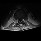
*Axial T1 C+ MR of the lumbar spine in a patient with Guillain-Barré syndrome (GBS) shows the characteristic ventral cauda equina (CE) nerve root enhancement and slight thickening.*
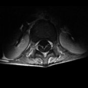
*Axial T1 C+ MR of the lumbar spine in a patient with Guillain-Barré syndrome (GBS) shows the characteristic ventral cauda equina (CE) nerve root enhancement and slight thickening.*
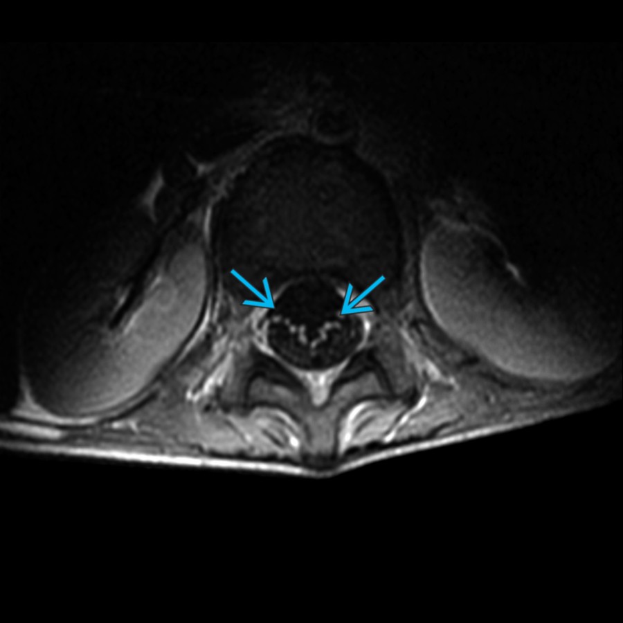
*Axial T1 C+ MR of the lumbar spine in a patient with Guillain-Barré syndrome (GBS) shows the characteristic ventral cauda equina (CE) nerve root enhancement and slight thickening.*

*Axial T1 C+ MR of the lumbar spine in a patient with Guillain-Barré syndrome (GBS) shows the characteristic ventral cauda equina (CE) nerve root enhancement and slight thickening.*

*Axial T1 C+ MR of the lumbar spine in a patient with Guillain-Barré syndrome (GBS) shows the characteristic ventral cauda equina (CE) nerve root enhancement and slight thickening.*
 CE. Also note enhancement of the pial surface of the distal cord/conus
  . GBS is an immune-mediated peripheral nerves and nerve roots disorder, usually triggered by infections.](images/app.statdx.com_image_thumbnail_5d50656f-8e56-40a4-9ad8-54778d94348f_annotated_true_size_900_quality_90_e8431c7b_20251018T122501Z.jpg)
*Sagittal T1 C+ FS MR of the lumbar spine in a patient with GBS shows CE nerve root enhancement, more intense in ventral than dorsal CE. Also note enhancement of the pial surface of the distal cord/conus . GBS is an immune-mediated peripheral nerves and nerve roots disorder, usually triggered by infections.*
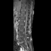
*Sagittal T1 C+ FS MR of the lumbar spine in a patient with GBS shows CE nerve root enhancement, more intense in ventral than dorsal CE. Also note enhancement of the pial surface of the distal cord/conus . GBS is an immune-mediated peripheral nerves and nerve roots disorder, usually triggered by infections.*
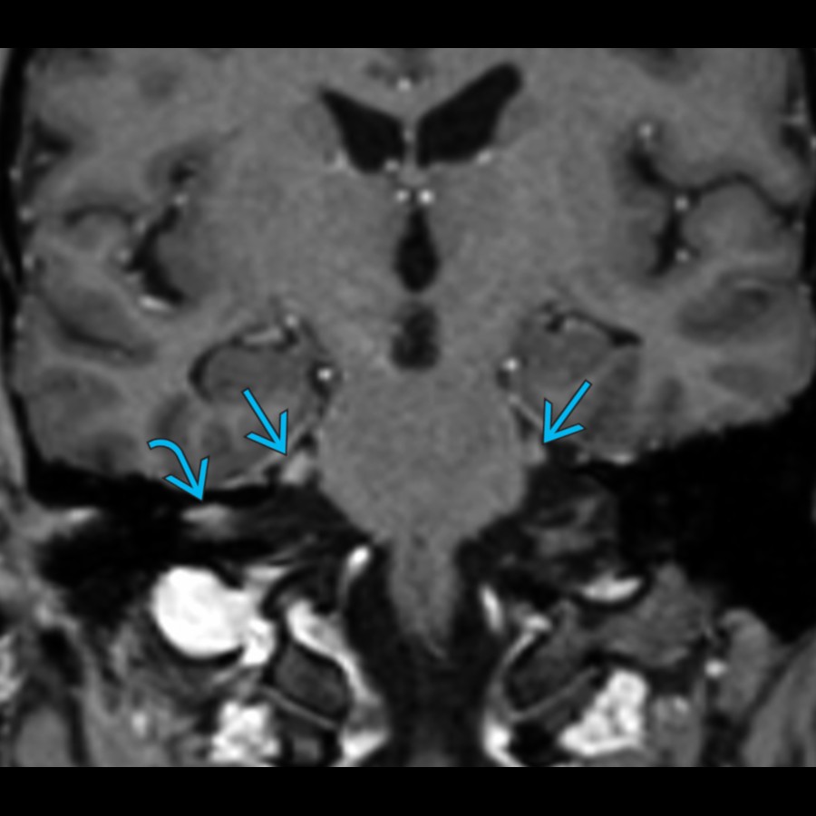
*Coronal angled T1 C+ MPRAGE MR reformat in a patient with Miller Fisher syndrome (MFS) shows mild thickening and enhancement of right facial (CNVII) and bilateral trigeminal (CNV) nerves.*
*Coronal angled T1 C+ MPRAGE MR reformat in a patient with Miller Fisher syndrome (MFS) shows mild thickening and enhancement of right facial (CNVII) and bilateral trigeminal (CNV) nerves.*

*Axial FLAIR MR in a patient with BBE shows hyperintense signal in the pons , middle cerebellar peduncles , and cerebellum . Both MFS and BBE may initially show ophthalmoplegia, ataxia, and areflexia, the differentiating feature being reduced consciousness and other brainstem signs in BBE later.*
*Axial FLAIR MR in a patient with BBE shows hyperintense signal in the pons , middle cerebellar peduncles , and cerebellum . Both MFS and BBE may initially show ophthalmoplegia, ataxia, and areflexia, the differentiating feature being reduced consciousness and other brainstem signs in BBE later.*
### Additional Images
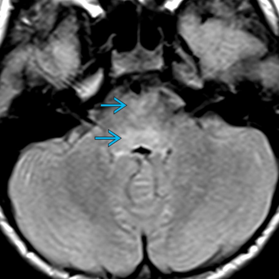
*Axial FLAIR MR in a patient with Bickerstaff brainstem encephalitis (BBE) shows abnormal hyperintense signal in the pons . Both MFS and BBE initially present with ophthalmoplegia, ataxia and areflexia; the differentiating feature being development of brainstem dysfunction in BBE later (reduced consciousness and pyramidal tract signs).*
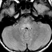
*Axial FLAIR MR in a patient with Bickerstaff brainstem encephalitis (BBE) shows abnormal hyperintense signal in the pons . Both MFS and BBE initially present with ophthalmoplegia, ataxia and areflexia; the differentiating feature being development of brainstem dysfunction in BBE later (reduced consciousness and pyramidal tract signs).*
. GBS is an immune-mediated peripheral nerves and nerve roots disorder, usually triggered by infections.](images/app.statdx.com_image_thumbnail_5d50656f-8e56-40a4-9ad8-54778d94348f_annotated_true_size_900_quality_90_e8431c7b_20251018T122501Z.jpg)
*Sagittal T1 C+ FS MR of the lumbar spine in a patient with GBS shows CE nerve root enhancement, more intense in ventral than dorsal CE. Also note enhancement of the pial surface of the distal cord/conus . GBS is an immune-mediated peripheral nerves and nerve roots disorder, usually triggered by infections.*

*Sagittal T1 C+ FS MR of the lumbar spine in a patient with GBS shows CE nerve root enhancement, more intense in ventral than dorsal CE. Also note enhancement of the pial surface of the distal cord/conus . GBS is an immune-mediated peripheral nerves and nerve roots disorder, usually triggered by infections.*

*Coronal angled T1 C+ MPRAGE MR reformat in a patient with Miller Fisher syndrome (MFS) shows mild thickening and enhancement of right facial (CNVII) and bilateral trigeminal (CNV) nerves.*

*Coronal angled T1 C+ MPRAGE MR reformat in a patient with Miller Fisher syndrome (MFS) shows mild thickening and enhancement of right facial (CNVII) and bilateral trigeminal (CNV) nerves.*

*Axial FLAIR MR in a patient with BBE shows hyperintense signal in the pons , middle cerebellar peduncles , and cerebellum . Both MFS and BBE may initially show ophthalmoplegia, ataxia, and areflexia, the differentiating feature being reduced consciousness and other brainstem signs in BBE later.*

*Axial FLAIR MR in a patient with BBE shows hyperintense signal in the pons , middle cerebellar peduncles , and cerebellum . Both MFS and BBE may initially show ophthalmoplegia, ataxia, and areflexia, the differentiating feature being reduced consciousness and other brainstem signs in BBE later.*
### Additional Images

*Axial FLAIR MR in a patient with Bickerstaff brainstem encephalitis (BBE) shows abnormal hyperintense signal in the pons . Both MFS and BBE initially present with ophthalmoplegia, ataxia and areflexia; the differentiating feature being development of brainstem dysfunction in BBE later (reduced consciousness and pyramidal tract signs).*

*Axial FLAIR MR in a patient with Bickerstaff brainstem encephalitis (BBE) shows abnormal hyperintense signal in the pons . Both MFS and BBE initially present with ophthalmoplegia, ataxia and areflexia; the differentiating feature being development of brainstem dysfunction in BBE later (reduced consciousness and pyramidal tract signs).*