---
title: "Inner Ear Lesion, Adult"
docid: "e5bbf757-d77a-4546-a848-d1a1a64cb230"
authors:
- key: "d19354f3-7ff2-495a-ad3f-064122e45602"
value: "Bernadette L. Koch, MD"
breadcrumbs:
-
name: "Head and Neck"
slug: "head-and-neck"
treeNodeId: "ed24ed8c-5d57-4629-879b-447b82d2973d"
-
name: "Differential Diagnosis"
slug: "differential-diagnosis"
treeNodeId: "40d68862-8975-4dde-ac2b-ebc43ab0fb5c"
-
name: "Temporal Bone"
slug: "temporal-bone"
treeNodeId: "2f7dc690-4970-486f-abb6-ed24d99b7a04"
-
name: "Anatomically Based Differentials"
slug: "anatomically-based-differentials"
treeNodeId: "115faedf-cc6e-41cd-a155-665ce5b062ee"
-
name: "Inner Ear Lesion, Adult"
slug: "inner-ear-lesion-adult"
treeNodeId: null
category: "Head and Neck"
documentVersionId: "e47e6e6d-1bca-481c-9123-ff1abb4acc60"
imageCount: 18
lastUpdated: "06/24/24"
pageDescription: "Inner Ear Lesion, Adult"
pageKeywords: "Head and Neck, Differential Diagnosis, Temporal Bone, Anatomically Based Differentials, Inner Ear Lesion, Adult"
pageTitle: "Inner Ear Lesion, Adult | STATdx"
enhancedTitle: "Inner Ear Lesion, Adult"
type: "DDX"
references: true
breadcrumbs:
- "Head and Neck"
- "Differential Diagnosis"
- "Temporal Bone"
- "Anatomically Based Differentials"
- "Inner Ear Lesion, Adult"
---
# ESSENTIAL INFORMATION
- ## Key Differential Diagnosis Issues
- Differential encompasses all lesions, including normal variants, that may be found in inner ear region of adults
- Inner ear region in temporal bone includes all structures from medial wall of middle ear to lateral aspect of petrous apex
- ## Helpful Clues for Common Diagnoses
- **Semicircular Canal Dehiscence**
- Clinical clues: Tinnitus, dizziness, vertigo, & conductive hearing loss (CHL)
- Tullio phenomenon: Sound-induced vertigo &/or nystagmus
- Pathophysiology: Focal superior semicircular canal (SCC) dehiscence causes "3rd" window in inner ear
- Bone CT findings
- Superior SCC roof dehiscent ≥ 2 mm into middle cranial fossa dura
- Transverse oblique CT reformation best shows lesion
- Rarely, posterior SCC dehiscence
- **Fenestral Otosclerosis**
- Clinical clues: Young adult develops gradual increased CHL
- Early disease location
- Begins at anterior margin of oval window (fissula ante fenestram)
- Bone CT findings
- Lucent & sclerotic lesions along margins of oval & round windows
- MR findings
- Enhancing millimeter-sized foci along margins of oval & round windows
- **Labyrinthine Ossificans**
- Clinical clue: Profound sensorineural hearing loss (SNHL) following episode of meningitis
- Pathophysiology: Healing of suppurative labyrinthitis may lead to osteoneogenesis in inner ear fluid spaces
- Bone CT findings
- Ossific plaques impinge on inner ear fluid spaces
- MR findings
- High-resolution T2: Loss of fluid signal/encroachment on membranous labyrinthine fluid spaces
- **High Jugular Bulb**
- Clinical clue: Often asymptomatic normal variant, uncommonly cause of pulsatile tinnitus
- Bone CT findings
- Cephalad portion of jugular bulb projects to level of internal auditory canal (IAC)
- MR findings
- May simulate enhancing lesion posterior to IAC
- **Jugular Bulb Diverticulum**
- Clinical clue: Asymptomatic normal variant
- Bone CT findings
- Thumb-like projection off jugular bulb
- Most commonly projects superiorly behind IAC
- May project in any direction
- MR findings
- May simulate enhancing lesion posterior to IAC
- **I****nner Ear****Fracture**
- Bone CT findings
- Transverse or longitudinal fracture line traverses inner ear structures
- Pneumolabyrinth possible
- Otic capsule sparing vs. otic capsule violating classification better predicts complications, such as SNHL, CNVII injury, & CSF leak
- ± ossicular dislocations &/or fractures
- MR findings
- High T1 signal (blood) within inner ear fluid spaces
- ## Helpful Clues for Less Common Diagnoses
- **Cochlear Incomplete Partition Type II (IP-2)**
- Clinical clue: Congenital SNHL in child or young adult with cascading hearing loss pattern
- Key fact: Most common congenital imaging abnormality in adult SNHL
- Bone CT findings
- Deficient interscalar septum (ISS) between middle & apical cochlear turns
- Flattening of interscalar ridge (anchor point) between upper basal & upper middle cochlear turns
- Usually with enlarged bony vestibular aqueduct
- MR findings
- Deficient ISS between middle & apical cochlear turns
- Usually with enlarged endolymphatic sac/duct
- Literature suggests dysplastic osseous spiral lamina-basilar membrane neural complex may mimic ISS on high-resolution, 3D cisternographic MR images, resulting in underdiagnosed IP-2 anomalies
- Reformat images parallel to lateral SCC to measure distance X = distance between osseous spiral lamina-basilar membrane complex of upper basal turn & 1st linear signal void anterior to basilar membrane
- When 1st linear signal void = normal ISS, distance X < 1.2 mm
- When 1st linear signal void = dysplastic osseous spiral lamina-basilar membrane neural complex, distance X ≥ 1.2 mm
- **Facial Nerve Venous Malformation ("Hemangioma")**
- Clinical clues: Mimics Bell palsy
- Facial nerve (FN) paralysis; often with rapid onset
- Location/morphology: Geniculate fossa most common
- Poorly circumscribed; amorphous
- Vascular malformation, not hemangioma
- Bone CT findings
- Enlarged geniculate fossa
- As it increases, spreads along anterior surface of petrous apex
- ~ 50% show "honeycomb" ossific matrix
- MR findings
- Amorphous enhancing geniculate ganglion area mass
- **Facial Nerve Schwannoma**
- Clinical clues: 50% present with hearing loss
- FN symptoms often subtle or absent
- Location/morphology: Most commonly found in geniculate fossa
- Smooth, tubular expanding lesion
- Bone CT findings
- Focal or tubular enlargement intratemporal FN canal
- MR findings
- High-resolution T2: Hypointense soft tissue nodule replacing normal fluid signal
- C+ MR: Enhancing mass follows intratemporal FN
- **Labyrinthitis**
- Clinical clue: Acute-onset vertigo, hearing loss ± FN paralysis
- Bone CT findings
- Acute phase normal
- MR findings
- Normal or diffuse enhancement of inner ear fluid spaces
- **Intralabyrinthine Schwannoma**
- Clinical clue: 10-20 year history of gradual increased SNHL
- Bone CT findings
- Normal unless larger transmodiolar, transmacular, or transotic type present
- MR findings
- Focal enhancement within inner ear fluid space
- T2-hypointense nodule replacing normal fluid signal
- Recommended descriptive terms include location: Intracochlear, intravestibular, vestibulocochlear, transmodiolar, transmacular, & transotic options
- ## Helpful Clues for Rare Diagnoses
- **Cochlear Otosclerosis**
- Clinical clue: Bilateral mixed CHL & SNHL
- Bone CT findings
- Lucent foci in bony labyrinth surrounding cochlea
- MR findings
- Punctate or linear enhancing foci in otic capsule
- **Intralabyrinthine Hemorrhage**
- Clinical clue: Idiopathic, anticoagulant therapy, posttraumatic, sickle cell disease
- Bone CT findings
- Normal unless associated with trauma
- MR findings
- High T1 signal in inner ear fluid spaces
- **Endolymphatic Sac Tumor**
- Clinical clues: Associated with von Hippel-Lindau disease > sporadic
- Bone CT findings
- Permeative lytic destruction of posterior petrous temporal bone at expected location of vestibular aqueduct/endolymphatic sac
- Spiculated or coarse calcifications within tumor matrix
- Thin calcification seen along posterior margin
- MR findings
- T1 high signal foci from blood products trapped within tumor matrix
- Avidly enhancing tumor; T1 flow voids ("pepper") also common
## References
# Selected References
1. [Razskazovskiy V et al: Prevalence of cochlear-facial and other non-superior semicircular canal third window dehiscence on high-resolution temporal bone CT. AJNR Am J Neuroradiol. 44(11):1309-13, 2023](http://www.ncbi.nlm.nih.gov/pubmed/?term=37884302%5Bpmid%5D)
1. [Deng F et al: Diagnostic performance of conebeam CT pixel values in active fenestral otosclerosis. AJNR Am J Neuroradiol. 42(9):1667-70, 2021](http://www.ncbi.nlm.nih.gov/pubmed/?term=34140277%5Bpmid%5D)
1. [Ganeshan D et al: Tumors in von Hippel-Lindau syndrome: from head to toe-comprehensive state-of-the-art review. Radiographics. 38(3):849-66, 2018](http://www.ncbi.nlm.nih.gov/pubmed/?term=29601266%5Bpmid%5D)
1. [Conte G et al: MR imaging in sudden sensorineural hearing loss. Time to talk. AJNR Am J Neuroradiol. 38(8):1475-9, 2017](http://www.ncbi.nlm.nih.gov/pubmed/?term=28546251%5Bpmid%5D)
1. [Reinshagen KL et al: Measurement for detection of incomplete partition type II anomalies on MR imaging. AJNR Am J Neuroradiol. 38(10):2003-7, 2017](http://www.ncbi.nlm.nih.gov/pubmed/?term=28775060%5Bpmid%5D)
1. [Sparacia G et al: Diagnostic performance of reformatted isotropic thin-section helical CT images in the detection of superior semicircular canal dehiscence. Neuroradiol J. 30(3):216-21, 2017](http://www.ncbi.nlm.nih.gov/pubmed/?term=28627985%5Bpmid%5D)
1. [Bausch B et al: Characterization of endolymphatic sac tumors and von Hippel-Lindau disease in the international ELST registry. Head Neck. 38 Suppl 1:E673-9, 2015](http://www.ncbi.nlm.nih.gov/pubmed/?term=25867206%5Bpmid%5D)
1. [Kennedy TA et al: Imaging of temporal bone trauma. Neuroimaging Clin N Am. 24(3):467-86, viii, 2014](http://www.ncbi.nlm.nih.gov/pubmed/?term=25086807%5Bpmid%5D)
1. [Nevoux J et al: Management of endolymphatic sac tumors: sporadic cases and von Hippel-Lindau disease. Otol Neurotol. 35(5):899-904, 2014](http://www.ncbi.nlm.nih.gov/pubmed/?term=24662627%5Bpmid%5D)
1. [Young JY et al: Preoperative imaging of sensorineural hearing loss in pediatric candidates for cochlear implantation. Radiographics. 34(5):E133-49, 2014](http://www.ncbi.nlm.nih.gov/pubmed/?term=25208295%5Bpmid%5D)
1. [Nadgir RN et al: Superior semicircular canal dehiscence: congenital or acquired condition? AJNR Am J Neuroradiol. 32(5):947-9, 2011](http://www.ncbi.nlm.nih.gov/pubmed/?term=21393404%5Bpmid%5D)
1. [Benoit MM et al: Facial nerve hemangiomas: vascular tumors or malformations? Otolaryngol Head Neck Surg. 142(1):108-14, 2010](http://www.ncbi.nlm.nih.gov/pubmed/?term=20096233%5Bpmid%5D)
1. [Offiah CE et al: Imaging appearances of unusual conditions of the middle and inner ear. Br J Radiol. 81(966):504-14, 2008](http://www.ncbi.nlm.nih.gov/pubmed/?term=18316346%5Bpmid%5D)
1. [Salomone R et al: Sudden hearing loss caused by labyrinthine hemorrhage. Braz J Otorhinolaryngol. 74(5):776-9, 2008](http://www.ncbi.nlm.nih.gov/pubmed/?term=19082362%5Bpmid%5D)
1. [Tieleman A et al: Imaging of intralabyrinthine schwannomas: a retrospective study of 52 cases with emphasis on lesion growth. AJNR Am J Neuroradiol. 29(5):898-905, 2008](http://www.ncbi.nlm.nih.gov/pubmed/?term=18321986%5Bpmid%5D)
## Images
### Selected Images
 to best advantage by showing the entire extent of the dehiscence in a single image. Symptomatic patients typically present with sound-induced vertigo &/or nystagmus, i.e., Tullio phenomenon.](images/app.statdx.com_image_thumbnail_985891cf-6755-4520-bbcb-4c7e11988c4c_annotated_true_size_900_quality_90_ce6c22a339ee7fa1a3f66339764f3ed2354da9b7.jpg)
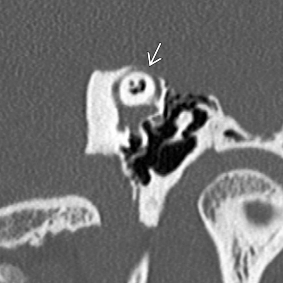
**Semicircular Canal Dehiscence**
*Transverse oblique bone CT reformation displays the dehiscent roof of the superior semicircular canal to best advantage by showing the entire extent of the dehiscence in a single image. Symptomatic patients typically present with sound-induced vertigo &/or nystagmus, i.e., Tullio phenomenon.*
**Semicircular Canal Dehiscence**
*Transverse oblique bone CT reformation displays the dehiscent roof of the superior semicircular canal to best advantage by showing the entire extent of the dehiscence in a single image. Symptomatic patients typically present with sound-induced vertigo &/or nystagmus, i.e., Tullio phenomenon.*
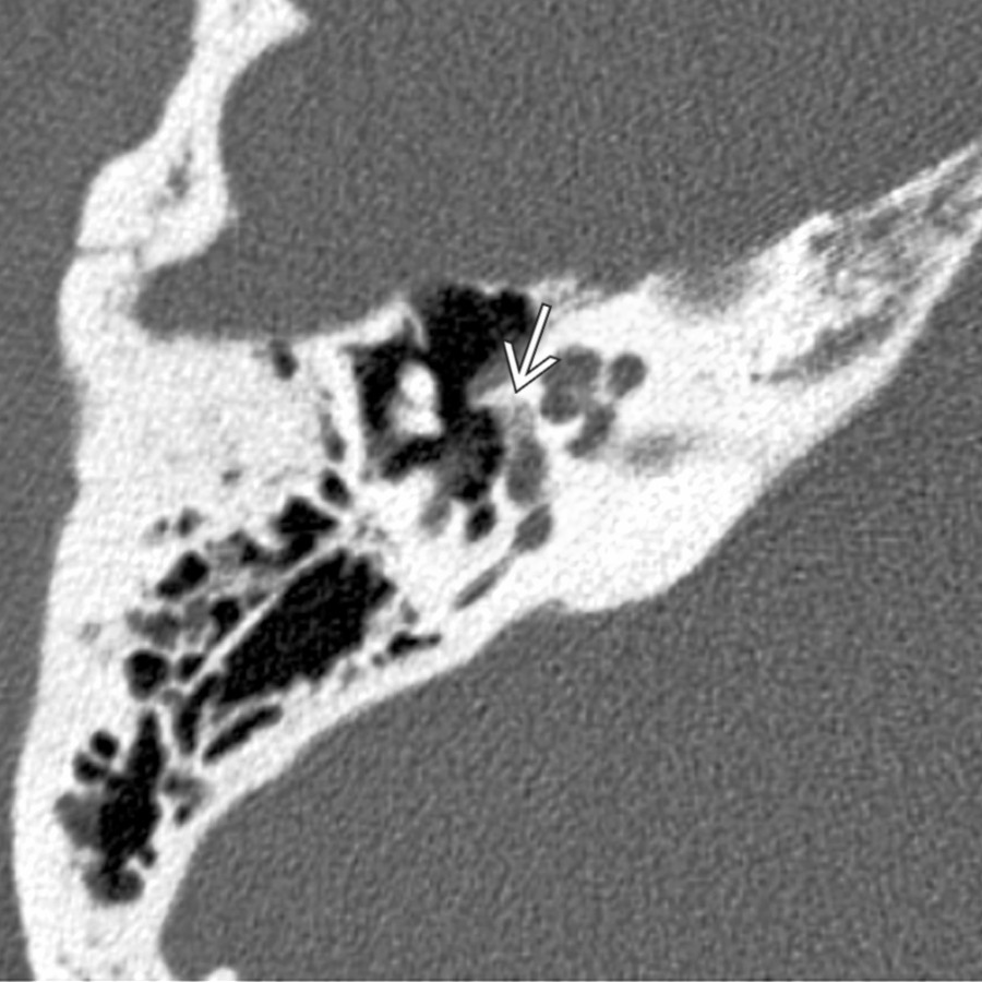
**Fenestral Otosclerosis**
*Axial bone CT demonstrates a subtle active otosclerotic plaque along the anterior margin of the oval window . This is the location of the fissula ante fenestram where fenestral otosclerosis begins.*
**Semicircular Canal Dehiscence**
*Transverse oblique bone CT reformation displays the dehiscent roof of the superior semicircular canal to best advantage by showing the entire extent of the dehiscence in a single image. Symptomatic patients typically present with sound-induced vertigo &/or nystagmus, i.e., Tullio phenomenon.*

**Semicircular Canal Dehiscence**
*Transverse oblique bone CT reformation displays the dehiscent roof of the superior semicircular canal to best advantage by showing the entire extent of the dehiscence in a single image. Symptomatic patients typically present with sound-induced vertigo &/or nystagmus, i.e., Tullio phenomenon.*

**Fenestral Otosclerosis**
*Axial bone CT demonstrates a subtle active otosclerotic plaque along the anterior margin of the oval window . This is the location of the fissula ante fenestram where fenestral otosclerosis begins.*
 . There was also partial opacification of the cochlea (not shown).](images/app.statdx.com_image_thumbnail_b2a9ad29-4d27-4db3-b98f-f651768b19d7_annotated_true_size_900_quality_90_b5f9c82e2490d02dce251c59436b5939758d64ef.jpg)
**Labyrinthine Ossificans**
*Axial bone CT in a patient with prior history of meningitis shows complete ossification of the vestibule and lateral semicircular canal . There was also partial opacification of the cochlea (not shown).*
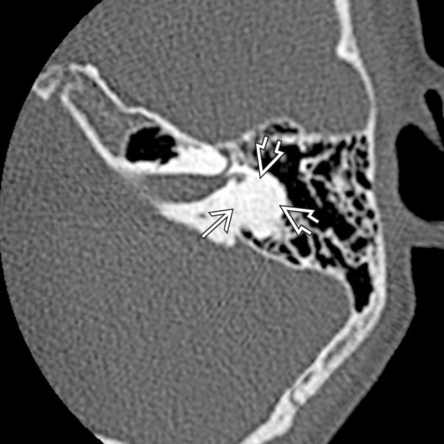
**Labyrinthine Ossificans**
*Axial bone CT in a patient with prior history of meningitis shows complete ossification of the vestibule and lateral semicircular canal . There was also partial opacification of the cochlea (not shown).*
 visible at the level of the internal auditory canal
  . The high jugular bulb abuts the bony vestibular aqueduct . Note that it does not enter the middle ear cavity as a dehiscent jugular bulb might.](images/app.statdx.com_image_thumbnail_e7dc8a77-f0ba-4016-9877-a9220474966a_annotated_true_size_900_quality_90_df68733466f6d94e662cc2369b9c30e7215b7a10.jpg)
**High Jugular Bulb**
*Axial bone CT shows a high jugular bulb visible at the level of the internal auditory canal . The high jugular bulb abuts the bony vestibular aqueduct . Note that it does not enter the middle ear cavity as a dehiscent jugular bulb might.*
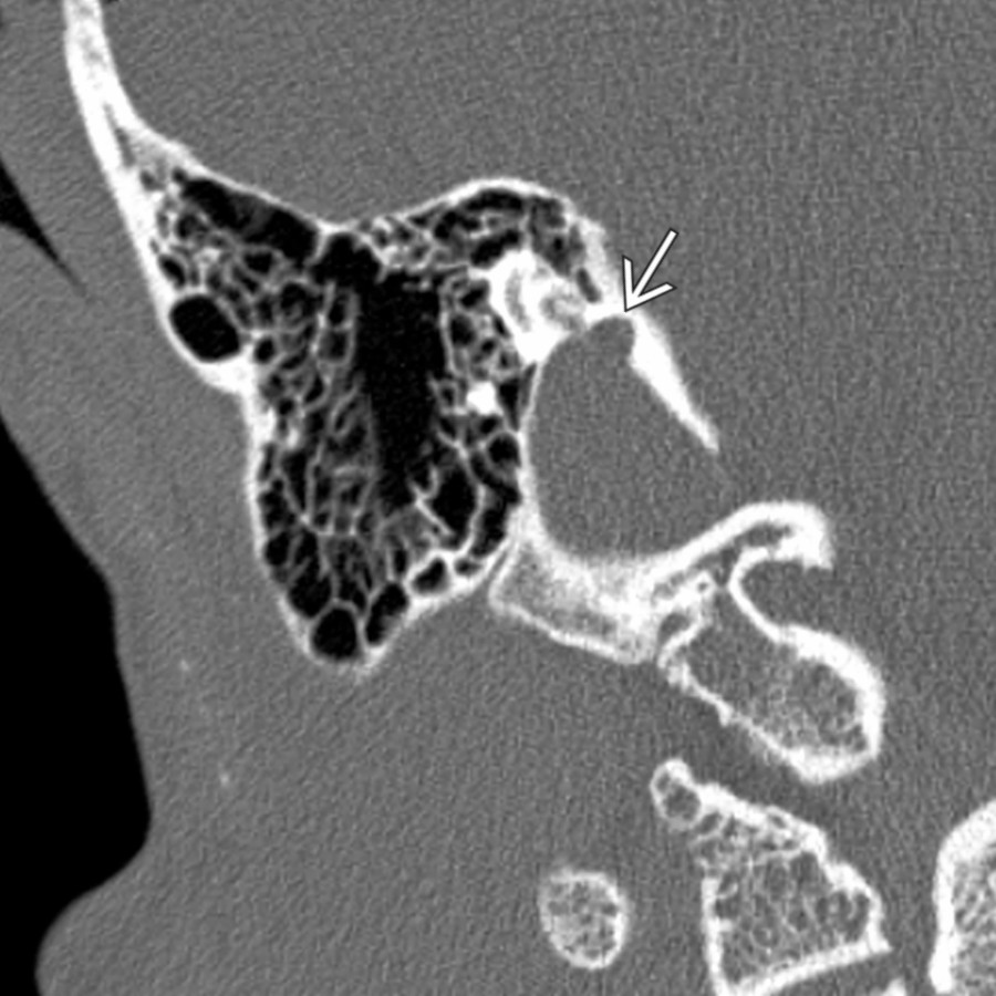
**Jugular Bulb Diverticulum**
*Coronal bone CT demonstrates a jugular bulb diverticulum as a superomedial projection off the top of the jugular bulb . These lesions are often asymptomatic but can be the cause of pulsatile tinnitus.*
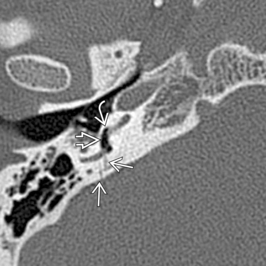
**Inner Ear Fracture**
*Axial bone CT in a patient with multiple skull base fractures secondary to a high-speed collision shows fractures traversing the left temporal bone, crossing the right posterior limb of the lateral semicircular canal with associated pneumolabyrinth in the vestibule and basal turn of the cochlea .*

**Cochlear Incomplete Partition Type II (IP-2)**
*Axial T2 MR shows a large endolymphatic sac along the posterior wall of the temporal bone. Note the associated cochlear dysplasia with modiolar deficiency and bulbous asymmetric scalar chambers . These patients present with a cascading pattern of progressive hearing loss.*

**Facial Nerve Venous Malformation ("Hemangioma")**
*Axial bone CT demonstrates a nonossifying, amorphous, geniculate ganglion lesion protruding into the anterior epitympanic recess. The anterior tympanic segment of CNVII is also shown.*

**Facial Nerve Schwannoma**
*Coronal T1 C+ FS MR reveals an enhancing well-circumscribed lesion of the geniculate ganglion . Notice that this ovoid facial nerve schwannoma sits just superior to the normal-appearing cochlea , the most common location for facial nerve schwannomas to occur.*

**Labyrinthitis**
*Coronal T1 C+ MR shows a diffusely enhancing cochlea along with facial nerve enhancement in the geniculate ganglion area . Clinical presentation included acute-onset vertigo and sensorineural hearing loss.*

**Intralabyrinthine Schwannoma**
*Axial T2 DRIVE image shows a very small intralabyrinthine hypointense schwannoma in the posterior aspect of the vestibule surrounded by otherwise normal hyperintense T2 signal intensity fluid. There was typical corresponding enhancement after contrast (not shown).*

**Cochlear Otosclerosis**
*Axial bone CT in a patient with severe cochlear and fenestral otosclerosis shows multifocal osteolytic foci surrounding the cochlea.*

**Intralabyrinthine Hemorrhage**
*Axial T1 MR reveals high-signal intralabyrinthine hemorrhage involving the cochlea , vestibule , and endolymphatic sac .*

**Endolymphatic Sac Tumor**
*Axial T1 MR shows a very large right endolymphatic sac tumor. The high-signal blood in the tumor matrix and broad involvement of the posterior temporal bone wall suggest this diagnosis. The majority of these lesions are associated with von Hippel-Lindau disease.*
### Additional Images
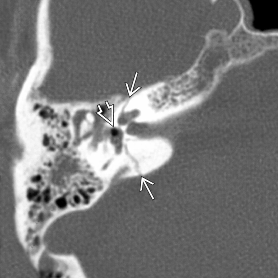
**Inner Ear Fracture**
*Axial bone CT reveals a transverse fracture traversing the inner ear structures , crossing obliquely from just anterior to the vestibular aqueduct to the vestibule and then anterolaterally to cross the cochlea. Notice the pneumolabyrinth in the vestibule indicating a perilymphatic fistula at the oval window level.*
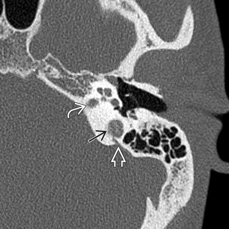
**High Jugular Bulb**
*Axial bone CT shows a high jugular bulb visible at the level of the internal auditory canal . The high jugular bulb abuts the bony vestibular aqueduct . Note that it does not enter the middle ear cavity as a dehiscent jugular bulb might.*

**Jugular Bulb Diverticulum**
*Coronal bone CT demonstrates a jugular bulb diverticulum as a superomedial projection off the top of the jugular bulb . These lesions are often asymptomatic but can be the cause of pulsatile tinnitus.*

**Inner Ear Fracture**
*Axial bone CT in a patient with multiple skull base fractures secondary to a high-speed collision shows fractures traversing the left temporal bone, crossing the right posterior limb of the lateral semicircular canal with associated pneumolabyrinth in the vestibule and basal turn of the cochlea .*

**Cochlear Incomplete Partition Type II (IP-2)**
*Axial T2 MR shows a large endolymphatic sac along the posterior wall of the temporal bone. Note the associated cochlear dysplasia with modiolar deficiency and bulbous asymmetric scalar chambers . These patients present with a cascading pattern of progressive hearing loss.*

**Facial Nerve Venous Malformation ("Hemangioma")**
*Axial bone CT demonstrates a nonossifying, amorphous, geniculate ganglion lesion protruding into the anterior epitympanic recess. The anterior tympanic segment of CNVII is also shown.*

**Facial Nerve Schwannoma**
*Coronal T1 C+ FS MR reveals an enhancing well-circumscribed lesion of the geniculate ganglion . Notice that this ovoid facial nerve schwannoma sits just superior to the normal-appearing cochlea , the most common location for facial nerve schwannomas to occur.*

**Labyrinthitis**
*Coronal T1 C+ MR shows a diffusely enhancing cochlea along with facial nerve enhancement in the geniculate ganglion area . Clinical presentation included acute-onset vertigo and sensorineural hearing loss.*

**Intralabyrinthine Schwannoma**
*Axial T2 DRIVE image shows a very small intralabyrinthine hypointense schwannoma in the posterior aspect of the vestibule surrounded by otherwise normal hyperintense T2 signal intensity fluid. There was typical corresponding enhancement after contrast (not shown).*

**Cochlear Otosclerosis**
*Axial bone CT in a patient with severe cochlear and fenestral otosclerosis shows multifocal osteolytic foci surrounding the cochlea.*

**Intralabyrinthine Hemorrhage**
*Axial T1 MR reveals high-signal intralabyrinthine hemorrhage involving the cochlea , vestibule , and endolymphatic sac .*

**Endolymphatic Sac Tumor**
*Axial T1 MR shows a very large right endolymphatic sac tumor. The high-signal blood in the tumor matrix and broad involvement of the posterior temporal bone wall suggest this diagnosis. The majority of these lesions are associated with von Hippel-Lindau disease.*
### Additional Images

**Inner Ear Fracture**
*Axial bone CT reveals a transverse fracture traversing the inner ear structures , crossing obliquely from just anterior to the vestibular aqueduct to the vestibule and then anterolaterally to cross the cochlea. Notice the pneumolabyrinth in the vestibule indicating a perilymphatic fistula at the oval window level.*
 and fenestral otosclerosis. The cochlear otospongiosis plaque initially appears to be a "3rd turn of the cochlea."](db4b27dc-274e-4f72-8592-6c2df676d7a8)
**Cochlear Otosclerosis**
*Axial bone CT shows a patient with severe cochlear and fenestral otosclerosis. The cochlear otospongiosis plaque initially appears to be a "3rd turn of the cochlea."*
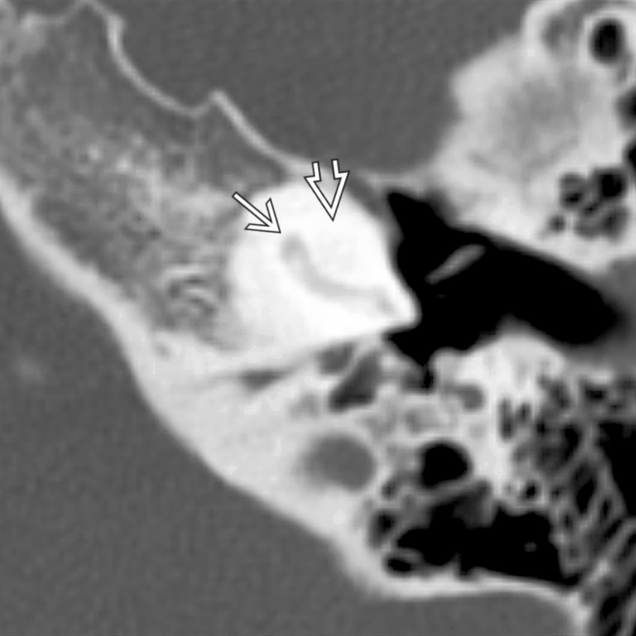
**Labyrinthine Ossificans**
*Axial bone CT shows severe labyrinthine ossificans. Notice that the basal turn of the cochlea is partially ossified but still identifiable whereas the 2nd turn is nearly invisible .*

**Intralabyrinthine Schwannoma**
*Axial T1 C+ FS MR shows a mildly enhancing schwannoma involving the posterolateral aspect of the basal turn of the cochlea . The lesion protrudes into the middle ear cavity. These patients typically present with gradual increase in sensorineural hearing loss.*
and fenestral otosclerosis. The cochlear otospongiosis plaque initially appears to be a "3rd turn of the cochlea."](db4b27dc-274e-4f72-8592-6c2df676d7a8)
**Cochlear Otosclerosis**
*Axial bone CT shows a patient with severe cochlear and fenestral otosclerosis. The cochlear otospongiosis plaque initially appears to be a "3rd turn of the cochlea."*

**Labyrinthine Ossificans**
*Axial bone CT shows severe labyrinthine ossificans. Notice that the basal turn of the cochlea is partially ossified but still identifiable whereas the 2nd turn is nearly invisible .*

**Intralabyrinthine Schwannoma**
*Axial T1 C+ FS MR shows a mildly enhancing schwannoma involving the posterolateral aspect of the basal turn of the cochlea . The lesion protrudes into the middle ear cavity. These patients typically present with gradual increase in sensorineural hearing loss.*