---
title: "Intramedullary Mass"
docid: "635c114e-6260-4e9d-a36a-368deeb05f05"
authors:
- key: "bee1f359-33fb-4cba-9e6b-ed1ca1842439"
value: "Jeffrey S. Ross, MD"
- key: "bf7af3a5-468c-48d0-8aea-4f6e808af979"
value: "Bryson Borg, MD"
breadcrumbs:
-
name: "Spine"
slug: "spine"
treeNodeId: "b337a156-914a-4696-a77c-af206720fab5"
-
name: "Differential Diagnosis"
slug: "differential-diagnosis"
treeNodeId: "ef5fd925-2033-4f3b-aa7c-640fef9aa956"
-
name: "Intramedullary"
slug: "intramedullary"
treeNodeId: "b72eef28-6a45-4665-8758-eb14b7ac4462"
-
name: "Anatomically Based Differentials"
slug: "anatomically-based-differentials"
treeNodeId: "3c8e44bc-84bc-455a-b832-a139b0d0a604"
-
name: "Intramedullary Mass"
slug: "intramedullary-mass"
treeNodeId: null
category: "Spine"
cmeTopicId: "6ba91891-8860-49fd-a3a6-ba09453efe41"
documentVersionId: "e4262d97-22fd-491c-8854-f05e2f9a215d"
imageCount: 32
lastUpdated: "02/14/23"
pageDescription: "Intramedullary Mass"
pageKeywords: "Spine, Differential Diagnosis, Intramedullary, Anatomically Based Differentials, Intramedullary Mass"
pageTitle: "Intramedullary Mass | STATdx"
enhancedTitle: "Intramedullary Mass"
type: "DDX"
references: true
breadcrumbs:
- "Spine"
- "Differential Diagnosis"
- "Intramedullary"
- "Anatomically Based Differentials"
- "Intramedullary Mass"
---
# ESSENTIAL INFORMATION
- ## Key Differential Diagnosis Issues
- MR ± contrast is best tool to evaluate intramedullary processes of cord
- Discovery of intramedullary cord lesion typically followed by imaging of remainder of neuraxis
- Infiltrative cord lesion: Image brain to potentially identify characteristic white matter lesion(s) of multiple sclerosis (MS)
- Discovery of intramedullary tumor typically accompanied by insidious onset of myelopathic symptoms (months or years)
- Patient with syrinx should be imaged at least once with contrast-enhanced MR to exclude cord neoplasm
- Hemorrhagic lesion: Think ependymoma, cavernous malformation, contusion
- ## Helpful Clues for Common Diagnoses
- **Multiple Sclerosis, Spinal Cord**
- Typically located eccentrically, not involving entire cord on axial imaging; relatively short in length (< 2 vertebral bodies)
- Enlargement of cord is unusual (6-14%)
- Dissemination in time: Some enhance, some do not
- 5-24% of patients with MS cord plaques may not have supratentorial disease at presentation
- **Neuromyelitis Optica Spectrum Disorder**
- Autoimmune astrocytopathic disease targeting water channel proteins (AQP4) with involvement of optic nerves and spinal cord with limited brain parenchymal involvement
- Longitudinally extensive transverse myelitis (LETM) (> 3 vertebral segments) T2 hyperintensity within cord
- Enhancement of optic nerves
- Brain lesions not typical for MS
- **Acute Disseminated Encephalitis, Spinal Cord**
- Clinical: Self-limited, monophasic demyelinating illness 5-14 days following viral infection or vaccination
- Usually indistinguishable from MS on imaging at single time point
- **Acute Transverse Myelitis, Idiopathic**
- Clinical: Acute-onset myelopathy, ascending or static loss of sensory and motor function in bilateral and symmetric distribution
- Infiltrative signal abnormality may extend above level of deficit; variable enhancement
- Mild fusiform enlargement may be present and simulate appearance of primary cord neoplasm
- **Ependymoma, Spinal Cord**
- Circumscribed, enhancing intramedullary mass; located centrally within cord
- Can show signs of necrosis (heterogeneity, cyst formation) and hemorrhage (hyperintense T1, susceptibility artifact, hemosiderin cap sign)
- Most common intramedullary neoplasm in adults
- 10% may show very little enhancement
- **Astrocytoma, Spinal Cord**
- Fusiform enlargement, infiltrative margins, long segment of involvement; no or variable enhancement
- Most commonly located in cervical and upper thoracic cord
- Uncommon/rare imaging features: Hemorrhage, necrosis, caudal location, exophytic growth, holocord involvement
- Cannot be reliably differentiated from ependymoma by imaging
- 2nd most common cord neoplasm in adults; most common cord neoplasm in children (60%)
- **Syringomyelia**
- Abnormal cystic cord lesion with surrounding gliosis; variable expansion of cord; focal or extensive; typically longitudinal
- Secondary to chronic insult/injury (cavitation) or to altered CSF dynamics in central canal of cord (technically termed hydromyelia, such as seen in Chiari 1 malformation)
- ## Helpful Clues for Less Common Diagnoses
- **Hemangioblastoma, Spinal Cord**
- Intensely enhancing, hypervascular tumor(s); usually located dorsally within cord
- Multiple lesions common (check posterior fossa)
- May or may not have associated syrinx, which can be disproportionately large relative to size of actual enhancing tumor
- Often with prominent serpiginous subarachnoid flow voids due to enlarged draining veins
- 70-90%**not**associated with von Hippel-Lindau
- **Intramedullary Arteriovenous Malformation**
- Hyperintense T2 signal in cord
- Tortuous vessels/flow voids on MR, hypervascularity on CT angiography
- **Infarction, Spinal Cord**
- Hyperintensity on T2WI, possibly with mild expansion
- Conus and variable thoracic cord involvement, cervical ischemia is atypical
- Most often associated with aortic pathology (dissection, thoracoabdominal aortic surgery), rarely with atherosclerotic disease or embolism
- Look for associated vertebral body infarction with geographic areas of abnormal increased T2 and decreased enhancement
- **Cavernous Malformation, Spinal Cord**
- Variable hyperintensity on T1, heterogeneously hyperintense on T2 with surrounding rim of susceptibility due to prior episodes of hemorrhage that blooms on gradient-echo sequences
- Rare enhancement; may have some surrounding edema if recent bleed
- **Metastases, Spinal Cord**
- Most common primary is lung, followed by breast
- Melanoma often shows intrinsic T1 hyperintensity
- **Lymphoma**
- Relatively low signal on T2WI
- Homogenous enhancement (like brain)
- May be extradural, intradural extramedullary or intramedullary
- ## Helpful Clues for Rare Diagnoses
- **Sarcoidosis**
- Mimic of other diseases; diagnosis usually preceded by known pulmonary or systemic involvement
- ## Other Essential Information
- Tumor associated syrinx (a.k.a., polar or satellite cysts): Intramedullary fluid collections rostral &/or caudal to tumor with nonenhancing margins
- More rostral tumor associated with higher likelihood of syrinx formation
## References
# Selected References
1. [Cooper D et al: Sarcoidosis. Emerg Med Clin North Am. 40(1):149-57, 2022](http://www.ncbi.nlm.nih.gov/pubmed/?term=34782085%5Bpmid%5D)
1. [Hussain I et al: Surgical management of intramedullary spinal cord tumors. Neurosurg Clin N Am. 31(2):237-49, 2020](http://www.ncbi.nlm.nih.gov/pubmed/?term=32147015%5Bpmid%5D)
1. [Freund P et al: MRI in traumatic spinal cord injury: from clinical assessment to neuroimaging biomarkers. Lancet Neurol. 18(12):1123-35, 2019](http://www.ncbi.nlm.nih.gov/pubmed/?term=31405713%5Bpmid%5D)
1. [Prasad S et al: What you need to know about AQP4, MOG, and NMOSD. Semin Neurol. 39(6):718-31, 2019](http://www.ncbi.nlm.nih.gov/pubmed/?term=31847043%5Bpmid%5D)
1. [Barnett Y et al: Conventional and advanced imaging in neuromyelitis optica. AJNR Am J Neuroradiol. 35(8):1458-66, 2014](http://www.ncbi.nlm.nih.gov/pubmed/?term=23764723%5Bpmid%5D)
1. [Mechtler LL et al: Spinal cord tumors: new views and future directions. Neurol Clin. 31(1):241-68, 2013](http://www.ncbi.nlm.nih.gov/pubmed/?term=23186903%5Bpmid%5D)
1. [Sato DK et al: Aquaporin-4 antibody-positive cases beyond current diagnostic criteria for NMO spectrum disorders. Neurology. 80(24):2210-6, 2013](http://www.ncbi.nlm.nih.gov/pubmed/?term=23677744%5Bpmid%5D)
1. [West TW et al: Acute transverse myelitis: demyelinating, inflammatory, and infectious myelopathies. Semin Neurol. 32(2):97-113, 2012](http://www.ncbi.nlm.nih.gov/pubmed/?term=22961185%5Bpmid%5D)
1. [Boström A et al: Surgery for spinal cord ependymomas: outcome and prognostic factors. Neurosurgery. 68(2):302-8; discussion 309, 2011](http://www.ncbi.nlm.nih.gov/pubmed/?term=21135741%5Bpmid%5D)
1. [Rodallec MH et al: Diagnostic imaging of solitary tumors of the spine: what to do and say. Radiographics. 28(4):1019-41, 2008](http://www.ncbi.nlm.nih.gov/pubmed/?term=18635627%5Bpmid%5D)
## Images
### Selected Images
 from the thoracic cord in this patient with multiple sclerosis. Cord expansion may occur in the acute phase but is uncommon.](images/app.statdx.com_image_thumbnail_0540de42-660c-4e5f-b494-f421de701eb0_annotated_true_size_900_quality_90_468bbe13.jpg)
**Multiple Sclerosis, Spinal Cord**
*Sagittal T2 MR shows focal areas of increased signal from the thoracic cord in this patient with multiple sclerosis. Cord expansion may occur in the acute phase but is uncommon.*

**Multiple Sclerosis, Spinal Cord**
*Sagittal T2 MR shows focal areas of increased signal from the thoracic cord in this patient with multiple sclerosis. Cord expansion may occur in the acute phase but is uncommon.*

**Neuromyelitis Optica Spectrum Disorder**
*Sagittal T2 MR shows a longitudinally extensive region of increased signal intensity within the expanded cervical cord. Neuromyelitis optica lesions typically are > 3 vertebral body segments in length.*

**Multiple Sclerosis, Spinal Cord**
*Sagittal T2 MR shows focal areas of increased signal from the thoracic cord in this patient with multiple sclerosis. Cord expansion may occur in the acute phase but is uncommon.*

**Multiple Sclerosis, Spinal Cord**
*Sagittal T2 MR shows focal areas of increased signal from the thoracic cord in this patient with multiple sclerosis. Cord expansion may occur in the acute phase but is uncommon.*

**Neuromyelitis Optica Spectrum Disorder**
*Sagittal T2 MR shows a longitudinally extensive region of increased signal intensity within the expanded cervical cord. Neuromyelitis optica lesions typically are > 3 vertebral body segments in length.*
 .](images/app.statdx.com_image_thumbnail_65acf236-3680-4c7c-ae92-a99433f647bc_annotated_true_size_900_quality_90_37c9d7a6.jpg)
**Ependymoma, Spinal Cord**
*Sagittal T1 MR shows expansion of the cervical cord with an isointense mass at the C3-C5 level with a large linear area of low-signal hemorrhage extending inferiorly within the cord .*

**Ependymoma, Spinal Cord**
*Sagittal T1 MR shows expansion of the cervical cord with an isointense mass at the C3-C5 level with a large linear area of low-signal hemorrhage extending inferiorly within the cord .*
 .](images/app.statdx.com_image_thumbnail_821eeb95-45b7-4f2a-b813-5939fcf43e8a_annotated_true_size_900_quality_90_cf865751.jpg)
**Ependymoma, Spinal Cord**
*Sagittal T2 MR shows heterogenous expansion of the cervical cord from the ependymoma nidus plus extension of hemorrhage inferiorly .*

**Syringomyelia**
*Sagittal T1 MR shows well-defined fluid signal intensity from the upper cervical cord in this patient with a syrinx and prior posterior fossa decompression for Chiari 1 malformation.*

**Contusion-Hematoma, Spinal Cord**
*Sagittal T2 MR shows a severe flexion injury at C6-C7 with a traumatic disc herniation and extensive contusion and hemorrhage within the cord . Note the edema extending superiorly and inferiorly from the injury site .*
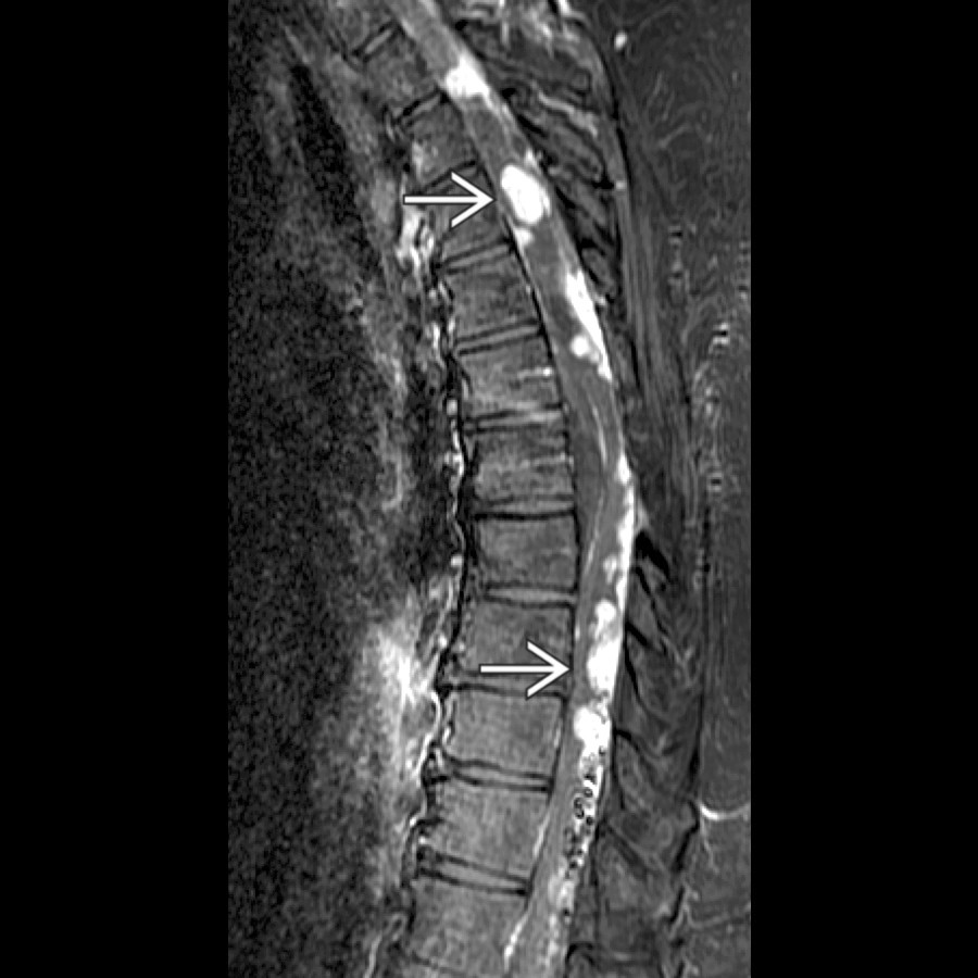
**Hemangioblastoma, Spinal Cord**
*Sagittal T1 C+ FS MR shows multiple enhancing masses along and within the thoracic cord in this patient with von Hippel-Lindau.*

**Intramedullary Arteriovenous Malformation**
*Sagittal T2 MR shows multiple prominent round flow voids from nidal aneurysms and venous varices in this patient with a large conus-type arteriovenous malformation (AVM).*

**Intramedullary Arteriovenous Malformation**
*Sagittal T1 MR shows a small, nonspecific focus of low signal intensity within the cervical cord at the C2-C3 level in this patient with a type II intramedullary AVM.*

**Intramedullary Arteriovenous Malformation**
*Sagittal T2 MR in this patient with a type II AVM shows the site of the nidus as well as linear areas of hemosiderin deposition with the cord from prior hemorrhage.*

**Infarction, Spinal Cord**
*Sagittal T2WI FSE MR shows ill-defined hyperintensity and mild enlargement of the conus medullaris in this patient with lower extremity paralysis following aortic aneurysm repair.*

**Ependymoma, Spinal Cord**
*Sagittal T2 MR shows heterogenous expansion of the cervical cord from the ependymoma nidus plus extension of hemorrhage inferiorly .*

**Syringomyelia**
*Sagittal T1 MR shows well-defined fluid signal intensity from the upper cervical cord in this patient with a syrinx and prior posterior fossa decompression for Chiari 1 malformation.*

**Contusion-Hematoma, Spinal Cord**
*Sagittal T2 MR shows a severe flexion injury at C6-C7 with a traumatic disc herniation and extensive contusion and hemorrhage within the cord . Note the edema extending superiorly and inferiorly from the injury site .*

**Hemangioblastoma, Spinal Cord**
*Sagittal T1 C+ FS MR shows multiple enhancing masses along and within the thoracic cord in this patient with von Hippel-Lindau.*

**Intramedullary Arteriovenous Malformation**
*Sagittal T2 MR shows multiple prominent round flow voids from nidal aneurysms and venous varices in this patient with a large conus-type arteriovenous malformation (AVM).*

**Intramedullary Arteriovenous Malformation**
*Sagittal T1 MR shows a small, nonspecific focus of low signal intensity within the cervical cord at the C2-C3 level in this patient with a type II intramedullary AVM.*

**Intramedullary Arteriovenous Malformation**
*Sagittal T2 MR in this patient with a type II AVM shows the site of the nidus as well as linear areas of hemosiderin deposition with the cord from prior hemorrhage.*

**Infarction, Spinal Cord**
*Sagittal T2WI FSE MR shows ill-defined hyperintensity and mild enlargement of the conus medullaris in this patient with lower extremity paralysis following aortic aneurysm repair.*
 with a classic heterogeneous hyperintense (popcorn) lesion surrounded by a hypointense hemosiderin rim.](images/app.statdx.com_image_thumbnail_4747283a-0449-4d70-9e2b-b75d3bee038f_annotated_true_size_900_quality_90_828d0b51.jpg)
**Cavernous Malformation, Spinal Cord**
*Sagittal T2 MR shows slightly expansile intramedullary lesion at the C2-C3 level with a classic heterogeneous hyperintense (popcorn) lesion surrounded by a hypointense hemosiderin rim.*

**Metastases, Spinal Cord**
*Sagittal T2WI STIR MR shows nonspecific expansion and hyperintensity within the cervical cord in this patient with metastatic disease.*

**Metastases, Spinal Cord**
*Sagittal T1 C+ MR shows a focal mass enhancing within the cervical cord in this patient with cord metastases .*

**Lymphoma**
*Sagittal T1 C+ MR shows nonspecific enlargement and enhancement of the cervical cord. The differential would include tumors (astrocytoma) and granulomatous disease. This patient has path-proven lymphoma.*

**Sarcoidosis**
*Sagittal T1 C+ MR shows an ovoid focus of irregular enhancement within the cervical cord at the C6 and C7 levels . This lesion was eventually diagnosed as sarcoidosis.*
### Additional Images

**Demyelinating Disease**
*Sagittal T1 C+ MR shows 3 foci of pathologic enhancement due to acute disseminated encephalomyelitis within the cervical cord in this patient with an acute illness following viral prodrome.*

**Cavernous Malformation, Spinal Cord**
*Sagittal T2 MR shows slightly expansile intramedullary lesion at the C2-C3 level with a classic heterogeneous hyperintense (popcorn) lesion surrounded by a hypointense hemosiderin rim.*

**Metastases, Spinal Cord**
*Sagittal T2WI STIR MR shows nonspecific expansion and hyperintensity within the cervical cord in this patient with metastatic disease.*

**Metastases, Spinal Cord**
*Sagittal T1 C+ MR shows a focal mass enhancing within the cervical cord in this patient with cord metastases .*

**Lymphoma**
*Sagittal T1 C+ MR shows nonspecific enlargement and enhancement of the cervical cord. The differential would include tumors (astrocytoma) and granulomatous disease. This patient has path-proven lymphoma.*

**Sarcoidosis**
*Sagittal T1 C+ MR shows an ovoid focus of irregular enhancement within the cervical cord at the C6 and C7 levels . This lesion was eventually diagnosed as sarcoidosis.*
### Additional Images

**Demyelinating Disease**
*Sagittal T1 C+ MR shows 3 foci of pathologic enhancement due to acute disseminated encephalomyelitis within the cervical cord in this patient with an acute illness following viral prodrome.*
 and mild enlargement of cervical cord due to multiple sclerosis (Devic subtype).](images/app.statdx.com_image_thumbnail_fc7d21f5-0197-4c0e-912d-7285454918a5_annotated_true_size_900_quality_90_041266dc.jpg)
**Demyelinating Disease**
*Sagittal T1 C+ MR shows patchy enhancement and mild enlargement of cervical cord due to multiple sclerosis (Devic subtype).*

**Ependymoma, Spinal Cord**
*Sagittal T1 C+ MR shows the typical appearance of ependymoma. Note a circumscribed, enhancing mass in the central cord with tumor syrinx.*

**Ependymoma, Spinal Cord**
*Sagittal T1 C+ MR shows an enhancing intraaxial mass with rostral and caudal syrinx and a satellite lesion enlarging the cervical cord. Note severe syringobulbia .*
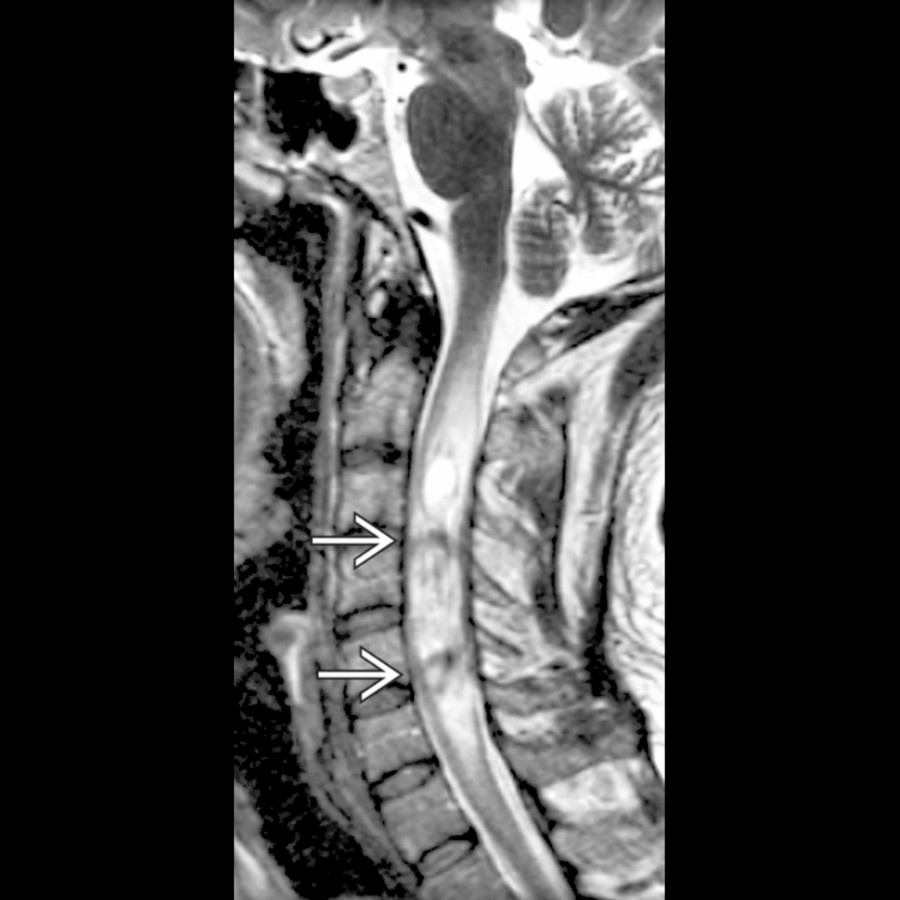
**Ependymoma, Spinal Cord**
*Sagittal T2 MR shows a complex cervical cord mass containing foci of susceptibility , demonstrating prior hemorrhage within an ependymoma.*

**Astrocytoma, Spinal Cord**
*Sagittal T2 MR shows an infiltrative lesion expanding the midcervical cord. C3-C5 decompressive laminectomy has been performed.*

**Astrocytoma, Spinal Cord**
*Sagittal T1 C+ MR shows an astrocytoma enlarging the cervical cord with patchy enhancement and infiltrative margins.*

**Syringomyelia**
*Sagittal T2 MR shows haustrated CSF signal mass in the central cord in a patient with a Chiari 1 malformation. Hypointense foci are due to flow artifact.*

**Hemangioblastoma, Spinal Cord**
*Sagittal T1 C+ MR shows holocord syrinx and 2 small dorsal enhancing lesions in this patient with multiple hemangioblastomas.*

**Hemangioblastoma, Spinal Cord**
*Sagittal T2 MR shows a nodular mass dorsally at the C4 level with a peripheral cyst . Syrinx enlarges the cord. The ring of hypointensity suggests sequelae of hemorrhage.*

**Intramedullary Arteriovenous Malformation**
*Sagittal T2 MR shows patchy hyperintensity within the cord due to intramedullary AVM. Note dorsal flow voids due to tortuous, dilatated vascularity .*

**Metastases, Spinal Cord**
*Sagittal T1 C+ FS MR shows focal intramedullary breast carcinoma metastasis enlarging the distal cord . Note vertebral metastasis with a mild compression fracture.*

**Sarcoidosis**
*Sagittal T1 C+ MR demonstrates extensive cord enlargement and intramedullary enhancement due to sarcoidosis.*

**Cysticercosis**
*Sagittal T2WI MR shows cystic thoracic cord mass in a patient with cysticercosis.*

**Lipoma, Spinal**
*Sagittal T1 MR shows a fusiform mass contacting the dorsal pial surface of the cord. The mass was isointense to subcutaneous fat on all pulse sequences.*

**Glioblastoma, Spinal Cord**
*Sagittal T1 C+ MR shows a large enhancing mass involving the upper thoracic cord with fusiform cord expansion.*
and mild enlargement of cervical cord due to multiple sclerosis (Devic subtype).](images/app.statdx.com_image_thumbnail_fc7d21f5-0197-4c0e-912d-7285454918a5_annotated_true_size_900_quality_90_041266dc.jpg)
**Demyelinating Disease**
*Sagittal T1 C+ MR shows patchy enhancement and mild enlargement of cervical cord due to multiple sclerosis (Devic subtype).*

**Ependymoma, Spinal Cord**
*Sagittal T1 C+ MR shows the typical appearance of ependymoma. Note a circumscribed, enhancing mass in the central cord with tumor syrinx.*

**Ependymoma, Spinal Cord**
*Sagittal T1 C+ MR shows an enhancing intraaxial mass with rostral and caudal syrinx and a satellite lesion enlarging the cervical cord. Note severe syringobulbia .*

**Ependymoma, Spinal Cord**
*Sagittal T2 MR shows a complex cervical cord mass containing foci of susceptibility , demonstrating prior hemorrhage within an ependymoma.*

**Astrocytoma, Spinal Cord**
*Sagittal T2 MR shows an infiltrative lesion expanding the midcervical cord. C3-C5 decompressive laminectomy has been performed.*

**Astrocytoma, Spinal Cord**
*Sagittal T1 C+ MR shows an astrocytoma enlarging the cervical cord with patchy enhancement and infiltrative margins.*

**Syringomyelia**
*Sagittal T2 MR shows haustrated CSF signal mass in the central cord in a patient with a Chiari 1 malformation. Hypointense foci are due to flow artifact.*

**Hemangioblastoma, Spinal Cord**
*Sagittal T1 C+ MR shows holocord syrinx and 2 small dorsal enhancing lesions in this patient with multiple hemangioblastomas.*

**Hemangioblastoma, Spinal Cord**
*Sagittal T2 MR shows a nodular mass dorsally at the C4 level with a peripheral cyst . Syrinx enlarges the cord. The ring of hypointensity suggests sequelae of hemorrhage.*

**Intramedullary Arteriovenous Malformation**
*Sagittal T2 MR shows patchy hyperintensity within the cord due to intramedullary AVM. Note dorsal flow voids due to tortuous, dilatated vascularity .*

**Metastases, Spinal Cord**
*Sagittal T1 C+ FS MR shows focal intramedullary breast carcinoma metastasis enlarging the distal cord . Note vertebral metastasis with a mild compression fracture.*

**Sarcoidosis**
*Sagittal T1 C+ MR demonstrates extensive cord enlargement and intramedullary enhancement due to sarcoidosis.*

**Cysticercosis**
*Sagittal T2WI MR shows cystic thoracic cord mass in a patient with cysticercosis.*

**Lipoma, Spinal**
*Sagittal T1 MR shows a fusiform mass contacting the dorsal pial surface of the cord. The mass was isointense to subcutaneous fat on all pulse sequences.*

**Glioblastoma, Spinal Cord**
*Sagittal T1 C+ MR shows a large enhancing mass involving the upper thoracic cord with fusiform cord expansion.*