---
title: "Left Ventricular Enlargement"
docid: "fbb972de-3e13-4c67-b7a4-f8901aa2efb8"
authors:
- key: "df804626-c042-4296-96e3-836a6da50fd6"
value: "Gregory Kicska, MD, PhD"
breadcrumbs:
-
name: "Cardiac"
slug: "cardiac"
treeNodeId: "fa90100b-619c-430e-8074-b5b9789bab39"
-
name: "Differential Diagnosis"
slug: "differential-diagnosis"
treeNodeId: "952326a0-b3ea-4a21-aa7a-d796cc9325ed"
-
name: "Left Ventricular Enlargement"
slug: "left-ventricular-enlargement"
treeNodeId: null
category: "Cardiac"
documentVersionId: "e8a63b4a-914d-475a-8dd1-cce5feaf7fec"
imageCount: 15
lastUpdated: "03/17/22"
pageDescription: "Left Ventricular Enlargement"
pageKeywords: "Cardiac, Differential Diagnosis, Left Ventricular Enlargement"
pageTitle: "Left Ventricular Enlargement | STATdx"
enhancedTitle: "Left Ventricular Enlargement"
type: "DDX"
breadcrumbs:
- "Cardiac"
- "Differential Diagnosis"
- "Left Ventricular Enlargement"
---
# ESSENTIAL INFORMATION
- ## Key Differential Diagnosis Issues
- Determination of LV chamber enlargement
- Radiographic
- Normal cardiothoracic ratio ≤ 0.5 on PA and ≤ 0.6 on AP at deep inspiration and proper positioning
- Expiratory and lordotic or rotated projections can change appearance of cardiac silhouette shape and size
- Leftward and downward displacement of left heart border
- LV extending 2 cm posterior to IVC border (Hoffman-Rigler sign) on lateral view
- Cross sectional
- LV volume may be measured qualitatively, not quantitatively, when only axial planes are available
- Reliable measurements require double oblique planes, usually short axis, and knowledge of phase within cardiac cycle
- Normal internal LV diameter at base is 3.9-5.3 cm for female and 4.2-5.9 cm for male patients
- 2-dimensional Simpson rule of discs in short axis or 3D auto-segmented are most reproducible
- Less reliable: Biplane method of Simpson rule and area length rule
- End-diastolic volume (EDV) > 170 mL in female and > 200 mL in male patients is indicative for enlargement
- EDV normalized by body surface area (EDV/BSA) are 2 standard deviations above mean if > 100 mL/m² in male and above 95 mL/m² in female patients
- Determination of LV wall thickness
- End-diastolic radial LV wall thickness > 1.2 cm is abnormal
- LV mass > 104 gm/m² in female or 119 gm/m² in male patients is specific for pathology
- Pitfalls
- Radiographic LV enlargement may be mimicked by pericardial effusion, expiration, poor lateral positioning or projection angle, or pericardial fat pad
- Misidentification of end diastole most frequent cause of erroneous left ventricular size measurement
- Cardiac volume may be affected by preimaging administration of β blockers or nitroglycerin
- ## Helpful Clues for Common Diagnoses
- **Heart Failure**
- Ischemic cardiomyopathy most common etiology, followed by diabetes and hypertension
- EF < 40%
- Multivessel coronary artery calcifications or stenosis
- Evidence of prior infarct, subendocardial fat
- If retrospective gated CT or MR performed, myocardium can be evaluated for evidence of hibernation
- Subendocardial or transmural delayed enhancement present in coronary artery distribution indicates ischemia
- If delayed enhancement excludes subendocardial layer, nonischemic etiologies should be considered
- **Aortic Regurgitation**
- Bicuspid valve or calcified aortic valve
- Incomplete coaptation of cusps during diastole
- Regurgitant jet present on bright-blood MR
- **Mitral Regurgitation**
- Mitral valve calcifications
- Dilated left atrium
- Isolated right upper lobe edema is rare manifestation resulting from regurgitant jet
- **Acute M****yocardial Infarction**
- Enlarged cardiac silhouette compared to recent prior
- Supporting clinical information, troponin leak, ECG changes, or typical chest pain
- ## Helpful Clues for Less Common Diagnoses
- **Patent Ductus Arteriosus**
- Initially, enlarged main pulmonary arteries; later, LV, LA, and ascending aortic enlargement
- LV enlargement with dilated ascending aorta in absence of valvular disease
- Best seen in gated CT or 3D MRA
- MR Qp:Qs ratio < 1:1
- **Coarctation of Aorta**
- Associated with bicuspid valve
- Hemodynamic narrowing represented by dilated intercostal collaterals
- Not to be confused with pseudocoarctation (tortuous arch without hemodynamic narrowing)
- Undiagnosed cases in adults often occur when narrowing distal to left subclavian take-off
- **Idiopathic Dilated Cardiomyopathy**
- Patients often < 60 years of age
- Diagnosis of exclusion
- Significant coronary artery occlusion or myocarditis to be excluded
- MR delayed enhancement present in ~ 40% of cases, most commonly mid-myocardial
- EF < 40% &/or fractional shortening < 25%
- **Hypertrophic Cardiomyopathy**
- LVOT view shows MR with systolic anterior motion of mitral valve leaflet
- Asymmetric septal, apical, and concentric variants exist
- In concentric variant, differential includes hypertensive heart disease/aortic stenosis, amyloidosis, and sarcoidosis
- Patchy mid myocardial enhancement in areas of LV thickening and RV insertion into LV
- **Amyloidosis**
- Patients typically > 65 years of age
- Increased LV wall thickness with poor or normal contractility
- Diffuse subendocardial perfusion defect
- Delayed enhancement inversion recovery sequences show equal relaxation times between blood pool and myocardium
- ## Helpful Clues for Rare Diagnoses
- **Athlete's Heart**
- Occurs in athletes who engage in prolonged aerobic activity
- End-diastolic wall thickness > 15 mm in young patient with dilated heart can be seen in athlete's heart
- LV volume will decrease following 3 months of deconditioning
- **Pregnancy-Induced Dilated Cardiomyopathy**
- Postpartum LV enlargement and hypokinesis
- Follow-up imaging in 3 months may show resolution
- **Alcohol-Induced Dilated Cardiomyopathy**
- Accompanying clinical history
- Follow-up imaging will show resolution if acute
## Images
### Selected Images
 , predominantly in an LAD distribution, representing prior infract.](images/app.statdx.com_image_thumbnail_92b7e257-4046-4d73-87aa-cae128d40108_annotated_true_size_900_quality_90_6011e427919c7c8306d0acaf4f4dbcfc4127f03e.jpg)
**Heart Failure**
*Coronal oblique NECT of ischemic heart failure shows LV enlargement with subepicardial fat , predominantly in an LAD distribution, representing prior infract.*
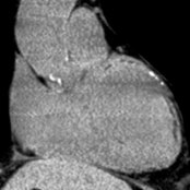
**Heart Failure**
*Coronal oblique NECT of ischemic heart failure shows LV enlargement with subepicardial fat , predominantly in an LAD distribution, representing prior infract.*

**Heart Failure**
*Short-axis inversion recovery MR through the LV mid-chamber shows dilated LV with late enhancement in a LAD distribution , compatible with ischemic cardiomyopathy.*

**Heart Failure**
*Four-chamber bright-blood MR in a patient with history of long, uncontrolled, standing hypertension shows a mildly dilated LV with diffuse wall thickening. This will eventually progress to an appearance indistinguishable from other dilated CM.*

**Heart Failure**
*Diastolic phase LVOT CECT shows markedly dilated LV without aortic valve disease. This patient had depressed EF and densely calcified coronary arteries, indicating ischemic cardiomyopathy.*

**Aortic Regurgitation**
*Coronal cine MR shows a turbulent jet originating at the aortic valve, directed toward the LV chamber .*

**Heart Failure**
*Coronal oblique NECT of ischemic heart failure shows LV enlargement with subepicardial fat , predominantly in an LAD distribution, representing prior infract.*

**Heart Failure**
*Coronal oblique NECT of ischemic heart failure shows LV enlargement with subepicardial fat , predominantly in an LAD distribution, representing prior infract.*

**Heart Failure**
*Short-axis inversion recovery MR through the LV mid-chamber shows dilated LV with late enhancement in a LAD distribution , compatible with ischemic cardiomyopathy.*

**Heart Failure**
*Four-chamber bright-blood MR in a patient with history of long, uncontrolled, standing hypertension shows a mildly dilated LV with diffuse wall thickening. This will eventually progress to an appearance indistinguishable from other dilated CM.*

**Heart Failure**
*Diastolic phase LVOT CECT shows markedly dilated LV without aortic valve disease. This patient had depressed EF and densely calcified coronary arteries, indicating ischemic cardiomyopathy.*

**Aortic Regurgitation**
*Coronal cine MR shows a turbulent jet originating at the aortic valve, directed toward the LV chamber .*
 due to mitral valve prolapse. The prolapsing leaflet is seen with a regurgitant jet directed at the septum.](images/app.statdx.com_image_thumbnail_fd3f4640-e4a3-4dbb-9f5c-7d70d022920d_annotated_true_size_900_quality_90_8b79474aeba8f8436eca1b315c24a31e1891eef7.jpg)
**Mitral Regurgitation**
*Systolic phase LVOT cine MR of mitral regurgitation shows low signal corresponding to regurgitation due to mitral valve prolapse. The prolapsing leaflet is seen with a regurgitant jet directed at the septum.*
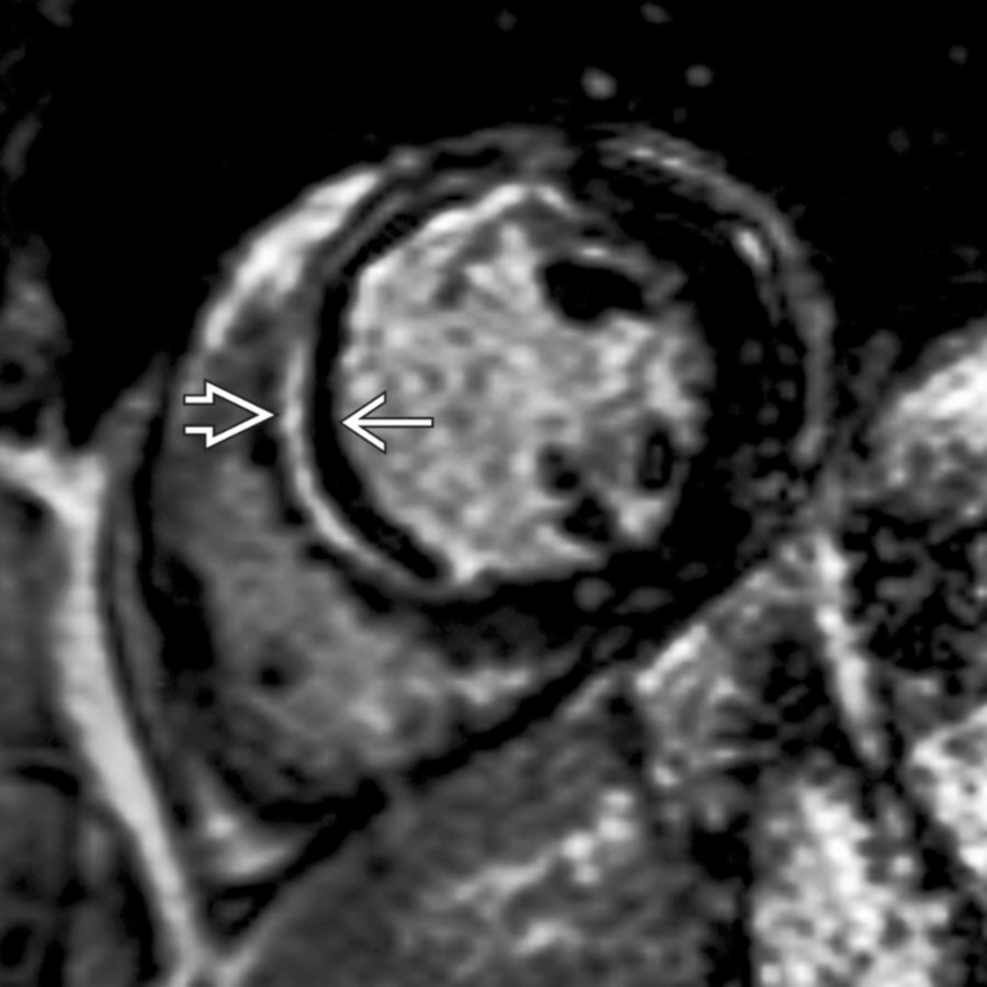
**Acute Myocardial Infarction**
*Short-axis inversion recovery FSE MR through the LV mid-chamber shows mid-myocardial LAD distribution late enhancement . Hypointense subendocardium indicates acute MI associated microvascular obstruction .*

**Patent Ductus Arteriosus**
*Four-chamber CTA shows dilation of the left atrium and left ventricle from chronic volume overload due to left to right shunting across the patent ductus arteriosus (not shown).*

**Patent Ductus Arteriosus**
*Axial oblique CTA shows a connection between the proximal descending aorta and the pulmonary artery, diagnostic of a patent ductus arteriosus. Left-to-right shunt resulted in LV enlargement.*

**Coarctation of Aorta**
*Sagittal T1 C+ FS MR shows focal narrowing distal to the left subclavian take-off . Presence of intercostal collaterals and LV enlargement indicated a hemodynamically significant stenosis, differentiating it from pseudocoarctation.*

**Dilated Cardiomyopathy**
*Axial NECT in a 41-year-old man with symptoms of heart failure shows LV dilation without CAD. Cardiomyopathy etiology was not found, and a diagnosis of idiopathic dilated cardiomyopathy was made.*

**Dilated Cardiomyopathy**
*Short-axis inversion recovery FSE MR shows septal mid-myocardial enhancement in a patient with dilated cardiomyopathy .*

**Hypertrophic Cardiomyopathy**
*Diastolic phase LVOT bright-blood cine MR of asymmetric variant hypertrophic cardiomyopathy shows asymmetric thickening of interventricular septum at base . Study should be interrogated for fibrosis and SAM.*

**Amyloidosis**
*Short-axis inversion recovery FSE MR through LV mid-chamber 10 minutes post contrast shows near-equal relaxation of blood pool and myocardium. This finding is caused by altered contrast concentration kinetics due to presence of amyloid protein.*
### Additional Images

**Aortic Regurgitation**
*Diastolic phase LVOT bright-blood cine MR of aortic regurgitation shows a turbulent jet originating at the aortic valve, directed toward the LV chamber .*
due to mitral valve prolapse. The prolapsing leaflet is seen with a regurgitant jet directed at the septum.](images/app.statdx.com_image_thumbnail_fd3f4640-e4a3-4dbb-9f5c-7d70d022920d_annotated_true_size_900_quality_90_8b79474aeba8f8436eca1b315c24a31e1891eef7.jpg)
**Mitral Regurgitation**
*Systolic phase LVOT cine MR of mitral regurgitation shows low signal corresponding to regurgitation due to mitral valve prolapse. The prolapsing leaflet is seen with a regurgitant jet directed at the septum.*

**Acute Myocardial Infarction**
*Short-axis inversion recovery FSE MR through the LV mid-chamber shows mid-myocardial LAD distribution late enhancement . Hypointense subendocardium indicates acute MI associated microvascular obstruction .*

**Patent Ductus Arteriosus**
*Four-chamber CTA shows dilation of the left atrium and left ventricle from chronic volume overload due to left to right shunting across the patent ductus arteriosus (not shown).*

**Patent Ductus Arteriosus**
*Axial oblique CTA shows a connection between the proximal descending aorta and the pulmonary artery, diagnostic of a patent ductus arteriosus. Left-to-right shunt resulted in LV enlargement.*

**Coarctation of Aorta**
*Sagittal T1 C+ FS MR shows focal narrowing distal to the left subclavian take-off . Presence of intercostal collaterals and LV enlargement indicated a hemodynamically significant stenosis, differentiating it from pseudocoarctation.*

**Dilated Cardiomyopathy**
*Axial NECT in a 41-year-old man with symptoms of heart failure shows LV dilation without CAD. Cardiomyopathy etiology was not found, and a diagnosis of idiopathic dilated cardiomyopathy was made.*

**Dilated Cardiomyopathy**
*Short-axis inversion recovery FSE MR shows septal mid-myocardial enhancement in a patient with dilated cardiomyopathy .*

**Hypertrophic Cardiomyopathy**
*Diastolic phase LVOT bright-blood cine MR of asymmetric variant hypertrophic cardiomyopathy shows asymmetric thickening of interventricular septum at base . Study should be interrogated for fibrosis and SAM.*

**Amyloidosis**
*Short-axis inversion recovery FSE MR through LV mid-chamber 10 minutes post contrast shows near-equal relaxation of blood pool and myocardium. This finding is caused by altered contrast concentration kinetics due to presence of amyloid protein.*
### Additional Images

**Aortic Regurgitation**
*Diastolic phase LVOT bright-blood cine MR of aortic regurgitation shows a turbulent jet originating at the aortic valve, directed toward the LV chamber .*