---
title: "Mass in Iliopsoas Compartment"
docid: "e39c8918-27cb-4dd6-9166-2b265b777a35"
authors:
- key: "c1df94ab-4a9f-44c4-add7-1f174fb9ac45"
value: "Siva P. Raman, MD"
breadcrumbs:
-
name: "Gastrointestinal"
slug: "gastrointestinal"
treeNodeId: "b52263f7-5978-4a22-a17d-7260e0033943"
-
name: "Differential Diagnosis"
slug: "differential-diagnosis"
treeNodeId: "a0fd80ff-6231-49d3-94b8-ea083449979d"
-
name: "Abdominal Wall"
slug: "abdominal-wall"
treeNodeId: "08db01f7-2961-47f7-954d-2a5fca7e707d"
-
name: "Anatomically Based Differentials"
slug: "anatomically-based-differentials"
treeNodeId: "1525b44f-9d47-4ff4-8330-693211bd5eb5"
-
name: "Mass in Iliopsoas Compartment"
slug: "mass-in-iliopsoas-compartment"
treeNodeId: null
category: "Gastrointestinal"
documentVersionId: "b98553dd-1557-4132-99d5-3304036dd914"
imageCount: 26
lastUpdated: "07/01/22"
pageDescription: "Mass in Iliopsoas Compartment"
pageKeywords: "Gastrointestinal, Differential Diagnosis, Abdominal Wall, Anatomically Based Differentials, Mass in Iliopsoas Compartment"
pageTitle: "Mass in Iliopsoas Compartment | STATdx"
enhancedTitle: "Mass in Iliopsoas Compartment"
type: "DDX"
references: true
breadcrumbs:
- "Gastrointestinal"
- "Differential Diagnosis"
- "Abdominal Wall"
- "Anatomically Based Differentials"
- "Mass in Iliopsoas Compartment"
---
# ESSENTIAL INFORMATION
- ## Key Differential Diagnosis Issues
- Iliopsoas compartment masses are much more likely to result from infection or hemorrhage than from tumor
- Iliopsoas pathology often due to spread from adjacent infection (such as spine) or adjacent tumor, rather than originating within iliopsoas itself
- ## Helpful Clues for Common Diagnoses
- **Iliopsoas****Hematoma**
- Iliopsoas is most common site for spontaneous retroperitoneal hemorrhage (usually due to bleeding diathesis or anticoagulation)
- Presence of multiple hematocrit levels within hematoma suggests coagulopathic hemorrhage
- Other causes include surgery, trauma, or extension of bleeding from adjacent structures (e.g., bleeding renal AML, ruptured abdominal aortic aneurysm, etc.)
- Appearance variable depending on age of hematoma
- Acute bleeding may simply appear as homogeneous enlargement of muscle ± hematocrit levels
- Chronic bleeds may appear hypodense and can be difficult to differentiate from abscess
- **Asymmetric Musculature (Mimic)**
- Iliopsoas muscles can be asymmetric in size, particularly in patients with unilateral leg amputation, paralysis, or lower extremity/spine arthritis
- **Secondary Infection/Abscess**
- Infection and abscess formation in iliopsoas typically due to spread of infection from contiguous structures
- Most commonly infectious spread from bone, kidney, and bowel (including appendix)
- Paraspinal psoas abscess should prompt careful search for infectious spondylitis (TB or pyogenic)
- Renal sources of iliopsoas infection include renal/perirenal abscess or xanthogranulomatous pyelonephritis
- Common bowel sources of iliopsoas infection include appendicitis, diverticulitis, or Crohn disease
- Often associated with other features of infection (fat stranding, blurring of fat planes, ectopic gas)
- ## Helpful Clues for Less Common Diagnoses
- **Retroperitoneal Fibrosis**
- Irregular soft tissue mass enveloping aorta, inferior vena cava, and ureters, which can involve adjacent psoas muscles
- Variable enhancement depending on stage with hyperenhancement in early stages of disease and hypoenhancement in later stages
- **Primary Infection**
- Iliopsoas compartment is rarely primary site of infection, except in immunocompromised patients (including HIV) and intravenous drug abusers
- Infection usually due to *Staphylococcus aureus*and mixed gram-negative organisms
- **Primary Neoplasm**
- Primary mesenchymal tumors (liposarcoma, fibrosarcoma, leiomyosarcoma, hemangiopericytoma, etc.) may rarely originate from iliopsoas compartment
- **Secondary Neoplasm**
- Hematogenous metastasis (e.g., lymphoma, melanoma) to iliopsoas very rare
- More commonly directly invaded by adjacent tumors (e.g., retroperitoneal sarcoma, lymphoma, neurogenic tumors, adjacent bone tumor, etc.)
- Plexiform neurofibroma (in neurofibromatosis) may involve psoas compartment
## References
# Selected References
1. [Mondie C et al: Retroperitoneal hematoma. StatPearls, 2021](http://www.ncbi.nlm.nih.gov/pubmed/?term=32644354%5Bpmid%5D)
1. [Improta L et al: Overview of primary adult retroperitoneal tumours. Eur J Surg Oncol. 46(9):1573-9, 2020](http://www.ncbi.nlm.nih.gov/pubmed/?term=32600897%5Bpmid%5D)
1. [Peisen F et al: Retroperitoneal fibrosis and its differential diagnoses: the role of radiological imaging. Rofo. 192(10):929-36, 2020](http://www.ncbi.nlm.nih.gov/pubmed/?term=32698236%5Bpmid%5D)
1. [Babic M et al: Infections of the spine. Infect Dis Clin North Am. 31(2):279-97, 2017](http://www.ncbi.nlm.nih.gov/pubmed/?term=28366222%5Bpmid%5D)
1. [Messiou C et al: Primary retroperitoneal soft tissue sarcoma: imaging appearances, pitfalls and diagnostic algorithm. Eur J Surg Oncol. 43(7):1191-8, 2017](http://www.ncbi.nlm.nih.gov/pubmed/?term=28057392%5Bpmid%5D)
1. [Sunga KL et al: Spontaneous retroperitoneal hematoma: etiology, characteristics, management, and outcome. J Emerg Med. 43(2):e157-61, 2012](http://www.ncbi.nlm.nih.gov/pubmed/?term=21911282%5Bpmid%5D)
1. [Singh AK et al: Neoplastic iliopsoas masses in oncology patients: CT findings. Abdom Imaging. 33(4):493-7, 2008](http://www.ncbi.nlm.nih.gov/pubmed/?term=17639377%5Bpmid%5D)
## Images
### Selected Images
 makes the hematomas more apparent. Hematocrit levels suggest coagulopathy as the underlying cause of the bleed.](images/app.statdx.com_image_thumbnail_0559bc1a-c9cb-40c7-8a58-17aae3d5f0dc_annotated_true_size_900_quality_90_1fa00e497cced470cff0064e2360a9675761b68c.jpg)
**Iliopsoas Hematoma**
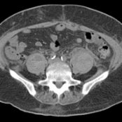
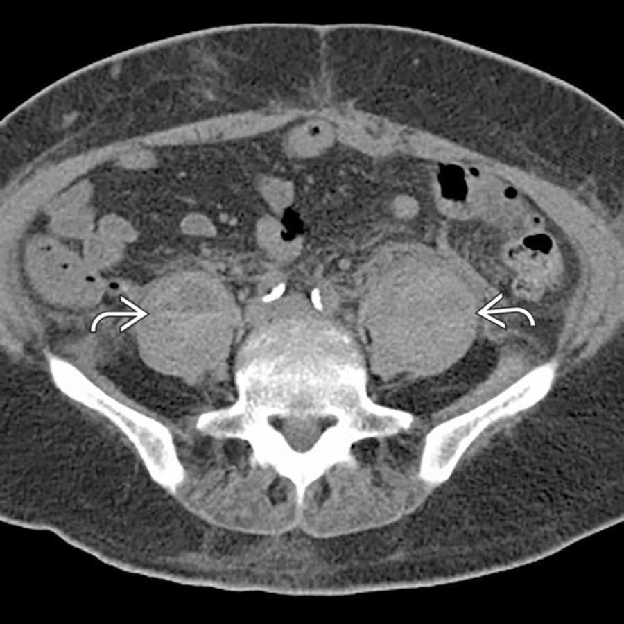
*Axial NECT shows enlargement of both psoas muscles due to coagulopathic hemorrhage. Although the hematoma is partly isodense to the muscle, the presence of hematocrit levels makes the hematomas more apparent. Hematocrit levels suggest coagulopathy as the underlying cause of the bleed.*
**Iliopsoas Hematoma**
*Axial NECT shows enlargement of both psoas muscles due to coagulopathic hemorrhage. Although the hematoma is partly isodense to the muscle, the presence of hematocrit levels makes the hematomas more apparent. Hematocrit levels suggest coagulopathy as the underlying cause of the bleed.*
**Iliopsoas Hematoma**
*Axial NECT shows enlargement of both psoas muscles due to coagulopathic hemorrhage. Although the hematoma is partly isodense to the muscle, the presence of hematocrit levels makes the hematomas more apparent. Hematocrit levels suggest coagulopathy as the underlying cause of the bleed.*

**Iliopsoas Hematoma**
*Axial NECT shows enlargement of both psoas muscles due to coagulopathic hemorrhage. Although the hematoma is partly isodense to the muscle, the presence of hematocrit levels makes the hematomas more apparent. Hematocrit levels suggest coagulopathy as the underlying cause of the bleed.*
 is atrophic due to a prior left leg amputation.](images/app.statdx.com_image_thumbnail_98fac04c-d23d-40f9-90d1-dcf5ce769092_annotated_true_size_900_quality_90_fa63483b1a8e8be37de5aaf71e418000308a86fa.jpg)
**Asymmetric Musculature (Mimic)**
*Axial CECT shows marked asymmetry of the psoas muscles. In this case, the left psoas muscle is atrophic due to a prior left leg amputation.*
**Secondary Infection/Abscess**
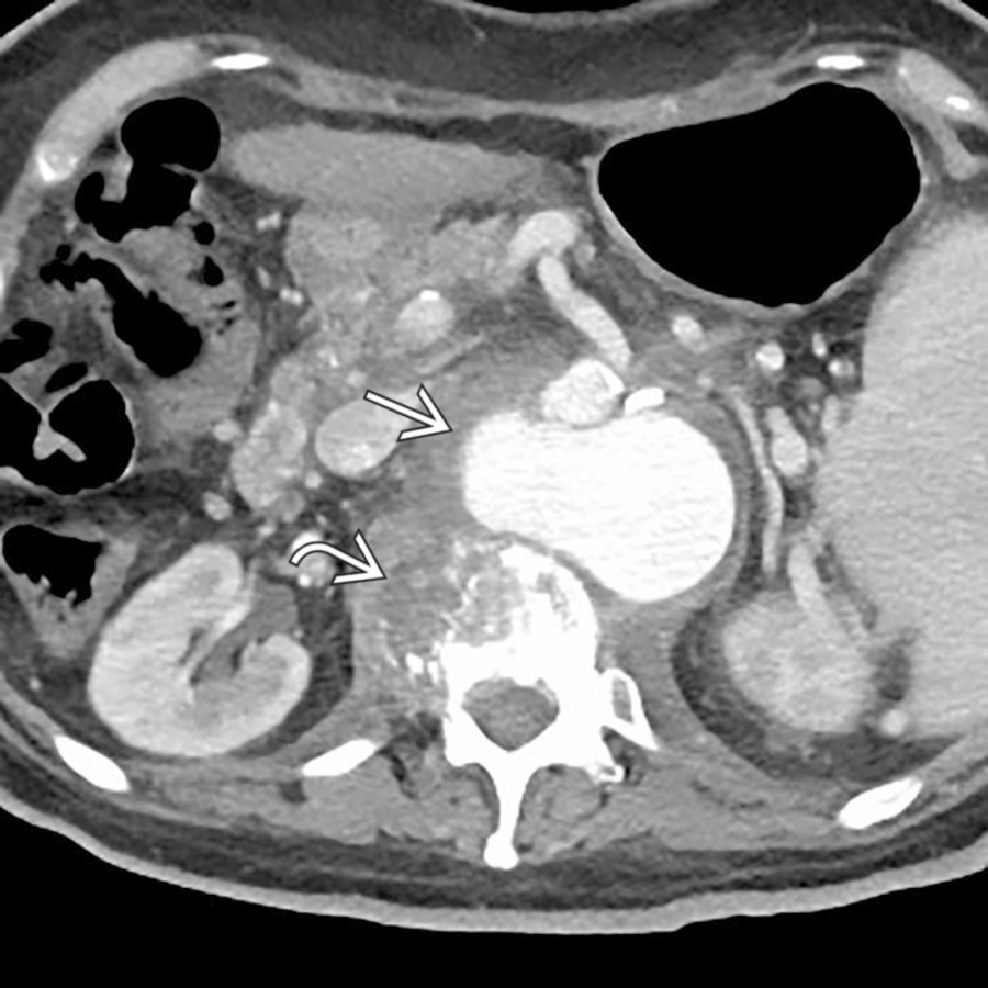
*Axial CECT shows lytic destruction of a vertebral body with an abscess extending into the right psoas muscle. Note the presence of a large mycotic aneurysm immediately anteriorly arising from the abdominal aorta.*
**Secondary Infection/Abscess**
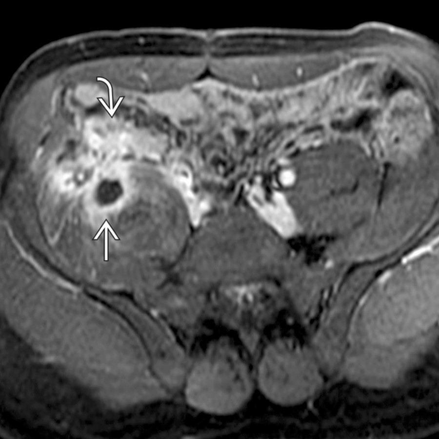
*Axial T1 C+ MR shows an abscess in the iliopsoas muscle directly contiguous with phlegmonous change in the right lower quadrant due to the patient's fistulizing Crohn disease.*
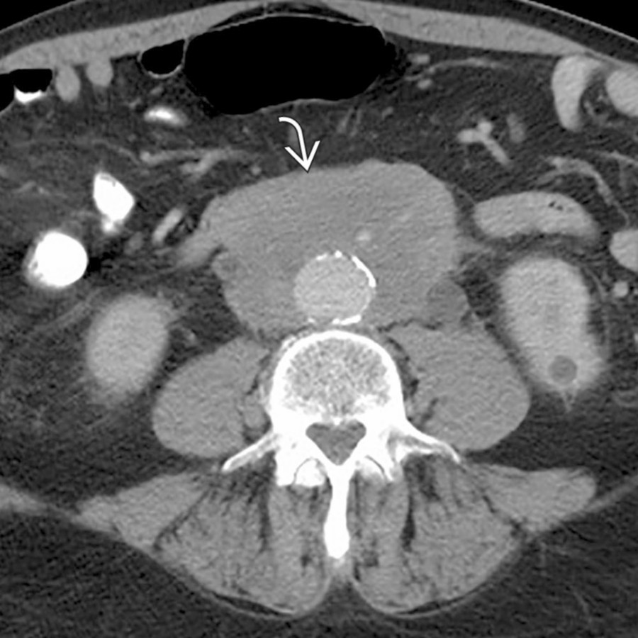
**Retroperitoneal Fibrosis**
*Axial CECT shows a rind of mass-like soft tissue encasing the aorta and inferior vena cava. While more mass-like than is typical, this was found to represent retroperitoneal fibrosis.*
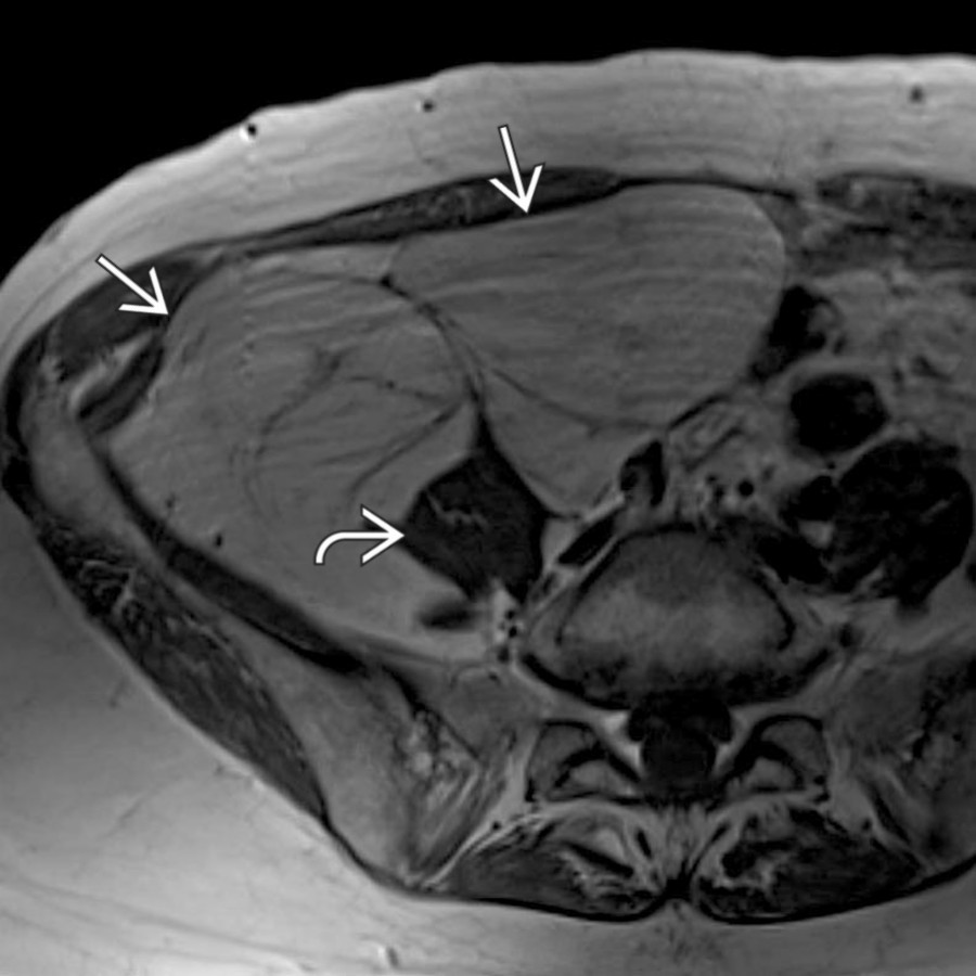
**Primary Neoplasm**
*Axial T1 MR shows a large, fat-containing mass occupying much of the right hemipelvis, with the right left psoas muscle appearing stretched and distorted as a result of the retroperitoneal tumor. This was found to be a well-differentiated liposarcoma at resection.*
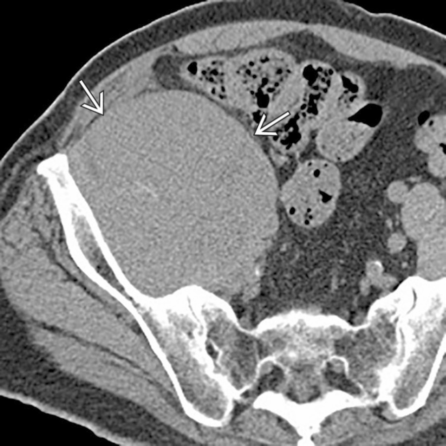
**Secondary Neoplasm**
*Axial CECT shows a large, homogeneous soft tissue mass involving the right psoas and iliopsoas musculature. There were multiple enlarged lymph nodes elsewhere (not shown), and this was found to represent non-Hodgkin lymphoma.*
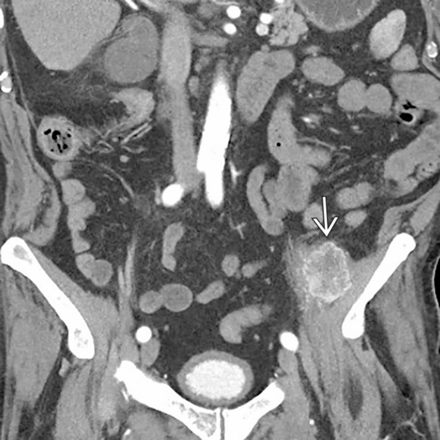
**Secondary Neoplasm**
*Coronal CECT shows an avidly enhancing mass with central necrosis arising in the left iliopsoas muscle, representing a metastasis related to the patient's known renal cell carcinoma.*
### Additional Images
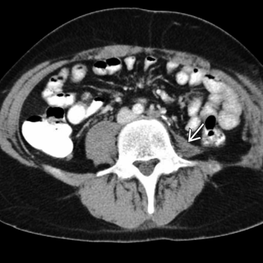
**Asymmetric Musculature (Mimic)**
*Axial CECT shows marked asymmetry of the psoas muscles. In this case, the left psoas muscle is atrophic due to a prior left leg amputation.*

**Secondary Infection/Abscess**
*Axial CECT shows lytic destruction of a vertebral body with an abscess extending into the right psoas muscle. Note the presence of a large mycotic aneurysm immediately anteriorly arising from the abdominal aorta.*

**Secondary Infection/Abscess**
*Axial T1 C+ MR shows an abscess in the iliopsoas muscle directly contiguous with phlegmonous change in the right lower quadrant due to the patient's fistulizing Crohn disease.*

**Retroperitoneal Fibrosis**
*Axial CECT shows a rind of mass-like soft tissue encasing the aorta and inferior vena cava. While more mass-like than is typical, this was found to represent retroperitoneal fibrosis.*

**Primary Neoplasm**
*Axial T1 MR shows a large, fat-containing mass occupying much of the right hemipelvis, with the right left psoas muscle appearing stretched and distorted as a result of the retroperitoneal tumor. This was found to be a well-differentiated liposarcoma at resection.*

**Secondary Neoplasm**
*Axial CECT shows a large, homogeneous soft tissue mass involving the right psoas and iliopsoas musculature. There were multiple enlarged lymph nodes elsewhere (not shown), and this was found to represent non-Hodgkin lymphoma.*

**Secondary Neoplasm**
*Coronal CECT shows an avidly enhancing mass with central necrosis arising in the left iliopsoas muscle, representing a metastasis related to the patient's known renal cell carcinoma.*
### Additional Images
 abutting the psoas compartment. Some portions of the mass have well-differentiated fatty components, characteristic of liposarcoma.](images/app.statdx.com_image_thumbnail_5a517824-72d3-427c-97fc-6a9c02d33c07_annotated_true_size_900_quality_90_f7e6e6cf4a239ec24ce706c3204e4baee97bab10.jpg)
**Primary Neoplasm**
*Axial CECT shows a huge retroperitoneal mass abutting the psoas compartment. Some portions of the mass have well-differentiated fatty components, characteristic of liposarcoma.*
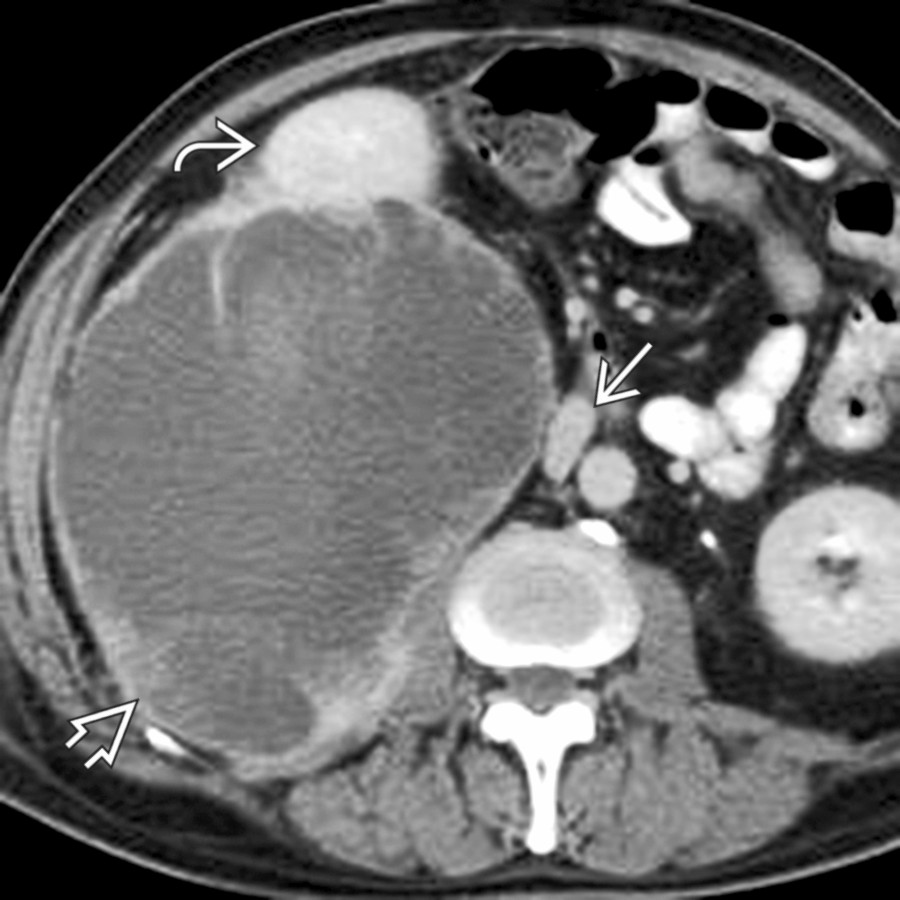
**Primary Neoplasm**
*Axial CECT shows a large, heterogeneous retroperitoneal mass , which displaces the right kidney anteriorly. Note medial displacement of the inferior vena cava (IVC) . Malignant fibrous histiocytoma.*
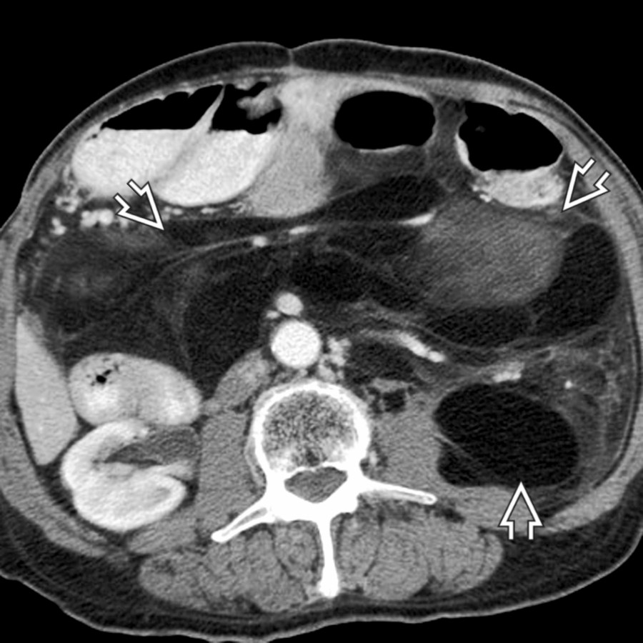
**Primary Neoplasm**
*Axial CECT shows a huge retroperitoneal mass abutting the psoas compartment. Some portions of the mass have well-differentiated fatty components, characteristic of liposarcoma.*

**Primary Neoplasm**
*Axial CECT shows a large, heterogeneous retroperitoneal mass , which displaces the right kidney anteriorly. Note medial displacement of the inferior vena cava (IVC) . Malignant fibrous histiocytoma.*
 and hematocrit sign
  , indicating coagulopathic bleeding.](images/app.statdx.com_image_thumbnail_e297411e-4578-474d-90d5-7a49fcb33643_annotated_true_size_900_quality_90_14ca7e04a6e3bb907d3520cae7476d29ea6f6264.jpg)
**Iliopsoas Hematoma**
*Axial CECT shows hemorrhage into multiple sites, including iliopsoas, with active bleeding and hematocrit sign , indicating coagulopathic bleeding.*
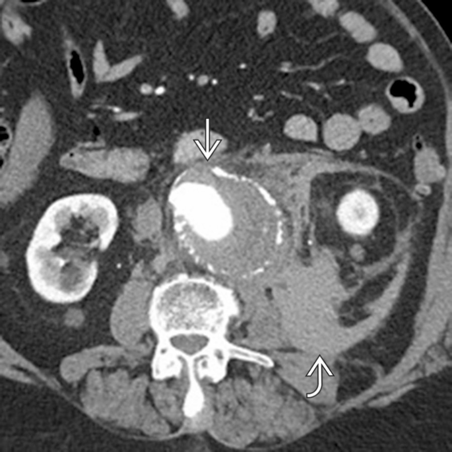
**Iliopsoas Hematoma**
*Axial CECT shows a large abdominal aortic aneurysm with extensive free hematoma tracking laterally in the retroperitoneum, including immediately adjacent to the psoas muscle, compatible with aneurysm rupture.*
**Iliopsoas Hematoma**
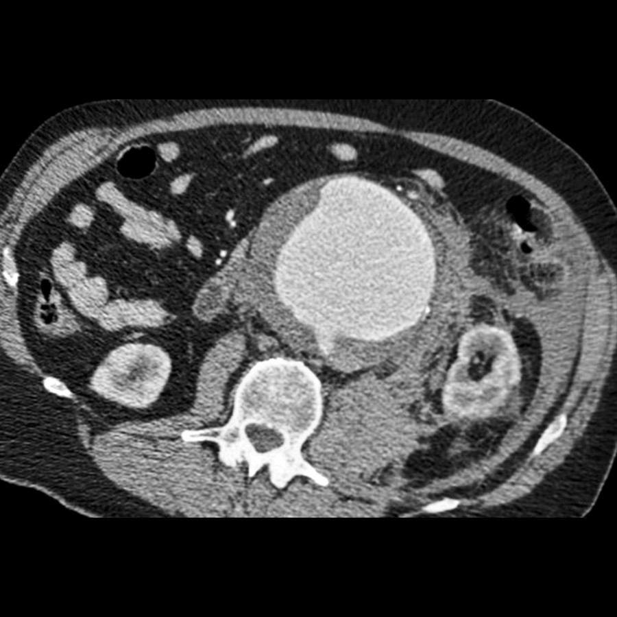
*Axial CECT shows a large abdominal aortic aneurysm that has ruptured into the psoas compartment and retroperitoneum. Blood dissects along the left psoas and throughout the retroperitoneal planes.*
**Iliopsoas Hematoma**
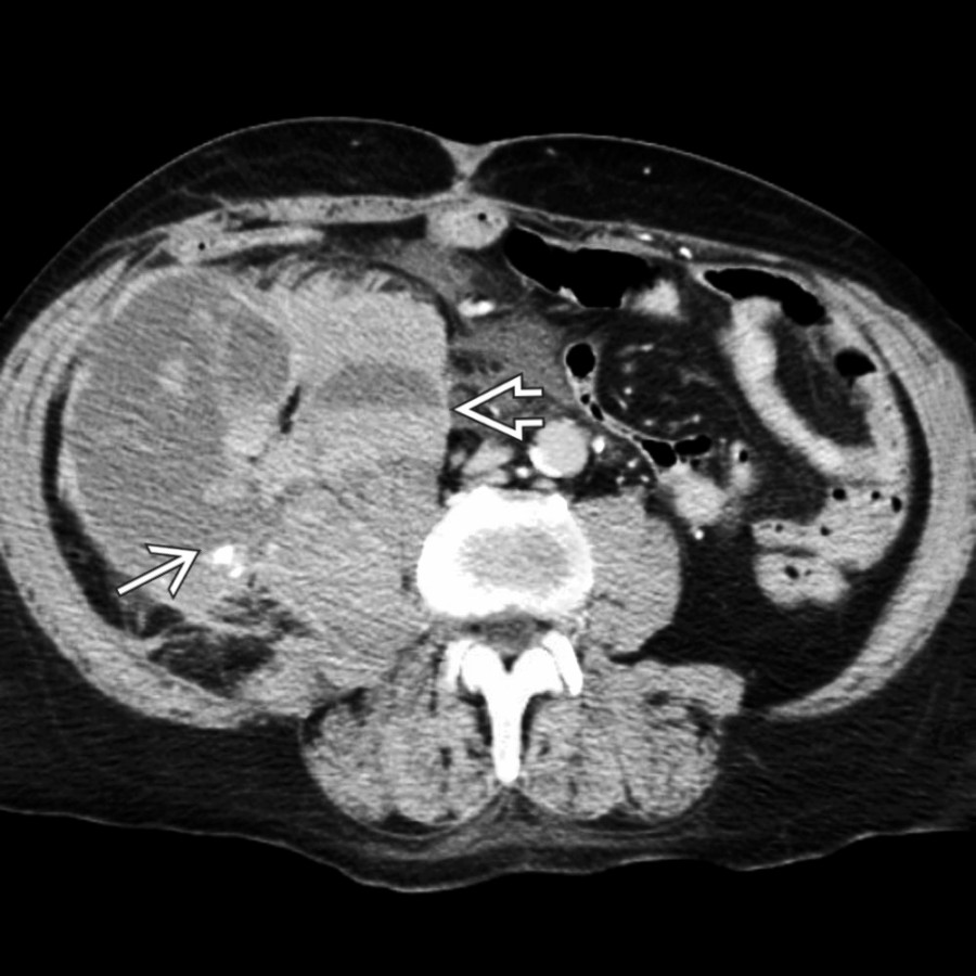
*Axial CECT shows a large retroperitoneal mass arising from the iliopsoas compartment. A focus of active bleeding and hematocrit levels confirm coagulopathic hemorrhage.*
**Retroperitoneal Fibrosis**
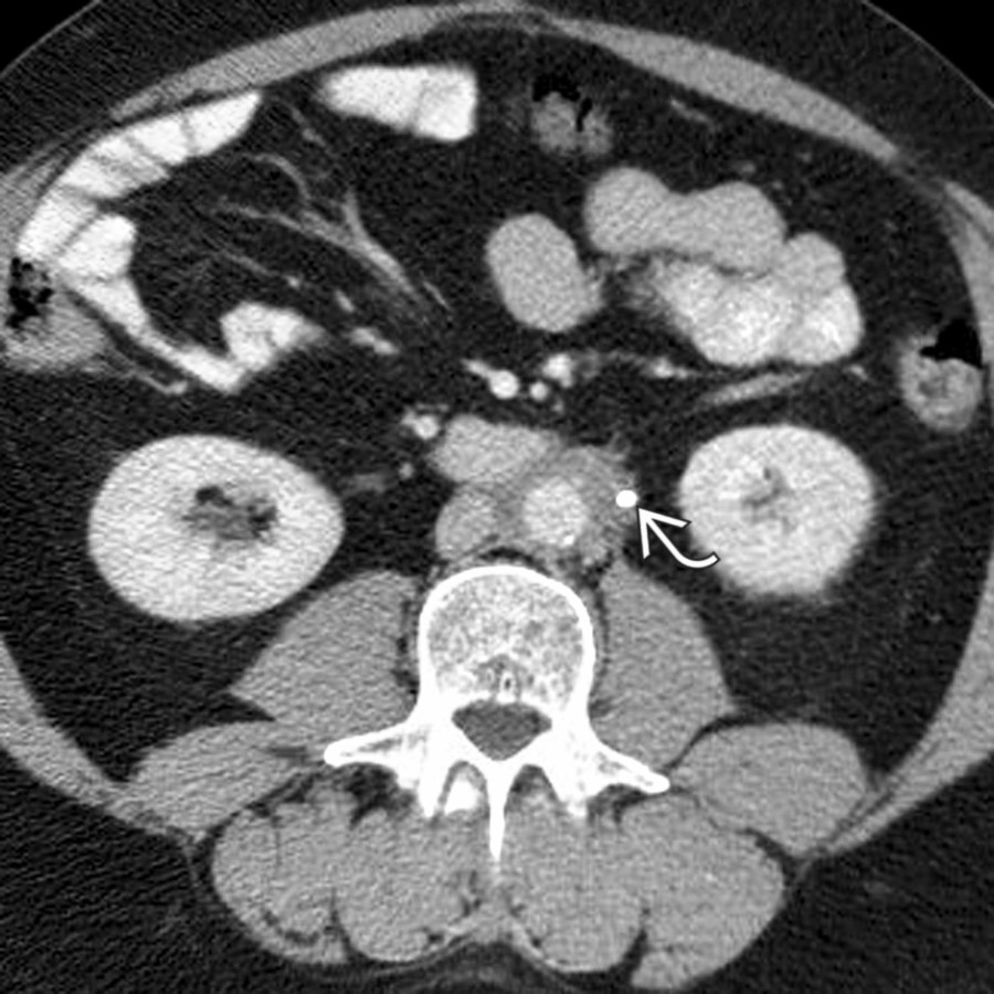
*Axial CECT shows mantle of soft tissue encasing aorta, IVC, and ureter (with stent ).*
**Secondary Infection/Abscess**
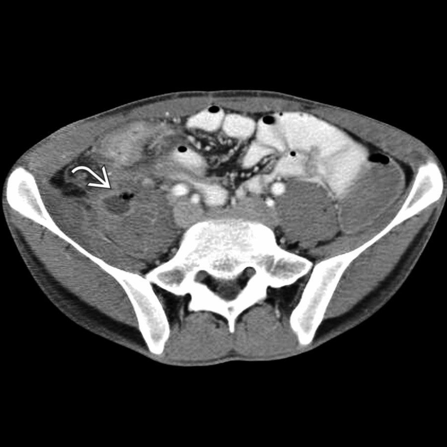
*Axial CECT shows a small collection of gas and fluid within the psoas muscle due to Crohn disease with a sinus tract from the terminal ileum.*
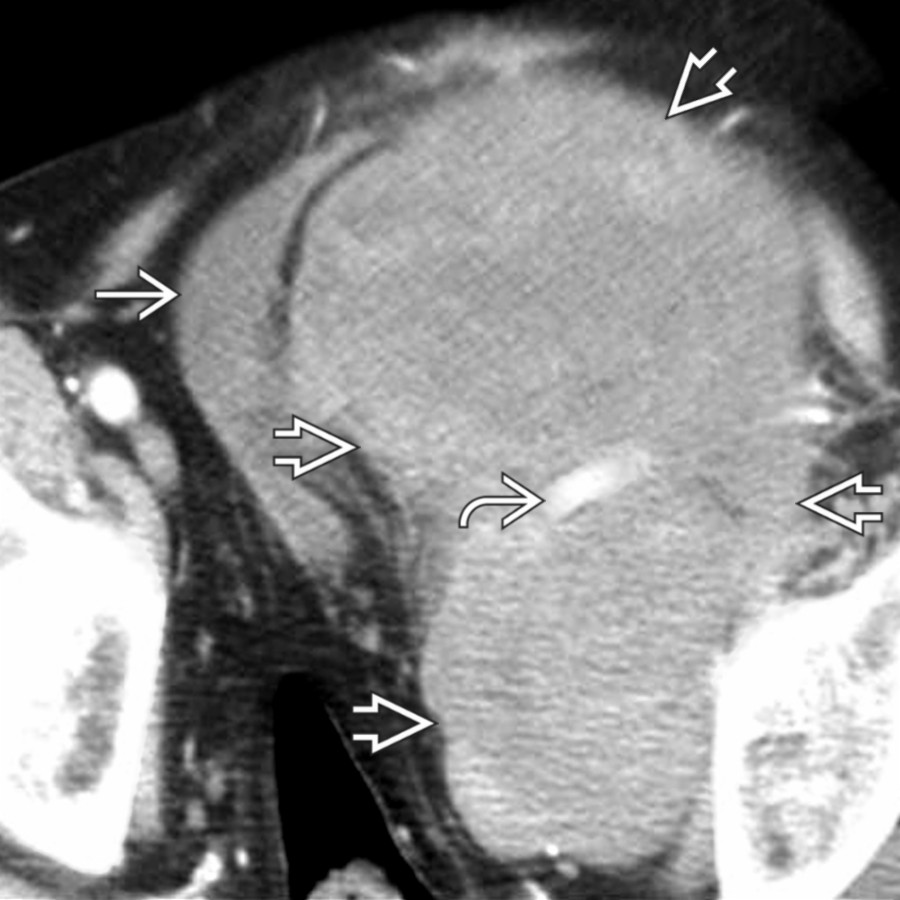
**Primary Neoplasm**
*Axial CECT shows a large, heterogeneous tumor , which displaces the bladder and external iliac artery . This is a leiomyosarcoma arising from the psoas muscle.*
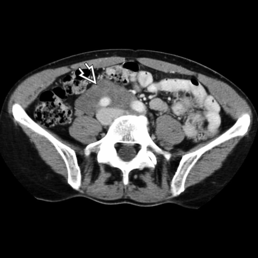
**Secondary Neoplasm**
*Axial CECT shows a soft tissue mass that extends along the aorta, IVC, and right psoas. Biopsy showed ganglioneuroma arising from the sympathetic neural chain.*

**Secondary Neoplasm**
*Axial CECT shows retroperitoneal , psoas compartment, and mesenteric lymphadenopathy in non-Hodgkin lymphoma.*
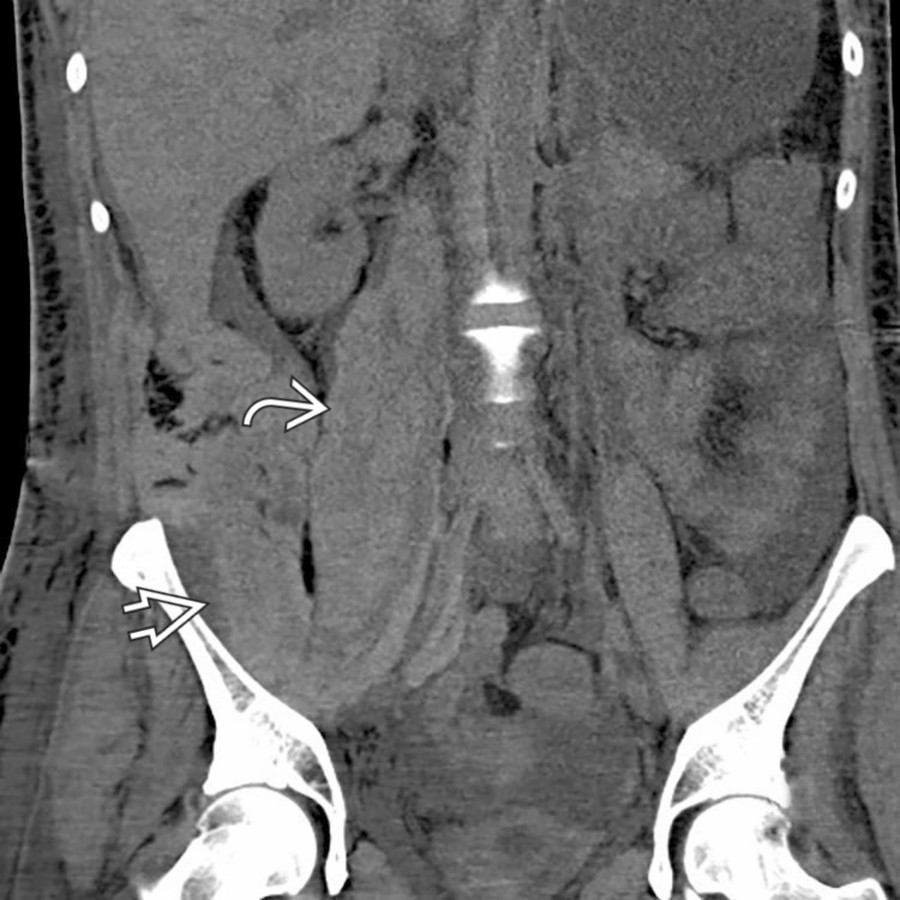
**Iliopsoas Hematoma**
*Coronal NECT shows a large hematoma extending from the right psoas muscle into the adjacent right iliopsoas muscle .*
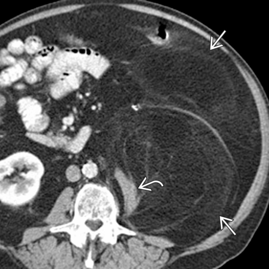
**Primary Neoplasm**
*Axial CECT shows a large, fat-containing mass occupying much of the left abdomen with the left psoas muscle appearing stretched and distorted as a result of the retroperitoneal tumor. This was found to be a well-differentiated liposarcoma at resection.*
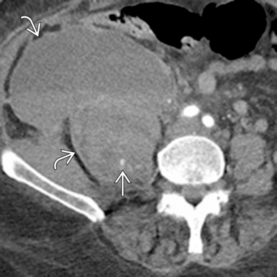
**Iliopsoas Hematoma**
*Axial CECT shows a large hematoma extending from the psoas muscle into the adjacent right abdomen. Note the presence of a tiny focus of active extravasation within the hematoma.*
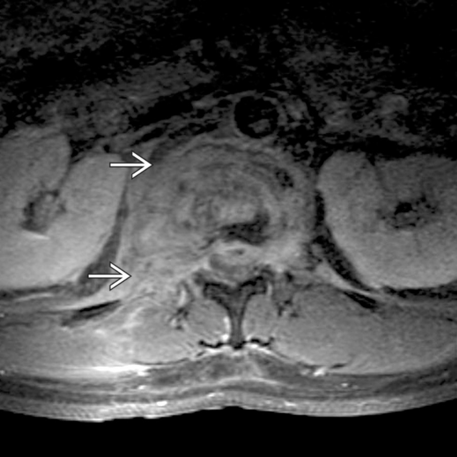
**Secondary Infection/Abscess**
*Axial T1 C+ FS MR at the level of the renal hilum shows predominantly right-sided enhancing paraspinal and epidural phlegmon , associated with discitis and vertebral osteomyelitis. The presence of psoas infection should always prompt careful appraisal of the spine.*
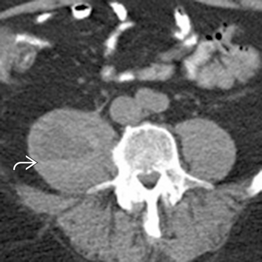
**Iliopsoas Hematoma**
*Axial NECT shows enlargement of the right psoas muscle due to coagulopathic hemorrhage. Although the hematoma is partly isodense to the muscle, the presence of a hematocrit level makes the bleed more apparent. Hematocrit levels suggest coagulopathy as the underlying cause of the bleed.*

**Secondary Neoplasm**
*Axial T1 C+ FS MR shows a heterogeneously enhancing mass expanding the right psoas muscle, found to be a neurogenic tumor (ganglioneuroma) at resection.*
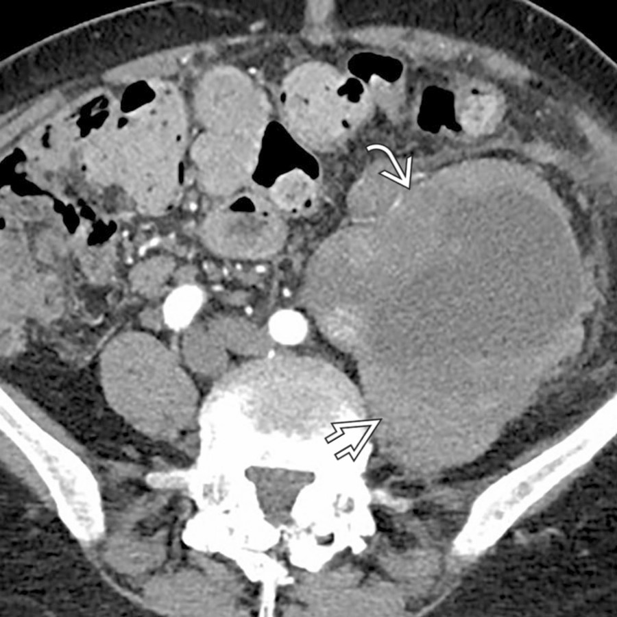
**Primary Neoplasm**
*Axial CECT shows a large mixed cystic and solid mass involving the left psoas muscle , representing a primary retroperitoneal leiomyosarcoma.*
, indicating coagulopathic bleeding.](images/app.statdx.com_image_thumbnail_e297411e-4578-474d-90d5-7a49fcb33643_annotated_true_size_900_quality_90_14ca7e04a6e3bb907d3520cae7476d29ea6f6264.jpg)
**Iliopsoas Hematoma**
*Axial CECT shows hemorrhage into multiple sites, including iliopsoas, with active bleeding and hematocrit sign , indicating coagulopathic bleeding.*

**Iliopsoas Hematoma**
*Axial CECT shows a large abdominal aortic aneurysm with extensive free hematoma tracking laterally in the retroperitoneum, including immediately adjacent to the psoas muscle, compatible with aneurysm rupture.*

**Iliopsoas Hematoma**
*Axial CECT shows a large abdominal aortic aneurysm that has ruptured into the psoas compartment and retroperitoneum. Blood dissects along the left psoas and throughout the retroperitoneal planes.*

**Iliopsoas Hematoma**
*Axial CECT shows a large retroperitoneal mass arising from the iliopsoas compartment. A focus of active bleeding and hematocrit levels confirm coagulopathic hemorrhage.*

**Retroperitoneal Fibrosis**
*Axial CECT shows mantle of soft tissue encasing aorta, IVC, and ureter (with stent ).*

**Secondary Infection/Abscess**
*Axial CECT shows a small collection of gas and fluid within the psoas muscle due to Crohn disease with a sinus tract from the terminal ileum.*

**Primary Neoplasm**
*Axial CECT shows a large, heterogeneous tumor , which displaces the bladder and external iliac artery . This is a leiomyosarcoma arising from the psoas muscle.*

**Secondary Neoplasm**
*Axial CECT shows a soft tissue mass that extends along the aorta, IVC, and right psoas. Biopsy showed ganglioneuroma arising from the sympathetic neural chain.*

**Secondary Neoplasm**
*Axial CECT shows retroperitoneal , psoas compartment, and mesenteric lymphadenopathy in non-Hodgkin lymphoma.*

**Iliopsoas Hematoma**
*Coronal NECT shows a large hematoma extending from the right psoas muscle into the adjacent right iliopsoas muscle .*

**Primary Neoplasm**
*Axial CECT shows a large, fat-containing mass occupying much of the left abdomen with the left psoas muscle appearing stretched and distorted as a result of the retroperitoneal tumor. This was found to be a well-differentiated liposarcoma at resection.*

**Iliopsoas Hematoma**
*Axial CECT shows a large hematoma extending from the psoas muscle into the adjacent right abdomen. Note the presence of a tiny focus of active extravasation within the hematoma.*

**Secondary Infection/Abscess**
*Axial T1 C+ FS MR at the level of the renal hilum shows predominantly right-sided enhancing paraspinal and epidural phlegmon , associated with discitis and vertebral osteomyelitis. The presence of psoas infection should always prompt careful appraisal of the spine.*

**Iliopsoas Hematoma**
*Axial NECT shows enlargement of the right psoas muscle due to coagulopathic hemorrhage. Although the hematoma is partly isodense to the muscle, the presence of a hematocrit level makes the bleed more apparent. Hematocrit levels suggest coagulopathy as the underlying cause of the bleed.*

**Secondary Neoplasm**
*Axial T1 C+ FS MR shows a heterogeneously enhancing mass expanding the right psoas muscle, found to be a neurogenic tumor (ganglioneuroma) at resection.*

**Primary Neoplasm**
*Axial CECT shows a large mixed cystic and solid mass involving the left psoas muscle , representing a primary retroperitoneal leiomyosarcoma.*