---
title: "Moyamoya"
docid: "c820f6bf-ddb6-4e75-b0ca-61263ed63b21"
authors:
- key: "5cff4116-3654-4b3a-bb75-5ebe0b8c9850"
value: "Anne G. Osborn, MD, FACR"
breadcrumbs:
-
name: "Brain"
slug: "brain"
treeNodeId: "6d8829f1-14d7-45af-8675-255189aa526a"
-
name: "Diagnosis"
slug: "diagnosis"
treeNodeId: "51c00394-446e-4a38-94af-d3b1d14d34e8"
-
name: "Pathology-Based Diagnoses"
slug: "pathology-based-diagnoses"
treeNodeId: "d9d3a8ed-f21b-4831-8c77-591a3500ef77"
-
name: "Stroke"
slug: "stroke"
treeNodeId: "12307683-f1ff-4823-a7d3-b10b40f9fd82"
-
name: "Nonatheromatous Vasculopathy"
slug: "nonatheromatous-vasculopathy"
treeNodeId: "ed2d2a03-ebd6-4a72-8608-effc92deb342"
-
name: "Moyamoya"
slug: "moyamoya"
treeNodeId: null
category: "Brain"
cmeTopicId: "6d619830-666a-4fbd-8e1c-9768a7d22b02"
documentVersionId: "61b569aa-0c21-432e-9ddc-9990f9d19651"
imageCount: 18
lastUpdated: "10/08/20"
pageDescription: "Moyamoya"
pageKeywords: "Brain, Diagnosis, Pathology-Based Diagnoses, Stroke, Nonatheromatous Vasculopathy, Moyamoya"
pageTitle: "Moyamoya | STATdx"
enhancedTitle: "Moyamoya"
type: "DX"
references: true
breadcrumbs:
- "Brain"
- "Diagnosis"
- "Pathology-Based Diagnoses"
- "Stroke"
- "Nonatheromatous Vasculopathy"
- "Moyamoya"
---
# KEY FACTS
- ## Terminology
- Progressive narrowing of distal ICA/proximal circle of Willis (COW) vessels with secondary collateralization
- Collateral vessels → cloud-like "puff of smoke" (moyamoya) at angiography
- Moyamoya disease (MMD) = primary (idiopathic) moyamoya
- Marked East-West gradient (more common in Japan, Korea)
- Moyamoya pattern of collaterals = secondary to many etiologies
- ## Imaging
- Best diagnostic clue: Attenuated COW with multiple tiny basal ganglia flow voids on MR
- Best imaging tool
- T1 MR C+/MRA
- DSA for delineating vascular details
- ## Pathology
- MMD
- RNF213 polymorphism in 95% of familial East Asian population, 79% of sporadic
- Correlated with early onset, severe form of MMD
- Secondary moyamoya (many causes)
- Syndromic (e.g., NF1), inflammatory states, prothrombotic states, premature aging, congenital mesenchymal defects, suprasellar irradiation in childhood
- ## Clinical Issues
- Bimodal age peaks
- 5-10 years and 2nd peak during 4th decade
- Most frequent cause of stroke in Asian children
- Presentation
- Children: Recurrent transient ischemic attacks (TIAs); hemorrhage rare
- Adults: TIAs, infarcts, hemorrhage (20%), migraines
# TERMINOLOGY
- ## Abbreviations
- Moyamoya disease (MMD)
- ## Synonyms
- Idiopathic progressive arteriopathy of childhood, spontaneous occlusion of circle of Willis (COW)
- ## Definitions
- Progressive bilateral stenosis or occlusion of terminal internal carotid artery (ICA)/proximal COW with abnormal vascular network at base of brain
- Compensatory development of collateral vascular network = "puff of smoke" (hazy network of basal collaterals)
- Primary (idiopathic) moyamoya = MMD
- More common in Japan, Korea
- Secondary (acquired) moyamoya
- Cranial radiation
- Atherosclerosis
- Neurofibromatosis (NF)
# IMAGING
- ## General Features
- Best diagnostic clue: Attenuated COW with multiple tiny basal ganglia (BG) flow voids on MR
- Location: Supraclinoid ICA/COW; anterior > > posterior circulation
- Size: Large vessel occlusion
- Morphology: "Puff of smoke" (moyamoya in Japanese)
- Cloud-like lenticulostriate and thalamostriate collaterals on angiography
- ## CT Findings
- ### NECT
- Children: 50-60% show anterior > posterior atrophy
- Can present with stroke (children) or intracranial (IC) hemorrhage (more common in adults)
- CECT
- Enhancing dots (enlarged lenticulostriate arteries) in BG
- Abnormal net-like vessels at base of brain
- CTA: Abnormal COW and net-like collaterals
- CT perfusion
- Depicts penumbra, infarct core in ischemic MMD
- Can quantify effect of revascularization therapies
- ## MR Findings
- T1WI: Multiple dot-like flow voids in BG
- T2WI
- ↑ signal in small vessel cortical and white matter infarcts
- Collateral vessels = net-like filling defects in basal cisterns
- FLAIR
- Bright sulci = leptomeningeal ivy sign
- Slow-flowing engorged pial vessels, thickened arachnoid membranes
- Correlates with ↓ cerebral vascular reserve
- T2* GRE
- Hemosiderin if prior hemorrhage
- Asymptomatic microbleeds occasionally seen in adults
- DWI: Very useful for "acute on chronic" disease
- T1WI C+
- Lenticulostriate collaterals → enhancing "dots" in BG and net-like thin vessels in cisterns
- Leptomeningeal enhancement (contrast-enhanced ivy sign) ↓ after effective bypass surgery
- MRA: Narrowed distal ICA and proximal COW vessels, ± synangiosis
- MRV: Some vasculopathies may also involve veins
- MRS: Lactate in acutely infarcted tissue
- NAA:Cr and Cho:Cr ratios frontal white matter improve/↑ after revascularization
- pMR: ↓ perfusion in deep hemispheric white matter, relative ↑ perfusion in posterior circulation
- Can also be helpful in evaluation of MMD following revascularization
- ## Ultrasonographic Findings
- Grayscale: Reduction of ICA lumen size
- Pulsed Doppler
- Doppler spectral waveforms in ICA show no flow (occluded) or high resistance (stenotic) flow pattern
- ↑ end-diastolic flow velocity, ↓ vascular resistance in external carotid artery (ECA) collaterals
- Color Doppler: Aliasing suggests stenoses
- Power Doppler: Contrast injection improves visualization of slow-flow stenotic vessels and collaterals
- ## Angiographic Findings
- Conventional
- Predominantly (not exclusively) anterior circulation
- Narrow proximal COW and ICA (earliest)
- Lenticulostriate and thalamoperforator collaterals (intermediate)
- Transdural and transosseous extracranial (EC)-IC collaterals (late)
- Dilatation of anterior choroidal artery + branches predicts adult hemorrhagic events
- ## Nuclear Medicine Findings
- PET: ↓ hemodynamic reserve capacity
- SPECT I-123-iomazenil: Neuronal density preserved if asymptomatic, ↓ if symptomatic
- ## Imaging Recommendations
- Best imaging tool: T1 C+ MR/MRA
- DSA for mapping collaterals, grading
- Protocol advice
- Contrast improves detection: Synangiosis, collaterals
- Catheter angiography defines anatomy of occlusions prior to bypass
- Diagnostic criteria: MR/MRA or catheter angiography
- Stenosis/occlusion of terminal ICA or proximal anterior cerebral artery (ACA) and middle cerebral artery (MCA)
- Abnormal vascular network/flow voids in BG
- Bilateral
- Unilateral findings in MCA = rete MCA anomaly
# DIFFERENTIAL DIAGNOSIS
- ## Ivy Sign
- Leptomeningeal metastases, subarachnoid hemorrhage, meningitis, ↑ inspired oxygen
- ## Punctate Foci in Basal Ganglia
- Cribriform lacunar state: No enhancement
- ## Severely Attenuated Circle of Willis
- Subarachnoid hemorrhage, meningitis, tumor encasement
- ## Rete Middle Cerebral Artery Anomaly
- Unilateral, not bilateral
- Weblike rete anomaly of MCA
- Twig-like horizontal MCA + branches
- May occur with aneurysm (50%)
- Occurs predominantly in Asians
# PATHOLOGY
- ## General Features
- ### Etiology
- MMD
- Ring finger protein 213 polymorphism in 95% of familial East Asian population, 79% of sporadic
- Correlated with early-onset, severe form of MMD
- Also recently reported in non-MMD disorders
- RNF213 vasculopathy spectrum (IC atherosclerosis, peripheral pulmonary artery stenosis, and rental artery stenosis)
- Secondary moyamoya
- Down syndrome, tuberous sclerosis, sickle cell disease, connective tissue disease, progeria, NF1
- NF1 with suprasellar tumor and radiation is disastrous
- Morning glory syndrome; syndromes with aneurysms, cardiac and ocular defects
- Inflammatory: CNS angiitis (of childhood), basal meningitis, atherosclerosis, head and neck infections
- Vasculopathies and prothrombotic states: XRT, Kawasaki, anticardiolipin antibody, factor V Leiden, polyarteritis nodosa, Behçet, SLE
- Epidemiology: MMD
- Incidence in Japan: 1:100,000
- Incidence in North America, Europe: 0.1:100,000
- 10-15% familial
- ## Staging, Grading, & Classification
- Staging criteria (after Suzuki)
- Stage 1: Narrowing of ICA bifurcation
- Stage 2: ACA, MCA, posterior cerebral artery (PCA) dilated
- Stage 3: Maximal basal collaterals; small ACA/MCA
- Stage 4: Fewer collaterals (vessels); small PCA
- Stage 5: Further ↓ collaterals; absent ACA/MCA/PCA
- Stage 6: Extensive ECA-pial collaterals
- ## Gross Pathologic & Surgical Features
- ↑ perforating (early) and ECA-ICA (late) collaterals in atrophic brain
- Hemorrhage (subarachnoid, intraventricular > parenchymal) adults
- ↑ saccular aneurysms (especially basilar in adults)
- ## Microscopic Features
- Intimal hyperplasia, medial layer thinness
- Excessive infolding, "waving" of internal elastic lamina
- Progressive narrowing, eventual obliteration of vessel lumen
- Periventricular pseudoaneurysms (cause of hemorrhage)
# CLINICAL ISSUES
- ## Presentation
- ### Most common signs/symptoms
- Children: Transient ischemic attacks (TIAs), alternating hemiplegia (exacerbated by crying), headache
- Adults: TIAs, cerebral infarct, or hemorrhage
- Hemorrhagic presentation more common in Asian adults
- ### Other signs/symptoms
- Children: Developmental delay, poor feeding, chorea
- ### Clinical profile
- Children more likely to have TIAs and to progress; adults more likely to infarct (but slower progression)
- Children more likely to have ipsilateral anterior plus posterior circulation involvement
- ## Demographics
- ### Age
- Bimodal age peaks (5-10 years and 2nd peak during 4th decade)
- Japan, Korea: 6 years > 35 years
- North America, Europe: 35 years > 6 years
- ### Sex
- M:F = 1:1.8; in familial cases, M:F = 1:5
- Most frequent cause of stroke in Asian children
- ## Natural History & Prognosis
- Progressive narrowing, collateralization, and ischemia
- Prognosis depends on etiology, ability to form collaterals, age/stage at diagnosis
- Pediatric cases usually advance to stage 5 within 10 years of onset
- Infantile moyamoya progresses faster
- Pediatric cases present with stroke
- Hemorrhagic moyamoya more common in older patients with large collateral vessels
- Has poorer outcome
- ## Treatment
- MMD
- Indirect bypass: Encephaloduroarteriomyosynangiosis (EDAMS) more effective in children
- 5-year risk of ipsilateral stroke post encephaloduroarteriosynangiosis (EDAS) = 15%
- Direct bypass: Superficial temporal artery-MCA (STA-MCA) more common in adults
- Combined EDAMS, STA-MCA
- Anticoagulation; correct/control prothrombotic states and inflammatory etiologies
- Hypertransfusion for sickle cell-related moyamoya
- Perivascular sympathectomy or superior cervical ganglionectomy (adults)
# DIAGNOSTIC CHECKLIST
- ## Consider
- Seek secondary causes of moyamoya
- ## Image Interpretation Pearls
- Enhanced asymmetric atrophy found on childhood CT, look for abnormal vascular pattern
- Adult moyamoya can present with IC hemorrhage
- ## Reporting Tips
- Successful revascularization = ↓ basal collaterals, ↑ flow in MCA branches, ↑ caliber of STA (direct bypass)
16877c2f-bb02-4c68-afaf-2bfadf0fb33f
## References
# Selected References
1. [Han Q et al: Quantitative analysis of revascularization in ischemic moyamoya disease via whole-brain computed tomography perfusion: a retrospective single-center study. Medicine (Baltimore). 99(7):e19168, 2020](http://www.ncbi.nlm.nih.gov/pubmed/?term=32049846%5Bpmid%5D)
1. [Ravindra VM et al: Preoperative computed tomography perfusion in pediatric moyamoya disease: a single-institution experience. J Neurosurg Pediatr. ePub, 2020](http://www.ncbi.nlm.nih.gov/pubmed/?term=31978885%5Bpmid%5D)
1. [Terrell D et al: Cerebral revascularization for moyamoya syndrome associated with sickle cell disease: a systematic review of the literature on the role of extracranial-intracranial bypass in treating neurologic manifestations of pediatric patients with sickle cell disease. World Neurosurg. 137:62-70, 2020](http://www.ncbi.nlm.nih.gov/pubmed/?term=32014541%5Bpmid%5D)
1. [Bang OY et al: Moyamoya disease and spectrums of RNF213 vasculopathy. Transl Stroke Res. 11(4):580-9, 2019](http://www.ncbi.nlm.nih.gov/pubmed/?term=31650369%5Bpmid%5D)
1. [Boulouis G et al: Nontraumatic pediatric intracerebral hemorrhage. Stroke. 50(12):3654-61, 2019](http://www.ncbi.nlm.nih.gov/pubmed/?term=31637968%5Bpmid%5D)
1. [Goyal P et al: Neuroimaging of pediatric arteriopathies. J Neuroimaging. 29(3):287-308, 2019](http://www.ncbi.nlm.nih.gov/pubmed/?term=30920080%5Bpmid%5D)
1. [Li J et al: Imaging of moyamoya disease and moyamoya syndrome: current status. J Comput Assist Tomogr. 43(2):257-63, 2019](http://www.ncbi.nlm.nih.gov/pubmed/?term=30589721%5Bpmid%5D)
1. [Liu ZW et al: Collateral circulation in moyamoya disease: a new grading system. Stroke. 50(10):2708-15, 2019](http://www.ncbi.nlm.nih.gov/pubmed/?term=31409266%5Bpmid%5D)
1. [Ravindran K et al: Surgical outcomes for pediatric moyamoya: a systematic review and meta-analysis. J Neurosurg Pediatr. 1-10, 2019](http://www.ncbi.nlm.nih.gov/pubmed/?term=31518973%5Bpmid%5D)
1. [Soun JE et al: Central nervous system vasculopathies. Radiol Clin North Am. 57(6):1117-31, 2019](http://www.ncbi.nlm.nih.gov/pubmed/?term=31582039%5Bpmid%5D)
1. [Acker G et al: Surgical management of moyamoya disease. Stroke. 49(2):476-82, 2018](http://www.ncbi.nlm.nih.gov/pubmed/?term=29343587%5Bpmid%5D)
1. [Cho KC et al: Rete middle cerebral artery anomalies: a unifying name, case series, and literature review. J Neurosurg. 131(2):453-61, 2018](http://www.ncbi.nlm.nih.gov/pubmed/?term=30074465%5Bpmid%5D)
1. [Huang S et al: Etiology and pathogenesis of moyamoya disease: an update on disease prevalence. Int J Stroke. 12(3):246-53, 2017](http://www.ncbi.nlm.nih.gov/pubmed/?term=28381201%5Bpmid%5D)
1. [Fujimura M et al: Moyamoya disease. Front Neurol Neurosci. 40:204-20, 2016](http://www.ncbi.nlm.nih.gov/pubmed/?term=27960175%5Bpmid%5D)
## Images
### Selected Images
 and strikingly enlarged lenticulostriate arteries
  coursing through basal ganglia. This is the puff of smoke (moyamoya) pattern.](images/app.statdx.com_image_thumbnail_f8a053e9-37d0-4b49-b29b-df7a88f86a26_annotated_true_size_900_quality_90_27837fe3723fbc371110af5310b071c54042de4d.jpg)
*Coronal graphic shows severe tapering of both distal internal carotid arteries (ICAs) and strikingly enlarged lenticulostriate arteries coursing through basal ganglia. This is the puff of smoke (moyamoya) pattern.*

*Coronal graphic shows severe tapering of both distal internal carotid arteries (ICAs) and strikingly enlarged lenticulostriate arteries coursing through basal ganglia. This is the puff of smoke (moyamoya) pattern.*
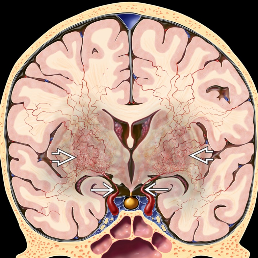
*Coronal graphic shows severe tapering of both distal internal carotid arteries (ICAs) and strikingly enlarged lenticulostriate arteries coursing through basal ganglia. This is the puff of smoke (moyamoya) pattern.*

*Coronal graphic shows severe tapering of both distal internal carotid arteries (ICAs) and strikingly enlarged lenticulostriate arteries coursing through basal ganglia. This is the puff of smoke (moyamoya) pattern.*
 within the ambient (circummesencephalic) cistern corresponding to collateral moyamoya vessels. Note the asymmetric atrophy.](images/app.statdx.com_image_thumbnail_79737443-73b4-4c68-ba89-ee52dc8575c4_annotated_true_size_900_quality_90_4a28b01279ce7df2974b4d48ebc50f18f4bf8a7d.jpg)
*Axial T2WI MR shows curvilinear net-like filling defects within the ambient (circummesencephalic) cistern corresponding to collateral moyamoya vessels. Note the asymmetric atrophy.*

*Axial T2WI MR shows curvilinear net-like filling defects within the ambient (circummesencephalic) cistern corresponding to collateral moyamoya vessels. Note the asymmetric atrophy.*
 . Note innumerable tortuous enlarged collaterals forming the puff of smoke appearance typical of MMD.](images/app.statdx.com_image_thumbnail_a1e6e2a8-db6b-43f9-af11-e51f0718819c_annotated_true_size_900_quality_90_a22aca2b208fe4bf19b0faa1b12dfd0986f6d174.jpg)
*Lateral internal carotid artery DSA in a 3-year-old child with moyamoya disease (MMD) shows near-total supraclinoid ICA stenosis . Note innumerable tortuous enlarged collaterals forming the puff of smoke appearance typical of MMD.*
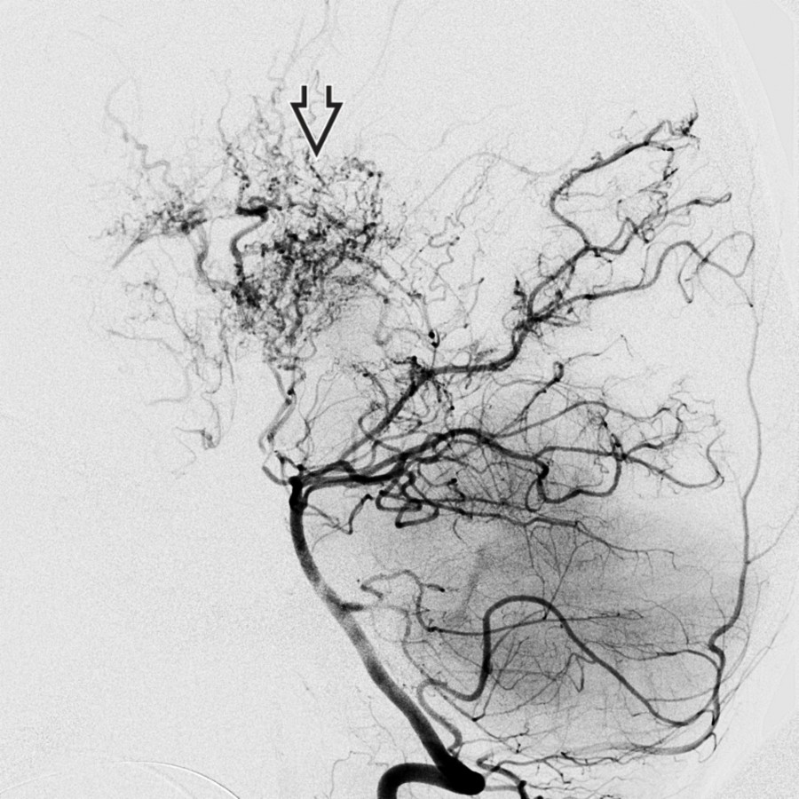
*Lateral DSA in the same patient shows the collaterals (moyamoya vessels) are supplied primarily by thalamoperforating and medial choroidal branches.*

*Lateral internal carotid artery DSA in a 3-year-old child with moyamoya disease (MMD) shows near-total supraclinoid ICA stenosis . Note innumerable tortuous enlarged collaterals forming the puff of smoke appearance typical of MMD.*

*Lateral DSA in the same patient shows the collaterals (moyamoya vessels) are supplied primarily by thalamoperforating and medial choroidal branches.*
 and around the midbrain . There is marked cortical atrophy along with enlarged temporal horns.](images/app.statdx.com_image_thumbnail_83f92ae8-be84-4a5d-9958-bbc9ab3afa23_annotated_true_size_900_quality_90_242953f7fd591c47bee93e3370a3e0095dc0b933.jpg)
*Axial T2WI MR in adult moyamoya shows attenuated, almost thread-like supraclinoid ICAs and middle cerebral arteries (MCAs) with numerous tiny collateral vessels in the suprasellar cistern and around the midbrain . There is marked cortical atrophy along with enlarged temporal horns.*
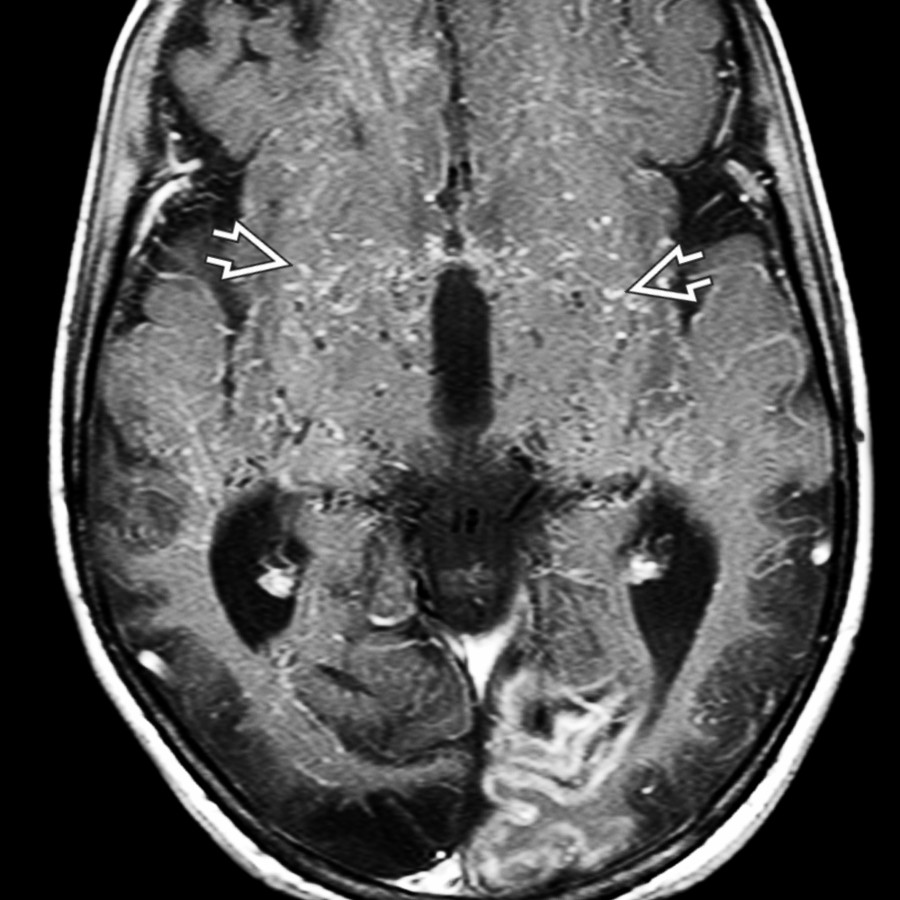
*Axial T1 C+ FS MR in the same patient shows contrast in innumerable small arterial collateral vessels in the basal ganglia and deep white matter , creating a puff of smoke appearance. (Courtesy H. Els, MD.)*

*Axial T2WI MR in a 23-year-old man with left hemisphere transient ischemic attacks (TIAs) shows the flow void of a normal right M1 MCA . The left MCA appears very hypoplastic and twig-like . There is an unusual web-like tangle of vessels seen within the sylvian fissure .*

*Submentovertex MR angiogram in the same patient shows a normal right MCA . The left MCA is thread-like , and its distal M3 and M4 branches are attenuated compared to the normal right side.*
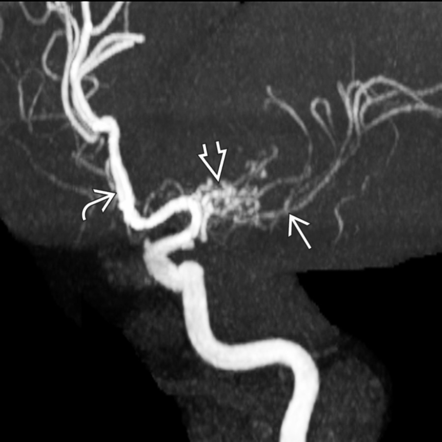
*Oblique view of the left internal carotid MR angiogram shows a normal anterior cerebral artery (ACA) , but the left MCA is severely attenuated with reduced distal branches. A web of numerous small, tangled vessels surrounds its M1 segment.*

*Oblique view of the left internal carotid DSA in the same patient shows the tangled web of vessels that surrounds the attenuated M1 MCA segment. This is thought to represent a rete MCA anomaly rather than segmental moyamoya.*
### Additional Images

*Axial FLAIR MR shows the ivy sign due to engorged vessels within sulci in another patient with moyamoya. The sulcal signal is so striking that this FLAIR scan resembles a T2WI.*
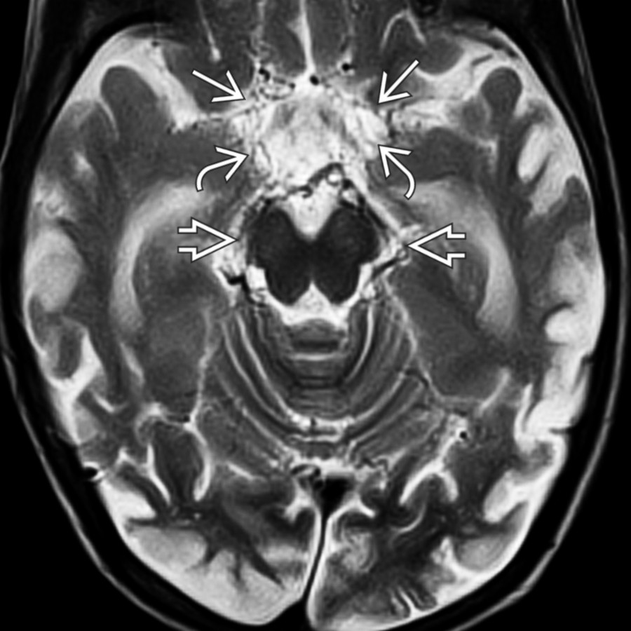
*Axial T2WI MR in adult moyamoya shows attenuated, almost thread-like supraclinoid ICAs and middle cerebral arteries (MCAs) with numerous tiny collateral vessels in the suprasellar cistern and around the midbrain . There is marked cortical atrophy along with enlarged temporal horns.*

*Axial T1 C+ FS MR in the same patient shows contrast in innumerable small arterial collateral vessels in the basal ganglia and deep white matter , creating a puff of smoke appearance. (Courtesy H. Els, MD.)*

*Axial T2WI MR in a 23-year-old man with left hemisphere transient ischemic attacks (TIAs) shows the flow void of a normal right M1 MCA . The left MCA appears very hypoplastic and twig-like . There is an unusual web-like tangle of vessels seen within the sylvian fissure .*

*Submentovertex MR angiogram in the same patient shows a normal right MCA . The left MCA is thread-like , and its distal M3 and M4 branches are attenuated compared to the normal right side.*

*Oblique view of the left internal carotid MR angiogram shows a normal anterior cerebral artery (ACA) , but the left MCA is severely attenuated with reduced distal branches. A web of numerous small, tangled vessels surrounds its M1 segment.*

*Oblique view of the left internal carotid DSA in the same patient shows the tangled web of vessels that surrounds the attenuated M1 MCA segment. This is thought to represent a rete MCA anomaly rather than segmental moyamoya.*
### Additional Images

*Axial FLAIR MR shows the ivy sign due to engorged vessels within sulci in another patient with moyamoya. The sulcal signal is so striking that this FLAIR scan resembles a T2WI.*
 with a puff of smoke appearance from collateral lenticulostriate vessels.](96491607-0164-4f15-95a4-68d2c36cb0f7)
*Lateral view, selective internal carotid angiogram, shows severe stenosis of the supraclinoid ICA with a puff of smoke appearance from collateral lenticulostriate vessels.*

*Lateral angiography, right ICA injection in the same patient, shows narrowing of the distal ICA and occlusion of the ACA and MCA with the puff of smoke appearance of enlarged lenticulostriate collaterals. Note the additional network of collaterals more posteriorly , likely thalamoperforators and posterior choroidal branches.*

*Lateral DSA of the vertebrobasilar circulation in the same patient shows a striking puff of smoke appearance caused by innumerable dilated collateral branches arising from the thalamostriate arteries.*

*Axial MRA shows occlusion of both distal ICAs , nonvisualization of MCAs and ACAs, and stenosis of posterior cerebral arteries (PCAs) in an 8 year old with hemiparetic migraines in this case of idiopathic arteriopathy of childhood.*

*Axial MRA in another patient with idiopathic progressive arteriopathy of childhood shows occluded supraclinoid ICAs . Note the bilateral synangiosis .*

*Axial T1WI MR shows right frontal and left temporaoccipital atrophy from remote ischemia. There are multiple small basal ganglia flow voids from lenticulostriate collaterals.*

*Axial T1WI C+ MR in the same patient shows multifocal white "dots" due to slow flow in multiple, enlarged lenticulostriate collaterals with intravascular enhancement.*
with a puff of smoke appearance from collateral lenticulostriate vessels.](96491607-0164-4f15-95a4-68d2c36cb0f7)
*Lateral view, selective internal carotid angiogram, shows severe stenosis of the supraclinoid ICA with a puff of smoke appearance from collateral lenticulostriate vessels.*

*Lateral angiography, right ICA injection in the same patient, shows narrowing of the distal ICA and occlusion of the ACA and MCA with the puff of smoke appearance of enlarged lenticulostriate collaterals. Note the additional network of collaterals more posteriorly , likely thalamoperforators and posterior choroidal branches.*

*Lateral DSA of the vertebrobasilar circulation in the same patient shows a striking puff of smoke appearance caused by innumerable dilated collateral branches arising from the thalamostriate arteries.*

*Axial MRA shows occlusion of both distal ICAs , nonvisualization of MCAs and ACAs, and stenosis of posterior cerebral arteries (PCAs) in an 8 year old with hemiparetic migraines in this case of idiopathic arteriopathy of childhood.*

*Axial MRA in another patient with idiopathic progressive arteriopathy of childhood shows occluded supraclinoid ICAs . Note the bilateral synangiosis .*

*Axial T1WI MR shows right frontal and left temporaoccipital atrophy from remote ischemia. There are multiple small basal ganglia flow voids from lenticulostriate collaterals.*

*Axial T1WI C+ MR in the same patient shows multifocal white "dots" due to slow flow in multiple, enlarged lenticulostriate collaterals with intravascular enhancement.*