---
title: "Multiinfarct Dementia"
docid: "3823c4d4-5e98-46da-a717-892fef54b382"
authors:
- key: "9d40c5b1-57d2-442c-9daf-8d8d9d53e24b"
value: "Akiva Mintz, MD, PhD, MHA, CFA"
- key: "bbc899b6-2885-44bb-a5b0-24eec7314d33"
value: "Bryan J. Neth, BS"
- key: "1f262abe-db83-4f18-99af-00bd3045cd4d"
value: "Marc Benayoun, MD, PhD"
breadcrumbs:
-
name: "Nuclear Medicine"
slug: "nuclear-medicine"
treeNodeId: "2406533f-6523-4211-841e-b92d6f8cf34e"
-
name: "Central Nervous System"
slug: "central-nervous-system"
treeNodeId: "bd6b5c36-69df-4f18-af9c-96cc24b52d8f"
-
name: "Neurodegeneration"
slug: "neurodegeneration"
treeNodeId: "f2b87cc7-926d-4915-8ec5-ca61a82e8bc9"
-
name: "Multiinfarct Dementia"
slug: "multiinfarct-dementia"
treeNodeId: null
category: "Nuclear Medicine"
cmeTopicId: "29e9ca9b-f22e-44aa-94a4-19393851339d"
documentVersionId: "2906e1eb-a93a-4cdb-8e7f-261c406e626c"
imageCount: 24
lastUpdated: "07/21/25"
pageDescription: "Multiinfarct Dementia"
pageKeywords: "Nuclear Medicine, Central Nervous System, Neurodegeneration, Multiinfarct Dementia"
pageTitle: "Multiinfarct Dementia | STATdx"
enhancedTitle: "Multiinfarct Dementia"
type: "DX"
references: true
breadcrumbs:
- "Nuclear Medicine"
- "Central Nervous System"
- "Neurodegeneration"
- "Multiinfarct Dementia"
---
# KEY FACTS
- ## Terminology
- Impairments in cognition and behavior affecting functional status due to pathologic changes resulting from various vascular insults throughout brain
- ## Imaging
- F-18 FDG PET may be used in differential diagnosis between vascular dementia and Alzheimer disease
- Glucose hypometabolism in multifocal (scattered) pattern of cortical with subcortical regions is indicative of vascular dementia
- Multifocal or unifocal hypometabolism involving cortical gray matter, subcortical white matter, and other structures
- Generally involves cerebral hemispheres, thalamus, basal ganglia, hippocampi
- Amyloid PET imaging does not demonstrate gray matter amyloid deposition in vascular dementia
- SPECT with Tc-99m HMPAO or Tc-99m ECD shows similar asymmetrically decreased perfusion
- ## Pathology
- Vascular-related lesions leading to loss of brain function
- Chronic small vessel insults > large vessel infarcts
- ## Clinical Issues
- Significant heterogeneity in clinical presentation depending on location, type, and size of vascular lesion
- Overt disease: Cognitive impairment due to clinically evident vascular event (i.e., stroke)
- Covert disease: Insidious process of vascular insults (clinically silent strokes)
- 2nd most common cause of dementia after Alzheimer disease
# TERMINOLOGY
- ## Definitions
- Impairments in cognition and behavior affecting functional status due to pathologic changes resulting from various vascular insults throughout brain
- Vascular cognitive impairment (VCI) is more comprehensive term incorporating vascular dementia (VaD) as well as other vascular-related cognitive impairment, including mild cognitive impairment due to vascular disease
# IMAGING
- ## General Features
- ### Best diagnostic clue
- Multifocal or unifocal infarcts involving cortical gray matter, subcortical white matter, and other structures
- Especially when correlates with other clinical findings
- Generally involve cerebral hemispheres, thalamus, basal ganglia, hippocampi
- Generally significant white matter involvement
- Bilateral > unilateral
- Size: Decreased due to atrophy
- Morphology: Multiple small &/or large vessel with lacunar infarcts
- ## Nuclear Medicine Findings
- F-18 FDG PET/CT
- Glucose hypometabolism in multifocal (scattered) pattern of cortical with subcortical regions
- Altered pattern depending on subtype (i.e., multifocal small vessel infarcts vs. large territory infarcts)
- Hypometabolism often in clinically affected areas: Correlates with other clinical findings
- May be used in differential diagnosis between VaD and Alzheimer disease (AD)
- AD pattern: Hypometabolism in bilateral parietotemporal with posterior cingulate cortices: Extension to frontal/occipital
- VaD may have hypometabolism in subcortical areas, which are spared in AD
- Amyloid PET without gray matter amyloid deposition
- May be used in differential diagnosis between VaD and AD
- SPECT with Tc-99m HMPAO or Tc-99m ECD shows similar asymmetric decreased perfusion
- ## Imaging Recommendations
- ### Best imaging tool
- F-18 FDG PET may aid in differential diagnosis
- MR to see vascular insults and look for potential reversible causes of dementia
- ### Protocol advice
- F-18 FDG PET
- Patient preparation
- Patient should fast, stop IV fluids containing dextrose, stop parenteral feeding for 4-6 hours
- Blood sugar should be < 150-200 mg/dL
- Patient should be placed in quiet, dimly lit room prior to and after injection for 30 min
- Radiopharmaceutical: 5-15 mCi F-18 FDG
- Dosimetry: Urinary bladder receives largest dose
- Image acquisition: 30-60 min after injection
# DIFFERENTIAL DIAGNOSIS
- [Alzheimer Disease](/document/alzheimer-disease/2aad3ac4-44fd-43e5-8e50-a86987483af3)
- Early F-18 FDG hypometabolism in parietotemporal and posterior cingulate cortices
- Later changes include frontal lobe
- Positive amyloid PET
- Atrophy of medial temporal lobe structures on MR/CT
- ## Dementia With Lewy Bodies
- Commonly presents with hallucinations, sleep disturbances, and parkinsonian motor features
- F-18 FDG PET hypometabolism in occipital cortex or striatal loss of dopamine transport
- [Normal-Pressure Hydrocephalus](/document/normal-pressure-hydrocephalus/834ccc3e-2116-4295-8408-0ac9a06bd2ff)
- Dilated ventricles on CT or MR
- Metabolic or perfusion reductions in lateral cortex (frontal and parietal), separation of caudate heads
- Reduced activity in periventricular WM and ventricular regions > expected for age
- ## Mixed Dementia
- Characteristic features of > 1 type of dementia
- Commonly includes AD and another pathology (such as VaD)
- ## Frontotemporal Dementia
- Commonly presents with personality and behavioral changes
- Atroph of frontal and anterior temporal lobes
- F-18 FDG PET hypometabolism primarily in frontal and anterior temporal lobes
- [Huntington Disease](/document/huntington-disease/4abd9bb7-7d47-4308-a71f-9985fdb23c2c)
- Major reduction in basal ganglia metabolism and perfusion (worse in caudate)
- Cortical reductions (late)
- ## Posttraumatic Dementia
- Pattern of abnormality on functional imaging variable: Depends on severity, directional forces at injury
- Chronic subdural may cause significant asymmetry
- Deceleration injury may cause anterior frontal and temporal tip reductions
- Atrophic pattern may occur; worse with increasing severity of injury
- Focal reduction correlates with encephalomalacia and other cortical abnormalities on CT or MR
- Basal ganglia, thalamic abnormalities may be present
- ## Drug-Related Dementia
- Cocaine and amphetamine abuse: Random small focal defects throughout brain
- Swiss cheese pattern on FDG PET and SPECT
- Alcohol abuse: May demonstrate frontal lobe decrease
- ## Autoimmune Dementia
- Vasculitis pattern in systemic lupus; frontal watershed abnormalities
- Small cortical defects mainly frontal lobe
- [Creutzfeldt-Jakob Disease](/document/creutzfeldt-jakob-disease-cjd/e1b27954-6591-4bb0-a659-b13790492620)
- Rapidly fatal, prion-related disease with impairments in cognition and behavioral changes
- Diffusion-weighted imaging hyperintensity in striatum, cingulum, and neocortex
- ## Causes of Reversible Dementia
- Normal-pressure hydrocephalus, vitamin B12 deficiency, hypothyroidism, depression, mass lesions, infections (neurosyphilis, HIV), trauma (chronic subdurals)
# PATHOLOGY
- ## General Features
- Vascular-related lesions leading to loss of brain function
- Chronic small vessel insults > large vessel infarcts
- Areas of infarction (focal or multifocal) with associated atrophy
- ## Risk Factors
- History of myocardial infarction/coronary artery disease, stroke/transient ischemic attack (TIA)
- Atherosclerosis, hypertension, hyperlipidemia, atrial fibrillation
- Diabetes, obesity, smoking, advanced age
# CLINICAL ISSUES
- ## Presentation
- Significant heterogeneity in clinical presentation depending on location, type, and size of vascular lesion
- Impairment in at least 1 cognitive domain leading to decline in functional status
- Executive function and attention more commonly initially affected than in AD
- Slow motor/information processing
- Language difficulties
- Changes in behavior and personality
- Depression and anxiety
- Other: Seizures, bladder incontinence, gait disturbance, and additional focal abnormalities
- Overt disease: Cognitive impairment due to clinically evident vascular event [large territory infarct(s)]
- > 60% of stroke patients (dementia in ~ 30% of cases)
- Covert disease: Insidious process of vascular insults (clinically silent with extensive lacunar infarcts)
- ## Demographics
- 2nd most common cause of dementia after AD
- > 10% of dementia cases
- Age: Incidence increases with age
- Sex: M > F
# DIAGNOSTIC CHECKLIST
- ## Image Interpretation Pearls
- Heterogeneous F-18 FDG activity without any specific pattern suggests VaD
- Lesions can include basal ganglia and other areas typically spared in other diseases
- MR correlation helpful
18eb7ef1-b90d-42fa-ae9c-fe185e01420c
## References
# Selected References
1. Minoshima S et al: FDG PET imaging evaluation of neurodegenerative dementias. In Cross DJ et al: Molecular Imaging of Neurodegenerative Disorders. Springer. 57-72, 2023
1. [Chang Wong E et al: Vascular cognitive impairment and dementia. Continuum (Minneap Minn). 28(3):750-80, 2022](http://www.ncbi.nlm.nih.gov/pubmed/?term=35678401%5Bpmid%5D)
1. [Minoshima S et al: (18)F-FDG PET imaging in neurodegenerative dementing disorders: insights into subtype classification, emerging disease categories, and mixed dementia with copathologies. J Nucl Med. 63(Suppl 1):2S-12S, 2022](http://www.ncbi.nlm.nih.gov/pubmed/?term=35649653%5Bpmid%5D)
1. [Nestor PJ et al: Clinical utility of FDG-PET for the differential diagnosis among the main forms of dementia. Eur J Nucl Med Mol Imaging. 45(9):1509-25, 2018](http://www.ncbi.nlm.nih.gov/pubmed/?term=29736698%5Bpmid%5D)
1. [Sawyer DM et al: Top-down systematic approach to interpretation of FDG-PET for dementia. Clin Nucl Med. 43(6):e212-4, 2018](http://www.ncbi.nlm.nih.gov/pubmed/?term=29659399%5Bpmid%5D)
1. [Heiss WD et al: PET imaging in the differential diagnosis of vascular dementia. J Neurol Sci. 322(1-2):268-73, 2012](http://www.ncbi.nlm.nih.gov/pubmed/?term=23043907%5Bpmid%5D)
1. [Román G et al: Contribution of neuroimaging to the diagnosis of Alzheimer's disease and vascular dementia. Arch Med Res. 43(8):671-6, 2012](http://www.ncbi.nlm.nih.gov/pubmed/?term=23142262%5Bpmid%5D)
## Images
### Selected Images
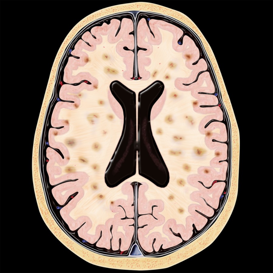
*Axial graphic shows multifocal infarcts involving the cortical gray matter and subcortical white matter bilaterally.*
*Axial graphic shows multifocal infarcts involving the cortical gray matter and subcortical white matter bilaterally.*
*Axial graphic shows multifocal infarcts involving the cortical gray matter and subcortical white matter bilaterally.*
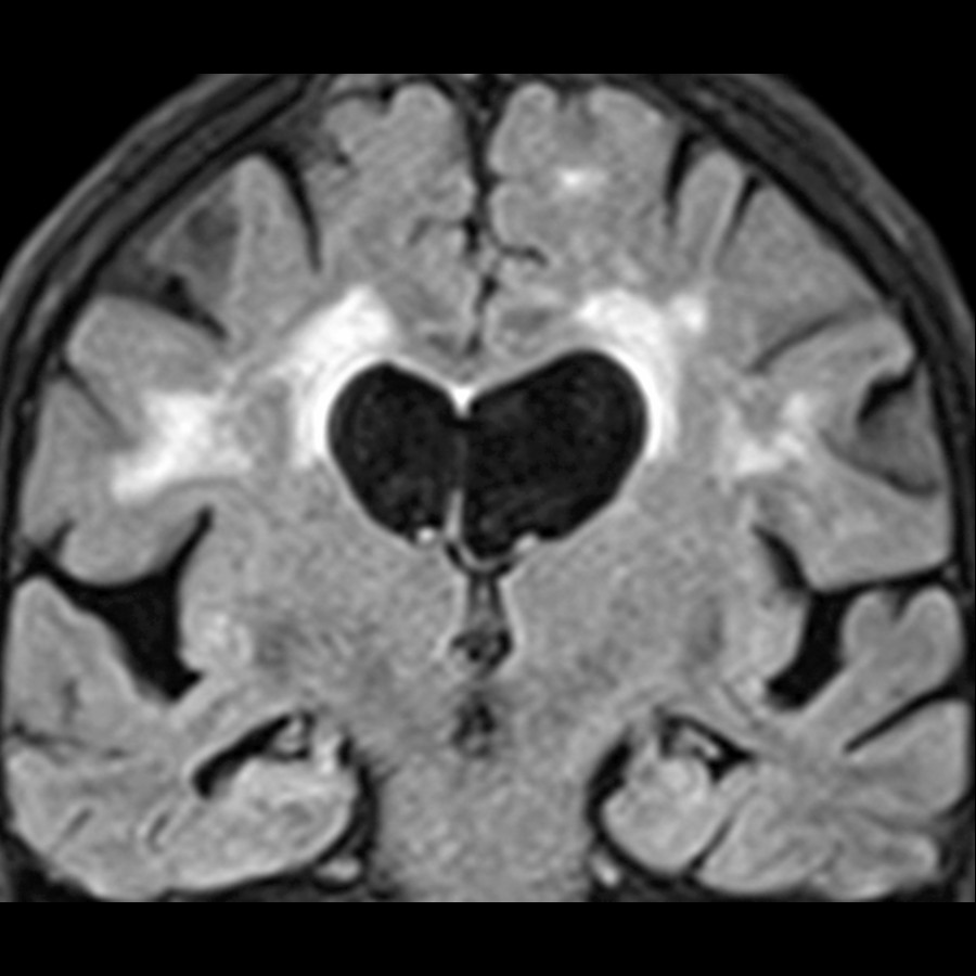
*Coronal FLAIR MR of a 72-year-old woman demonstrates FLAIR signal abnormality in the periventricular and subcortical white matter (leukoaraiosis). This finding is consistent with a small vessel ischemic etiology.*
 , the right occipital lobe
  and the cingulate gyrus
  , consistent with multiinfarct dementia.](images/app.statdx.com_image_thumbnail_5f66168d-7754-460f-9e67-f9e5338828e7_annotated_true_size_900_quality_90_e93c4d26b79a5d5f933d169bd4b8835bc499bff1.jpg)
*Surface mapping of an FDG PET scan performed in a 96-year-old man demonstrates multiple areas of significant cortical abnormality in glucose metabolism, including the left frontal lobe , the right occipital lobe and the cingulate gyrus , consistent with multiinfarct dementia.*
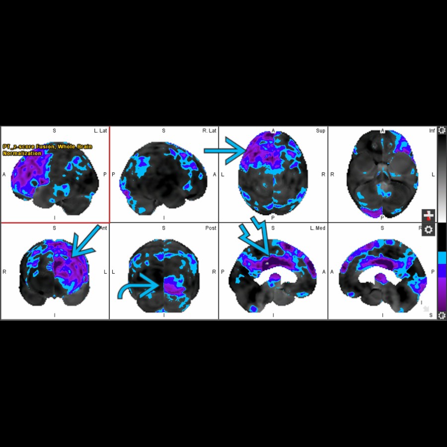
*Surface mapping of an FDG PET scan performed in a 96-year-old man demonstrates multiple areas of significant cortical abnormality in glucose metabolism, including the left frontal lobe , the right occipital lobe and the cingulate gyrus , consistent with multiinfarct dementia.*
 and globally decreased F-18 FDG uptake, also consistent with vascular dementia (VaD).](images/app.statdx.com_image_thumbnail_d2ae652b-f028-4e9e-9cac-3b360345030e_annotated_true_size_900_quality_90_492fa70c1930f37903e3700341b443818a337817.jpg)
*Axial F-18 FDG PET in a patient presenting with dementia shows more unilateral areas of hypometabolism and globally decreased F-18 FDG uptake, also consistent with vascular dementia (VaD).*
### Additional Images
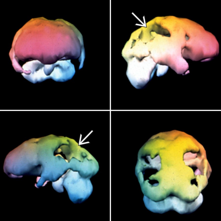
*Alzheimer dementia is shown. Note the parietal and posterior temporal reductions and sparing of occipital and frontal lobes.*
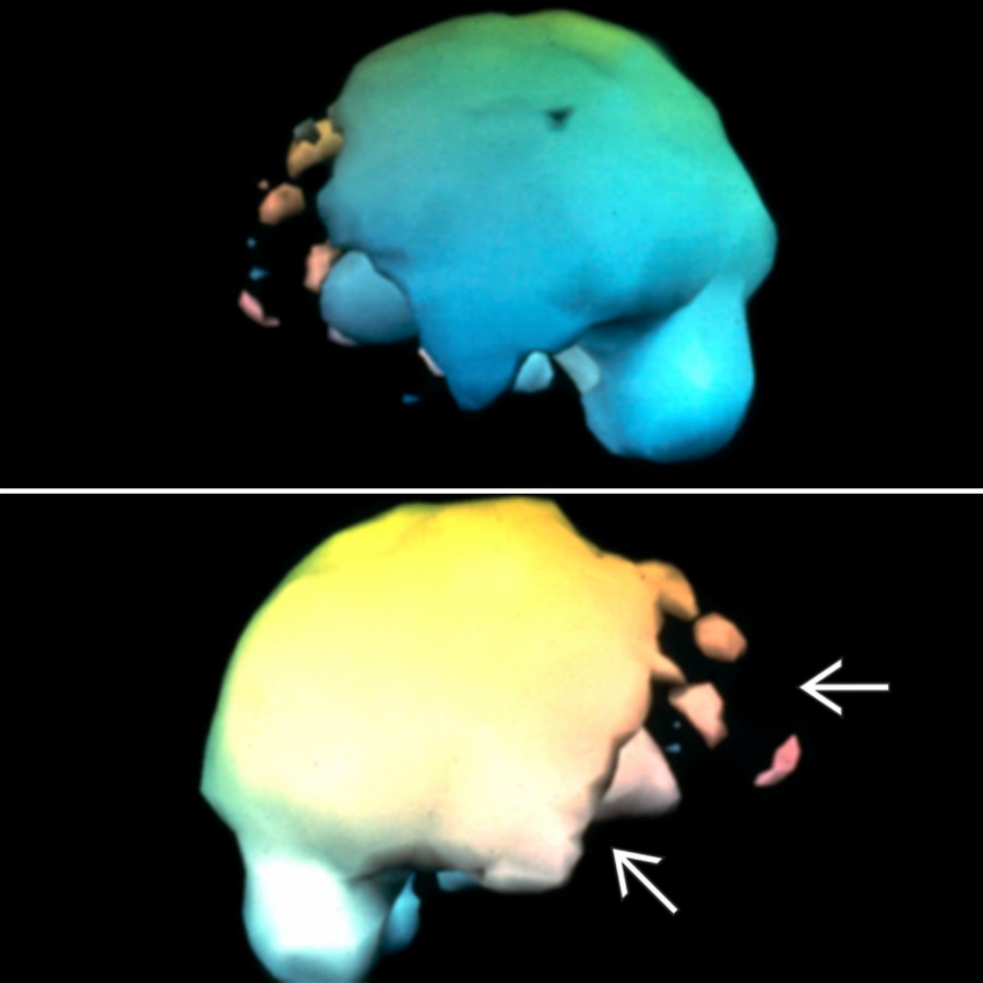
*Frontotemporal dementia (FTD) is shown. Note the frontal and anterior temporal reductions and sparing of parietal, posterior temporal, and occipital regions.*
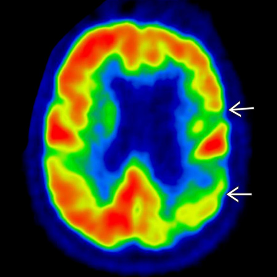
*Axial F-18 FDG PET in a patient presenting with dementia shows more unilateral areas of hypometabolism and globally decreased F-18 FDG uptake, also consistent with vascular dementia (VaD).*
### Additional Images

*Alzheimer dementia is shown. Note the parietal and posterior temporal reductions and sparing of occipital and frontal lobes.*

*Frontotemporal dementia (FTD) is shown. Note the frontal and anterior temporal reductions and sparing of parietal, posterior temporal, and occipital regions.*
 is also involved.](images/app.statdx.com_image_thumbnail_cae51cd3-ac69-4862-9d82-17078b4400c9_annotated_true_size_900_quality_90_631ae1b35164ac73c19d8d7005109487a4e6639f.jpg)
*Axial FDG PET in a patient with Lewy body disease shows parietal and posterior temporal reduction similar to Alzheimer disease (AD) , but occipital cortex is also involved.*
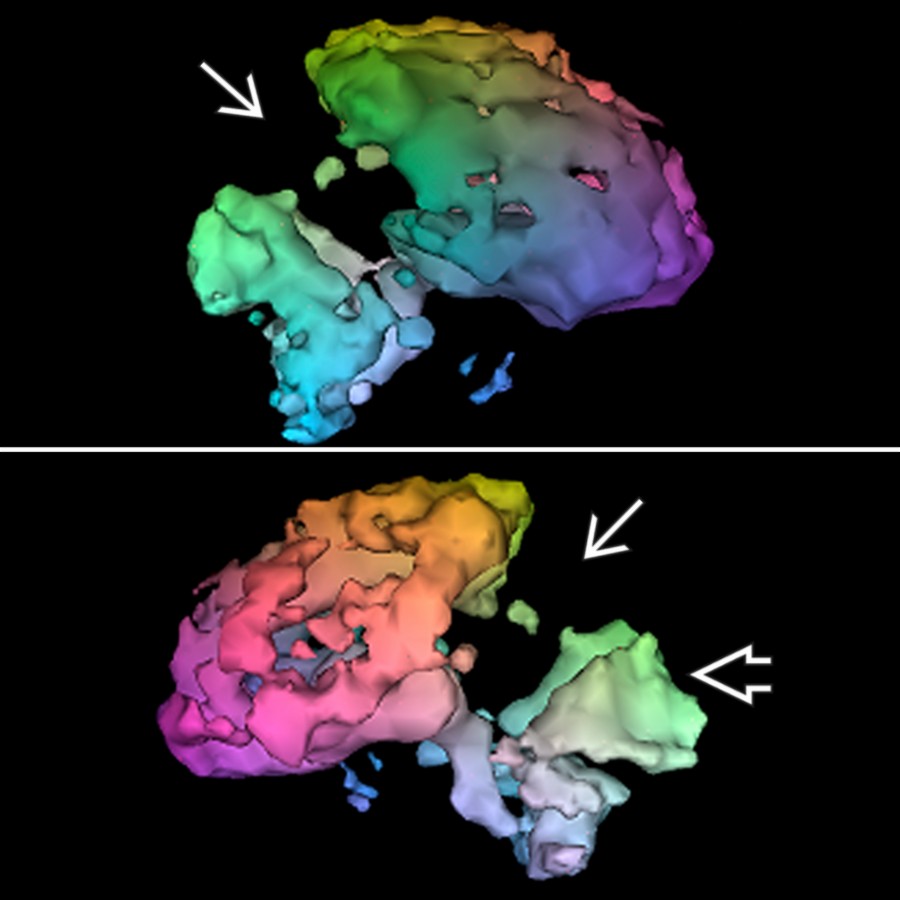
*Surface-rendered Tc-99m ECD SPECT in the same patient demonstrates severe reductions of parietal, temporal , and occipital cortex .*

*Axial FDG PET in a case of autopsy-proven Creutzfeldt-Jakob disease (CJD) shows multiple cortical defects .*

*Surface-rendered Tc-99m ECD SPECT shows a pattern similar to AD ; however, clinical course was that of a rapid progressive dementia ending in death within 12 months of onset.*

*Axial Tc-99m ECD SPECT in a patient with clinical progressive supranuclear palsy (PSP) presentation shows severe frontal lobe decrease and mild reduction in caudate heads .*

*Surface-rendered Tc-99m ECD SPECT demonstrates severe frontal decrease relative to parietal and occipital cortex .*

*Axial Tc-99m ECD SPECT (baseline) in a patient with early FTD shows frontal atrophy and mild reduction in perfusion .*

*Axial Tc-99m ECD SPECT in the same patient (18 months post baseline) demonstrates significant decrease of frontal lobe activity from baseline study , consistent with worsening dementia.*

*Surface-rendered Tc-99m ECD SPECT of the same patient (baseline) demonstrates mild frontal lobe findings .*

*Surface-rendered Tc-99m ECD SPECT 18 months post baseline demonstrates worsening .*

*Axial FDG PET in a patient with Huntington disease and mild dementia shows severe reduction in basal ganglia .*

*Surface-rendered Tc-99m ECD SPECT in the same patient shows mild frontal and parietal reductions .*

*Surface-rendered Tc-99m ECD SPECT in a patient with normal pressure hydrocephalus (NPH) shows severe frontal and parietal defects with preservation of vertex .*

*Axial FDG PET in a patient with Lewy body disease shows parietal and posterior temporal reduction similar to Alzheimer disease (AD) , but occipital cortex is also involved.*

*Surface-rendered Tc-99m ECD SPECT in the same patient demonstrates severe reductions of parietal, temporal , and occipital cortex .*

*Axial FDG PET in a case of autopsy-proven Creutzfeldt-Jakob disease (CJD) shows multiple cortical defects .*

*Surface-rendered Tc-99m ECD SPECT shows a pattern similar to AD ; however, clinical course was that of a rapid progressive dementia ending in death within 12 months of onset.*

*Axial Tc-99m ECD SPECT in a patient with clinical progressive supranuclear palsy (PSP) presentation shows severe frontal lobe decrease and mild reduction in caudate heads .*

*Surface-rendered Tc-99m ECD SPECT demonstrates severe frontal decrease relative to parietal and occipital cortex .*

*Axial Tc-99m ECD SPECT (baseline) in a patient with early FTD shows frontal atrophy and mild reduction in perfusion .*

*Axial Tc-99m ECD SPECT in the same patient (18 months post baseline) demonstrates significant decrease of frontal lobe activity from baseline study , consistent with worsening dementia.*

*Surface-rendered Tc-99m ECD SPECT of the same patient (baseline) demonstrates mild frontal lobe findings .*

*Surface-rendered Tc-99m ECD SPECT 18 months post baseline demonstrates worsening .*

*Axial FDG PET in a patient with Huntington disease and mild dementia shows severe reduction in basal ganglia .*

*Surface-rendered Tc-99m ECD SPECT in the same patient shows mild frontal and parietal reductions .*

*Surface-rendered Tc-99m ECD SPECT in a patient with normal pressure hydrocephalus (NPH) shows severe frontal and parietal defects with preservation of vertex .*
 .](b2483042-f995-4199-ab60-52d0e7c6144d)
*In-111 DTPA cisternogram (24 hours) in the same patient shows abnormal ventricular activity .*

*Axial Tc-99m ECD SPECT in a patient with multiinfarct dementia shows multiple infarcts of the frontal and parietal cortex .*

*Surface-rendered Tc-99m ECD SPECT in the same patient shows asymmetric cortical findings .*

*Surface-rendered Tc-99m ECD SPECT in a patient with history of cocaine abuse and early dementia shows diffuse cortical findings.*

*Surface-rendered Tc-99m ECD SPECT in a patient with history of methamphetamine abuse and early dementia is shown.*
.](b2483042-f995-4199-ab60-52d0e7c6144d)
*In-111 DTPA cisternogram (24 hours) in the same patient shows abnormal ventricular activity .*

*Axial Tc-99m ECD SPECT in a patient with multiinfarct dementia shows multiple infarcts of the frontal and parietal cortex .*

*Surface-rendered Tc-99m ECD SPECT in the same patient shows asymmetric cortical findings .*

*Surface-rendered Tc-99m ECD SPECT in a patient with history of cocaine abuse and early dementia shows diffuse cortical findings.*

*Surface-rendered Tc-99m ECD SPECT in a patient with history of methamphetamine abuse and early dementia is shown.*