---
title: "Normal Aging Brain"
docid: "2a315550-b2ea-4afe-a2ef-f93a2209f276"
authors:
- key: "a25c450b-3d34-4f64-bba3-cc0834813df6"
value: "Miral D. Jhaveri, MD, MBA"
breadcrumbs:
-
name: "Brain"
slug: "brain"
treeNodeId: "6d8829f1-14d7-45af-8675-255189aa526a"
-
name: "Diagnosis"
slug: "diagnosis"
treeNodeId: "51c00394-446e-4a38-94af-d3b1d14d34e8"
-
name: "Pathology-Based Diagnoses"
slug: "pathology-based-diagnoses"
treeNodeId: "d9d3a8ed-f21b-4831-8c77-591a3500ef77"
-
name: "Acquired Toxic/Metabolic/Degenerative Disorders"
slug: "acquired-toxicmetabolicdegenerativ-"
treeNodeId: "ba3cfeaf-64d9-4117-91e8-d2ce58783fc5"
-
name: "Dementias and Degenerative Disorders"
slug: "dementias-and-degenerative-disorde-"
treeNodeId: "6381104d-7a4c-4be5-bb19-3cd90837d547"
-
name: "Normal Aging Brain"
slug: "normal-aging-brain"
treeNodeId: null
category: "Brain"
cmeTopicId: "5933cd1e-de83-43b9-b172-894d762b8787"
documentVersionId: "7789baf5-4f7f-4519-b217-90bd5c560838"
imageCount: 31
lastUpdated: "09/29/20"
pageDescription: "Normal Aging Brain"
pageKeywords: "Brain, Diagnosis, Pathology-Based Diagnoses, Acquired Toxic/Metabolic/Degenerative Disorders, Dementias and Degenerative Disorders, Normal Aging Brain"
pageTitle: "Normal Aging Brain | STATdx"
enhancedTitle: "Normal Aging Brain"
type: "DX"
references: true
breadcrumbs:
- "Brain"
- "Diagnosis"
- "Pathology-Based Diagnoses"
- "Acquired Toxic/Metabolic/Degenerative Disorders"
- "Dementias and Degenerative Disorders"
- "Normal Aging Brain"
---
# KEY FACTS
- ## Terminology
- ↓ overall brain volume with advancing age
- Reflected in relative ↑ CSF spaces
- ## Imaging
- Broad spectrum of "normal" on imaging in elderly
- "Successfully aging brain"
- Smooth, thin, periventricular, high signal rim on FLAIR is normal
- White matter hyperintensities (WMHs) absent/few
- ↓ total brain volume
- Selective atrophy of white matter (not gray matter) predominates
- ± punctate hippocampal Ca⁺⁺
- Enlarged perivascular (Virchow-Robin) spaces
- WMHs ↑ in number/size after 50 years
- Focal high signal intensity in splenium of corpus callosum
- GRE/SWI
- Increasing mineralization of basal ganglia with age
- "Black line" in visual, motor/sensory cortex
- Microbleeds are relatively common in aging patients
- Lower prevalence than in cerebral amyloid disease & Alzheimer disease
- ## Top Differential Diagnoses
- Mild cognitive impairment
- Alzheimer disease
- Sporadic subcortical arteriosclerotic encephalopathy
- Vascular dementia
- Frontotemporal lobar degeneration
- ## Clinical Issues
- WMHs correlate with age, silent stroke, hypertension, female sex
- ## Diagnostic Checklist
- Cannot predict cognitive function from CT/MR
- If brain volume loss appears disproportionate to age, look for potential neurodegenerative or systemic causes
# TERMINOLOGY
- ## Definitions
- ↓ overall brain volume with advancing age
- Reflected in relative ↑ CSF spaces
# IMAGING
- ## General Features
- ### Best diagnostic clue
- "Successfully aging brain"
- Thin, periventricular, high-signal rim
- Absent/few white matter hyperintensities (WMHs)
- Mild shrinkage of selected cerebellar regions
- ### Location
- Selective atrophy of white matter (WM) predominates, not gray matter (GM)
- Striatum (primarily caudate nucleus, putamen)
- ### Size
- ↓ total brain volume
- Absolute striatal size
- Caudate ↓ linearly with age
- Putamen remains relatively stable
- Relative striatal size (ratio of absolute size:total brain volume)
- Caudate remains relatively stable
- Putamen ↑ linearly with age
- ### Morphology
- Brain tissue ↓, CSF volume ↑
- Reflects overall WM volume loss > focal WMHs
- Rounded appearance of dilated ventricles, sulci ↑
- Strong correlation between WM volume and CSF volume: Measure of overall brain atrophy
- ## CT Findings
- ### NECT
- Enlarged ventricles, widened cortical sulci
- Patchy periventricular low densities
- ± symmetrical, punctate calcifications in globi pallidi (GP)
- ± curvilinear vascular Ca⁺⁺
- ± punctate hippocampal Ca⁺⁺
- ### CECT
- No parenchymal enhancement
- ## MR Findings
- ### T1WI
- Mild but symmetric ventricular enlargement, proportionate prominence of subarachnoid spaces
- Mild but significant age-related shrinkage of
- Posterior vermis (lobules 6, 7, and 8-10)
- Cerebellar hemispheres
- Apparent age invariance of anterior vermis, ventral pons
- Enlarged perivascular (Virchow-Robin) spaces
- Common in aging, considered reflection of cerebral small vessel disease
- Isointense to CSF on all sequences
- Conform to course of penetrating arteries
- Round/oval/curvilinear
- Smooth, well-defined margins
- Bilateral, often symmetrical; usually no mass effect
- ↑ in number, size (> 2 mm) with age
- Can be found in most areas
- Midbrain, hippocampi, basal ganglia (BG), & centrum semiovale
- Tend to cluster around anterior commissure
- Inferior 1/3 of putamen, external capsule
- ### T2WI
- Focal/confluent periventricular WMHs
- Number/size ↑ after 50 years; ~ universal after 65 years
- Only rough correlation with cognitive function
- Significant overlap with dementias
- Infarct-like T2-hyperintense lesions
- Seen in 1/3 of asymptomatic patients > 65 years
- 70% < 10 mm
- Mostly in BG, thalami
- Probably represent clinically silent lacunar infarcts
- ### FLAIR
- Smooth, thin, periventricular hyperintense rim is normal
- Focal high signal intensity in splenium of corpus callosum
- BG and thalamic foci
- Perivascular spaces suppress
- Lacunar infarcts hyperintense
- ### T2* GRE
- SWI: Increasing mineralization of BG with age
- Normal in GP, abnormal in thalamus
- Can see linear "waves" or conglomerate mineralization in GP
- Putaminal hypointensity less prominent until 8th decade
- Microbleeds are common in aging brain
- SWI demonstrates microbleeds in 20% of patients > 60 years and in 60-70% of patients > 80 years
- Microbleeds are relatively common in aging patients
- Lower prevalence than in cerebral amyloid disease & Alzheimer disease
- BG, brainstem, and cerebellar microbleeds indicative of chronic hypertensive encephalopathy
- Lobar and cortical microbleeds typical of amyloid angiopathy
- SWI: "Black line" in visual, motor/sensory cortex
- Common, normal in older patients
- ### DWI
- Small but significant ↑ water diffusibility
- ADC ↑
- DTI: Loss of fractional anisotropy in normal-appearing WM
- ### T1WI C+
- Age-related WMHs do not enhance
- If enhancing, consider acute/subacute lacunar infarct or metastases
- ### MRS
- Metabolite distribution varies among different brain regions
- Choline (Cho) content ↑ with aging
- Creatine (Cr) ↑ with aging
- N-acetyl aspartate (NAA) ↓ in cortex, centrum semiovale, temporal lobes
- ## Nuclear Medicine Findings
- ### PET
- Metabolic alterations common
- Global, regional changes in cerebral blood flow (CBF)
- Gradual ↓ in regional CBF of GM, WM
- Particularly in frontal lobes
- Age-related shift from anterior to posterior cortical metabolism
- Putamen receives primarily posterior cortical input
- Caudate receives relatively more anterior cortical input
- Relative glucose metabolic rate (rGMR) measured by FDG PET
- With age, rGMR ↑ in putamen and ↓ in caudate
- ↓ pre-/postsynaptic dopamine markers in BG
- Tc-99m HMPAO SPECT, Xe-133 inhalation show regional, global reduction in CBF
- ## Imaging Recommendations
- ### Best imaging tool
- MR with FLAIR, DWI, T2* GRE/SWI
# DIFFERENTIAL DIAGNOSIS
- ## Mild Cognitive Impairment
- Overlap with normal on standard imaging studies
- Associated with ↓ size and number of regions of brain activation in response to memory tasks despite normal-appearing brain on conventional MR
- Higher calculated hippocampal ADCs (not visible)
- Subtle hypoperfusion, hypometabolism in parahippocampal regions, cingulum, thalamus
- ↓ NAA
- [Alzheimer Disease](/document/alzheimer-disease/f71f5cf5-b1af-4c6d-b145-b4c10eec7b58)
- Parietal and temporal cortical atrophy
- Striking volume loss in hippocampi, entorhinal cortex
- Often coexisting microvascular disease, WMHs
- Striking temporoparietal hypometabolism, hypoperfusion
- ↓ NAA, ↑ myoinositol (mI)
- [Sporadic Subcortical Arteriosclerotic Encephalopathy](/document/chronic-hypertensive-encephalopathy/1afc1f3f-203d-4cdf-8d49-2283cb13d6db)
- Associated with hypertension
- Numerous WMHs (overlap with normal)
- Multiple lacunar infarcts
- Lenticular nuclei, pons, thalamus, internal capsule, and caudate nuclei
- Diffuse, confluent regions of periventricular WM involvement (leukoaraiosis)
- [Vascular Dementia](/document/vascular-dementia/f59dab57-c511-4369-8fcc-592421a4b8d1)
- Hyperintense lesions on T2WI and focal atrophy suggestive of chronic infarcts
- [Frontotemporal Lobar Degeneration](/document/frontotemporal-lobar-degeneration/49510d0e-acf7-45cb-9eb1-53f8193b0b6d)
- Asymmetric frontal, anterior temporal atrophy
- T2 hyperintensity in frontotemporal WM
- Dilated subarachnoid space over frontal lobes signifying atrophy
- ↓ metabolic activity in frontotemporal cortices
# PATHOLOGY
- ## General Features
- ### Etiology
- Previous conception of aging: Substantial cortical neuronal loss with age
- New: Predominant neuroanatomic changes
- WM alterations, subcortical neuronal loss
- Reduction in cell size > cell number
- Neuronal dysfunction rather than loss of neurons/synapses
- ↓ neuronal viability or function associated with accelerated membrane degradation &/or ↑ glial cell numbers
- Loss of synapses and dendritic pruning in selected areas rather than globally
- Some investigators consider accumulation of neurofibrillary tangles (NFTs) may be responsible for memory loss associated with aging
- ### Genetics
- Clearly affect aging of brain
- Apolipoprotein E (*APOE*) and 6 novel risk-associated single nucleotide polymorphisms (SNPs) on chromosome 17q25 associated with brain pathology in aging
- ## Gross Pathologic & Surgical Features
- Widened sulci, proportionate large ventricles
- Minor thinning of cortical mantle, predominant changes in subcortical WM
- ## Microscopic Features
- Degeneration of neurons and oligodendrocytes
- ↓ myelinated fibers in subcortical WM
- ↑ extracellular space, gliosis
- Iron deposition in GP, putamen
- WM capillaries lose pericytes, have thinner endothelium
- Dilated perivascular spaces of Virchow-Robin
- Extension of subarachnoid space that accompanies penetrating vessels into brain to level of capillaries
- Senile plaques
- Extracellular amyloid deposits in cerebral GM
- Lewy bodies
- Intraneuronal clumps of α-synuclein and ubiquitin proteins
- Found in 5-10% of cognitively intact individuals
- NFTs
- Tau phosphorylation, mitochondrial dysfunction may precede full NFT formation
- NFTs appear in small numbers in entorhinal and transentorhinal cortices early in aging (patients ~ 60 years)
- NFTs may induce neural dysfunction, destruction of synapses, and, eventually, neuronal death
# CLINICAL ISSUES
- ## Presentation
- ### Most common signs/symptoms
- Normal cognitive function
- Mild cognitive impairment correlates with ↑ risk of Alzheimer disease
- ## Demographics
- ### Age
- > 60 years
- ### Sex
- Differences in striatal size
- Relatively constant across lifespan in men
- Variable across lifespan in women: Smaller in women aged 50-70 years than in men
- Differences in rGMR
- Caudate: Higher rGMR in women than men
- Putamen: Equal rGMR in women and men
- Greater dopamine transporters in caudate in women
- ### Epidemiology
- WMHs correlate with age, silent stroke, hypertension, female sex
- ## Natural History & Prognosis
- Parenchymal volume ↓, CSF spaces ↑ progressively
- WMHs progressively ↑ with age
# DIAGNOSTIC CHECKLIST
- ## Consider
- Striatum may mediate age-associated cognitive decline
- ↓ volume, functional activity with age
- ## Image Interpretation Pearls
- Broad spectrum of "normal" on imaging in elderly
- Cannot predict cognitive function from CT/MR
- If brain volume loss appears disproportionate to age, look for potential neurodegenerative or systemic causes
a71a0c8f-d105-4815-9edf-3fc113d5acc4
## References
# Selected References
1. [Dubost F et al: Enlarged perivascular spaces in brain MRI: automated quantification in four regions. Neuroimage. 185:534-44, 2019](http://www.ncbi.nlm.nih.gov/pubmed/?term=30326293%5Bpmid%5D)
1. [Jonkman LE et al: Normal Aging Brain Collection Amsterdam (NABCA): a comprehensive collection of postmortem high-field imaging, neuropathological and morphometric datasets of non-neurological controls. Neuroimage Clin. 22:101698, 2019](http://www.ncbi.nlm.nih.gov/pubmed/?term=30711684%5Bpmid%5D)
1. [Walker L et al: Neurodegenerative diseases and ageing. Subcell Biochem. 91:75-106, 2019](http://www.ncbi.nlm.nih.gov/pubmed/?term=30888650%5Bpmid%5D)
1. [de Brouwer EJM et al: Hippocampal calcifications: risk factors and association with cognitive function. Radiology. 288(3):815-20, 2018](http://www.ncbi.nlm.nih.gov/pubmed/?term=29893650%5Bpmid%5D)
1. [Haller S et al: Cerebral microbleeds: imaging and clinical significance. Radiology. 287(1):11-28, 2018](http://www.ncbi.nlm.nih.gov/pubmed/?term=29558307%5Bpmid%5D)
1. [Xekardaki A et al: Neuropathological changes in aging brain. Adv Exp Med Biol. 821:11-7, 2015](http://www.ncbi.nlm.nih.gov/pubmed/?term=25416106%5Bpmid%5D)
1. [van Velsen EF et al: Brain cortical thickness in the general elderly population: the Rotterdam Scan Study. Neurosci Lett. 550:189-94, 2013](http://www.ncbi.nlm.nih.gov/pubmed/?term=23831346%5Bpmid%5D)
1. [Poels MM et al: Arterial stiffness and cerebral small vessel disease: the Rotterdam Scan Study. Stroke. 43(10):2637-42, 2012](http://www.ncbi.nlm.nih.gov/pubmed/?term=22879099%5Bpmid%5D)
1. [Ikram MA et al: The Rotterdam Scan Study: design and update up to 2012. Eur J Epidemiol. 26(10):811-24, 2011](http://www.ncbi.nlm.nih.gov/pubmed/?term=22002080%5Bpmid%5D)
1. [Poels MM et al: Incidence of cerebral microbleeds in the general population: the Rotterdam Scan Study. Stroke. 42(3):656-61, 2011](http://www.ncbi.nlm.nih.gov/pubmed/?term=21307170%5Bpmid%5D)
1. [Ni JM et al: Regional diffusion changes of cerebral grey matter during normal aging--a fluid-inversion prepared diffusion imaging study. Eur J Radiol. 75(2):134-8, 2010](http://www.ncbi.nlm.nih.gov/pubmed/?term=19443158%5Bpmid%5D)
1. [Williams LR et al: Clinical correlates of cerebral white matter hyperintensities in cognitively normal older adults. Arch Gerontol Geriatr. 50(2):127-31, 2010](http://www.ncbi.nlm.nih.gov/pubmed/?term=19356807%5Bpmid%5D)
1. [Salat DH et al: Regional white matter volume differences in nondemented aging and Alzheimer's disease. Neuroimage. 44(4):1247-58, 2009](http://www.ncbi.nlm.nih.gov/pubmed/?term=19027860%5Bpmid%5D)
1. [Galluzzi S et al: Aging. Neurol Sci. 29 Suppl 3:296-300, 2008](http://www.ncbi.nlm.nih.gov/pubmed/?term=18941717%5Bpmid%5D)
1. [Gruber S et al: Metabolic changes in the normal ageing brain: consistent findings from short and long echo time proton spectroscopy. Eur J Radiol. 68(2):320-7, 2008](http://www.ncbi.nlm.nih.gov/pubmed/?term=17964104%5Bpmid%5D)
1. [Harder SL et al: Mineralization of the deep gray matter with age: a retrospective review with susceptibility-weighted MR imaging. AJNR Am J Neuroradiol. 29(1):176-83, 2008](http://www.ncbi.nlm.nih.gov/pubmed/?term=17989376%5Bpmid%5D)
1. [Kövari E et al: Cortical microinfarcts and demyelination significantly affect cognition in brain aging. Stroke. 35(2):410-4, 2004](http://www.ncbi.nlm.nih.gov/pubmed/?term=14707236%5Bpmid%5D)
1. [Brickman AM et al: Striatal size, glucose metabolic rate, and verbal learning in normal aging. Brain Res Cogn Brain Res. 17(1):106-16, 2003](http://www.ncbi.nlm.nih.gov/pubmed/?term=12763197%5Bpmid%5D)
## Images
### Selected Images
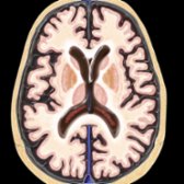
*Axial graphic depicts a normally aging brain in an 80-year-old patient. Note the widening of sulci & ventricles in the absence of any brain parenchymal abnormalities.*
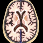
*Axial graphic depicts a normally aging brain in an 80-year-old patient. Note the widening of sulci & ventricles in the absence of any brain parenchymal abnormalities.*
*Axial graphic depicts a normally aging brain in an 80-year-old patient. Note the widening of sulci & ventricles in the absence of any brain parenchymal abnormalities.*
*Axial graphic depicts a normally aging brain in an 80-year-old patient. Note the widening of sulci & ventricles in the absence of any brain parenchymal abnormalities.*
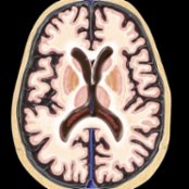
*Axial graphic depicts a normally aging brain in an 80-year-old patient. Note the widening of sulci & ventricles in the absence of any brain parenchymal abnormalities.*
 & subtle hyperintensity in the splenium
  of corpus callosum is common & normal. In addition, there are few white matter FLAIR hyperintensities
  .](images/app.statdx.com_image_thumbnail_11947b5d-e12f-4106-af97-1347d785f408_size_168_quality_85_771096ac_20251014T204549Z.jpg)
*Axial FLAIR MR in a 78-year-old man shows prominence of the ventricles & sulci due to age-related volume loss. Smooth, thin, periventricular hyperintense rim & subtle hyperintensity in the splenium of corpus callosum is common & normal. In addition, there are few white matter FLAIR hyperintensities .*
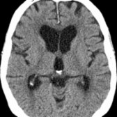
*Axial NECT in a 83-year-old man demonstrates mild sulcal enlargement & mild ventriculomegaly . The white matter appears relatively normal with subtle periventricular hypodensities.*
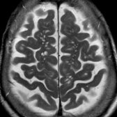
*Axial T2 MR in a 84-year-old woman demonstrates numerous enlarged perivascular spaces in the centrum semiovale bilaterally . Note prominence of the cortical sulci due to age-related volume loss. Enlarged perivascular spaces are commonly seen in the midbrain, hippocampi, basal ganglia, & centrum semiovale.*
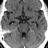
*Axial NECT in a 82-year-old patient shows subtle calcifications in the region of the hippocampal formations bilaterally .*
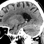
*Sagittal NECT in the same patient demonstrates punctate calcification in the region of the tail of the hippocampus . Note normal calcification in the choroid plexus within the atrium of the lateral ventricle. Hippocampal calcifications are seen with greater prevalence over the age of 50.*
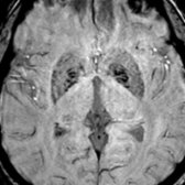
*Axial SWI MR in a 76-year-old patient demonstrates horizontal linear "waves" of mineralization in the globus pallidi , a normal finding in the aging brain. There is less prominent hypointensity in the putamina due to iron deposition.*
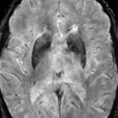
*Axial SWI MR in an 85-year-old patient demonstrates marked fairly symmetric hypointensity in the basal ganglia related to normal mineralization with age. Putaminal hypointensity on SWI is less prominent until 8th decade.*
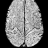
*Axial SWI MR in an 83-year-old woman demonstrates linear hypointensity along the cortical motor area referred to as cortical pencil lining. This relates to natural age-related iron accumulation in healthy brains.*
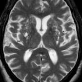
*Axial T2 MR in 82-year-old man demonstrates enlarged perivascular spaces in the basal ganglia bilaterally giving a cribriform appearance. With advancing age, perivascular spaces are found with increasing frequency & larger apparent size (> 2 mm).*
### Additional Images
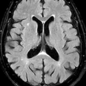
*Axial FLAIR MR in a 78-year-old man shows prominence of the ventricles & sulci due to age-related volume loss. Smooth, thin, periventricular hyperintense rim & subtle hyperintensity in the splenium of corpus callosum is common & normal. In addition, there are few white matter FLAIR hyperintensities .*

*Axial NECT in a 83-year-old man demonstrates mild sulcal enlargement & mild ventriculomegaly . The white matter appears relatively normal with subtle periventricular hypodensities.*

*Axial T2 MR in a 84-year-old woman demonstrates numerous enlarged perivascular spaces in the centrum semiovale bilaterally . Note prominence of the cortical sulci due to age-related volume loss. Enlarged perivascular spaces are commonly seen in the midbrain, hippocampi, basal ganglia, & centrum semiovale.*

*Axial NECT in a 82-year-old patient shows subtle calcifications in the region of the hippocampal formations bilaterally .*

*Sagittal NECT in the same patient demonstrates punctate calcification in the region of the tail of the hippocampus . Note normal calcification in the choroid plexus within the atrium of the lateral ventricle. Hippocampal calcifications are seen with greater prevalence over the age of 50.*

*Axial SWI MR in a 76-year-old patient demonstrates horizontal linear "waves" of mineralization in the globus pallidi , a normal finding in the aging brain. There is less prominent hypointensity in the putamina due to iron deposition.*

*Axial SWI MR in an 85-year-old patient demonstrates marked fairly symmetric hypointensity in the basal ganglia related to normal mineralization with age. Putaminal hypointensity on SWI is less prominent until 8th decade.*

*Axial SWI MR in an 83-year-old woman demonstrates linear hypointensity along the cortical motor area referred to as cortical pencil lining. This relates to natural age-related iron accumulation in healthy brains.*

*Axial T2 MR in 82-year-old man demonstrates enlarged perivascular spaces in the basal ganglia bilaterally giving a cribriform appearance. With advancing age, perivascular spaces are found with increasing frequency & larger apparent size (> 2 mm).*
### Additional Images
 .](images/app.statdx.com_image_thumbnail_5fd4a977-5e2b-4e3a-92e5-eb062a73a31a_size_168_quality_85_804ad9ba_20251014T204549Z.jpg)
*Axial T2WI MR shows atrophy & white matter changes of aging .*
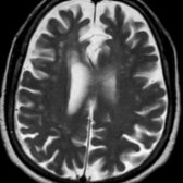
*Axial T2WI MR shows atrophy & white matter changes of aging .*
 , & fasciculi nigrae
  .](images/app.statdx.com_image_thumbnail_b504b9ca-3242-4005-aaa6-746a383c7666_size_168_quality_85_915188ee_20251014T204549Z.jpg)
*Axial T2* SWI MR reveals normal mineralization of the red nucleus , substantia nigra , & fasciculi nigrae .*
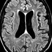
*Axial FLAIR MR in an elderly patient shows dilated ventricles, wide cortical sulci, & periventricular white matter hyperintensity.*
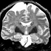
*Coronal T2WI MR in an elderly patient shows marked hypointensity in both lenticular nuclei & wide cortical sulci.*
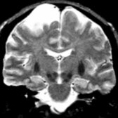
*Coronal T2WI MR in the same individual shows hypointense putamina & normal hippocampal size despite loss of brain cortex.*
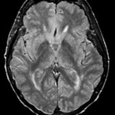
*Axial PD FSE intermediate MR in a 79-year-old patient without cognitive impairment shows mild periventricular white matter hyperintensities.*
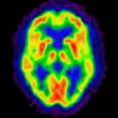
*FDG PET in a normal 83-year-old shows normal metabolism in the brain cortex, basal ganglia, & thalami. (Courtesy N. Foster, MD & the University of Michigan PET Center.)*
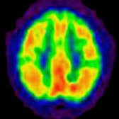
*FDG PET in the same patient shows essentially normal glucose metabolism (depicted in red & yellow) in the cerebral cortex, except for small regions of decreased metabolism.*
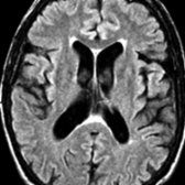
*Axial FLAIR shows mild periventricular hyperintensity & mild enlargement of the ventricles & sulci in a 65-year-old man. No focal hyperintensities are seen in the hemispheric white matter; cortical thickness & signal intensity are normal.*
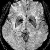
*Axial T2* SWI MR demonstrates hypointensity in the lentiform nuclei, particularly related to the globus pallidi .*
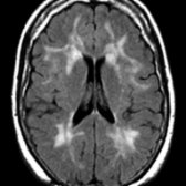
*Axial FLAIR MR demonstrates confluent hyperintense white matter changes that may occur during normal aging.*
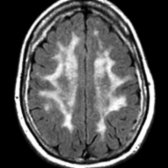
*Axial FLAIR MR in the same patient demonstrates prominent subcortical white matter hyperintensity .*
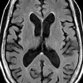
*Axial FLAIR MR in a 79-year-old man shows enlargement of ventricles & sulci due to age-related volume loss. Smooth, thin, periventricular hyperintense rim & subtle hyperintensity in the splenium of corpus callosum is common & normal.*
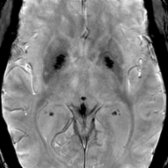
*Axial SWI in a 67-year-old woman shows striking hypointensity in the globi pallidi with less prominent hypointensity in the putamina due to iron deposition.*
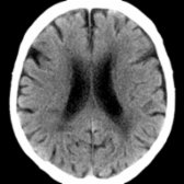
*Axial NECT in an 85-year-old patient without cognitive impairment shows wide sulci & lateral ventricles, as well as moderate periventricular hypodense white matter.*
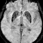
*Axial T2* SWI MR demonstrates marked hypointensity in the basal ganglia related to normal mineralization with age.*
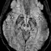
*Axial T2* SWI MR reveals normal mineralization of the red nucleus , substantia nigra , & fasciculi nigrae .*

*Axial FLAIR MR in an elderly patient shows dilated ventricles, wide cortical sulci, & periventricular white matter hyperintensity.*

*Coronal T2WI MR in an elderly patient shows marked hypointensity in both lenticular nuclei & wide cortical sulci.*

*Coronal T2WI MR in the same individual shows hypointense putamina & normal hippocampal size despite loss of brain cortex.*

*Axial PD FSE intermediate MR in a 79-year-old patient without cognitive impairment shows mild periventricular white matter hyperintensities.*

*FDG PET in a normal 83-year-old shows normal metabolism in the brain cortex, basal ganglia, & thalami. (Courtesy N. Foster, MD & the University of Michigan PET Center.)*

*FDG PET in the same patient shows essentially normal glucose metabolism (depicted in red & yellow) in the cerebral cortex, except for small regions of decreased metabolism.*

*Axial FLAIR shows mild periventricular hyperintensity & mild enlargement of the ventricles & sulci in a 65-year-old man. No focal hyperintensities are seen in the hemispheric white matter; cortical thickness & signal intensity are normal.*

*Axial T2* SWI MR demonstrates hypointensity in the lentiform nuclei, particularly related to the globus pallidi .*

*Axial FLAIR MR demonstrates confluent hyperintense white matter changes that may occur during normal aging.*

*Axial FLAIR MR in the same patient demonstrates prominent subcortical white matter hyperintensity .*

*Axial FLAIR MR in a 79-year-old man shows enlargement of ventricles & sulci due to age-related volume loss. Smooth, thin, periventricular hyperintense rim & subtle hyperintensity in the splenium of corpus callosum is common & normal.*

*Axial SWI in a 67-year-old woman shows striking hypointensity in the globi pallidi with less prominent hypointensity in the putamina due to iron deposition.*

*Axial NECT in an 85-year-old patient without cognitive impairment shows wide sulci & lateral ventricles, as well as moderate periventricular hypodense white matter.*

*Axial T2* SWI MR demonstrates marked hypointensity in the basal ganglia related to normal mineralization with age.*
 related to normal mineralization with age.](images/app.statdx.com_image_thumbnail_ae10c277-d1c0-4b33-835b-53f6d103b6a1_size_168_quality_85_0613b3de_20251014T204550Z.jpg)
*Axial T2* SWI MR demonstrates marked hypointensity in the basal ganglia related to normal mineralization with age.*
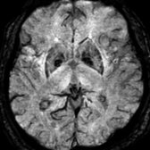
*Axial T2* SWI MR demonstrates horizontal linear "waves" of mineralization in the globus pallidi , a normal finding in the aging brain.*
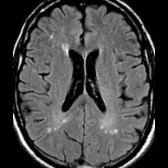
*Axial FLAIR in a 72-year-old man shows scattered white matter hyperintensities . Aging brains may demonstrate a few scattered nonconfluent white matter hyperintensities. Increased prevalence of white matter hyperintensities with cardiovascular risk factors such as diabetes & hyperlipidemia is shown.*
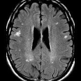
*Axial FLAIR in the same patient shows additional subcortical white matter hyperintensities .*
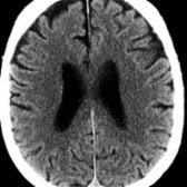
*Axial NECT demonstrates mild sulcal enlargement & mild ventriculomegaly in a 70-year-old patient. The white matter appears completely normal, without periventricular hypodensities or white matter lacunar infarcts.*
related to normal mineralization with age.](images/app.statdx.com_image_thumbnail_ae10c277-d1c0-4b33-835b-53f6d103b6a1_size_168_quality_85_0613b3de_20251014T204550Z.jpg)
*Axial T2* SWI MR demonstrates marked hypointensity in the basal ganglia related to normal mineralization with age.*

*Axial T2* SWI MR demonstrates horizontal linear "waves" of mineralization in the globus pallidi , a normal finding in the aging brain.*

*Axial FLAIR in a 72-year-old man shows scattered white matter hyperintensities . Aging brains may demonstrate a few scattered nonconfluent white matter hyperintensities. Increased prevalence of white matter hyperintensities with cardiovascular risk factors such as diabetes & hyperlipidemia is shown.*

*Axial FLAIR in the same patient shows additional subcortical white matter hyperintensities .*

*Axial NECT demonstrates mild sulcal enlargement & mild ventriculomegaly in a 70-year-old patient. The white matter appears completely normal, without periventricular hypodensities or white matter lacunar infarcts.*