---
title: "Quadrigeminal Cistern Mass"
docid: "828ddbbf-3daf-45b9-8dce-2ab3582fddaa"
authors:
- key: "f184750a-90b4-47a7-907b-23b05d70357a"
value: "Chang Yueh Ho, MD"
breadcrumbs:
-
name: "Brain"
slug: "brain"
treeNodeId: "6d8829f1-14d7-45af-8675-255189aa526a"
-
name: "Differential Diagnosis"
slug: "differential-diagnosis"
treeNodeId: "a7fdd139-664e-4bb8-8d18-400e4733ff60"
-
name: "Sella/Juxtasellar, Pineal Region"
slug: "sellajuxtasellar-pineal-region"
treeNodeId: "5e38b9c1-3137-47e3-aa83-1fc82cb4099a"
-
name: "Anatomically Based Differentials"
slug: "anatomically-based-differentials"
treeNodeId: "7a51b2ca-8fee-4c16-aff3-b7189f68ea60"
-
name: "Quadrigeminal Cistern Mass"
slug: "quadrigeminal-cistern-mass"
treeNodeId: null
category: "Brain"
documentVersionId: "f1b37d5a-a0fe-4dbb-9e53-9f3a204d6cec"
imageCount: 31
lastUpdated: "01/27/23"
pageDescription: "Quadrigeminal Cistern Mass"
pageKeywords: "Brain, Differential Diagnosis, Sella/Juxtasellar, Pineal Region, Anatomically Based Differentials, Quadrigeminal Cistern Mass"
pageTitle: "Quadrigeminal Cistern Mass | STATdx"
enhancedTitle: "Quadrigeminal Cistern Mass"
type: "DDX"
references: true
breadcrumbs:
- "Brain"
- "Differential Diagnosis"
- "Sella/Juxtasellar, Pineal Region"
- "Anatomically Based Differentials"
- "Quadrigeminal Cistern Mass"
---
# ESSENTIAL INFORMATION
- ## Key Differential Diagnosis Issues
- Quadrigeminal cistern (QC) lesions are smaller subset of pineal region masses
- Bounded by quadrigeminal plate, splenium, vermis, & tentorial margin
- Contents: Pia, pineal gland, caudal internal cerebral veins, vein of Galen, posterior cerebral artery, posteromedial choroidal arteries, CNIV
- Masses arising from QC itself (& its contents) < < those from nearby structures
- ## Helpful Clues for Common Diagnoses
- **Leptomeningeal Metastases**
- From primary CNS or non-CNS neoplasms
- Linear &/or nodular enhancing lesions
- Image entire neuraxis
- ## Helpful Clues for Less Common Diagnoses
- **Cyst of Velum Interpositum**
- Axial MR/CT shows triangular-shaped CSF space between bodies of lateral ventricles
- Follows CSF on all sequences
- Mimics arachnoid cyst of QC
- Splays & elevates fornix
- Inferiorly displaces internal cerebral veins
- **Medial Atrial Diverticulum**
- Massive hydrocephalus causes dehiscence of fornix → diverticula of inferior medial atrial wall
- Creates subarachnoid cyst that may herniate medial to occipital lobe into QC
- **Arachnoid Cyst**
- Sharply demarcated extraaxial cyst that follows CSF
- May be eccentric
- Elevates internal cerebral veins
- **Neurocysticercosis**
- Cystic lesion isointense to CSF; may see discrete, eccentric scolex
- Basal cistern cysts may be racemose
- **CNS Tuberculoma**
- Ring-enhancing abscess without central ↓ diffusion
- ## Helpful Clues for Rare Diagnoses
- **Lipoma**
- Well-delineated, lobulated, extraaxial mass with fat attenuation/intensity
- Ca⁺⁺ varies from none to extensive
- **Epidermoid Cyst**
- Lobulated, irregular, CSF-like mass
- Incomplete FLAIR suppression; diffusion restriction
- Surface ectoderm rests included with neural tube closure
- **Dermoid Cyst**
- Fat containing: MR fat suppression or negative HU
- May rupture with sulcal fat & chemical meningitis
- **Vein of Galen Malformation**
- Misnomer: Arteriovenous fistula draining into enlarged median prosencephalic vein
- Dilated arteries feeding into large midline venous pouch
- Look for prominent flow voids & phase artifact
## References
# Selected References
1. [Silva MA et al: Surgical management of quadrigeminal cistern arachnoid cysts: case series and literature review. J Neurosurg Pediatr. 29(4):427-34, 2022](http://www.ncbi.nlm.nih.gov/pubmed/?term=34996040%5Bpmid%5D)
1. [Smith AB et al: From the archives of the AFIP: lesions of the pineal region: radiologic-pathologic correlation. Radiographics. 30(7):2001-20, 2010](http://www.ncbi.nlm.nih.gov/pubmed/?term=21057132%5Bpmid%5D)
## Images
### Selected Images
 in the quadrigeminal cistern in this child with a primary tumor in the spinal cord. This was a glioneuronal tumor at resection.](images/app.statdx.com_image_thumbnail_e4f8b8b3-99fa-4f6f-8b62-e22e1cf97177_annotated_true_size_900_quality_90_74f5c5f7.jpg)
**Leptomeningeal Metastases**
*Axial T1 C+ MR shows abnormal leptomeningeal enhancement in the quadrigeminal cistern in this child with a primary tumor in the spinal cord. This was a glioneuronal tumor at resection.*

**Leptomeningeal Metastases**
*Axial T1 C+ MR shows abnormal leptomeningeal enhancement in the quadrigeminal cistern in this child with a primary tumor in the spinal cord. This was a glioneuronal tumor at resection.*

**Leptomeningeal Metastases**
*Axial T1 C+ MR shows abnormal leptomeningeal enhancement in the quadrigeminal cistern in this child with a primary tumor in the spinal cord. This was a glioneuronal tumor at resection.*

**Leptomeningeal Metastases**
*Axial T1 C+ MR shows abnormal leptomeningeal enhancement in the quadrigeminal cistern in this child with a primary tumor in the spinal cord. This was a glioneuronal tumor at resection.*
 .](images/app.statdx.com_image_thumbnail_a42409a3-5856-41bf-8b70-08f230be8087_annotated_true_size_900_quality_90_e1cb7a0c.jpg)
**Medial Atrial Diverticulum**
*Sagittal T2 MR in a child with hydrocephalus shows markedly enlarged ventricles. The left lateral ventricle herniates through the medial wall of the atria, through the quadrigeminal plate cistern and compresses the cerebellum inferiorly below the tentorium .*

**Arachnoid Cyst**
*Sagittal T2 MR shows a large cyst centered in the quadrigeminal cistern with mass effect on the cerebellum inferiorly , which elevates the flow voids of the internal cerebral veins .*

**Neurocysticercosis**
*Axial T1 C+ MR shows abnormal leptomeningeal enhancement in the right suprasellar cistern with nonenhancing racemose cysts in the left ambient and quadrigeminal plate cistern. This patient had a history of neurocysticercosis.*

**CNS Tuberculoma**
*Axial T1 C+ MR shows multiple ring-enhancing lesions in the parenchyma and basal cisterns. Note the nodular enhancement in the ambient and quadrigeminal plate cistern in this patient with CNS tuberculosis.*

**Lipoma**
*Axial T2 FS MR shows a tectal mass projecting into the quadrigeminal plate cistern with signal loss . This is consistent with a tectal lipoma. Intracranial lipomas are typically midline and can be associated with malformations, particularly the corpus callosum.*

**Epidermoid Cyst**
*Axial DWI trace MR shows a hyperintense mass in the left quadrigeminal cistern . This followed CSF on T1 and T2 but had incomplete FLAIR suppression. Epidermoid cysts are from inclusion of surface ectoderm during neural tube closure with midline predominance.*

**Medial Atrial Diverticulum**
*Sagittal T2 MR in a child with hydrocephalus shows markedly enlarged ventricles. The left lateral ventricle herniates through the medial wall of the atria, through the quadrigeminal plate cistern and compresses the cerebellum inferiorly below the tentorium .*

**Arachnoid Cyst**
*Sagittal T2 MR shows a large cyst centered in the quadrigeminal cistern with mass effect on the cerebellum inferiorly , which elevates the flow voids of the internal cerebral veins .*

**Neurocysticercosis**
*Axial T1 C+ MR shows abnormal leptomeningeal enhancement in the right suprasellar cistern with nonenhancing racemose cysts in the left ambient and quadrigeminal plate cistern. This patient had a history of neurocysticercosis.*

**CNS Tuberculoma**
*Axial T1 C+ MR shows multiple ring-enhancing lesions in the parenchyma and basal cisterns. Note the nodular enhancement in the ambient and quadrigeminal plate cistern in this patient with CNS tuberculosis.*

**Lipoma**
*Axial T2 FS MR shows a tectal mass projecting into the quadrigeminal plate cistern with signal loss . This is consistent with a tectal lipoma. Intracranial lipomas are typically midline and can be associated with malformations, particularly the corpus callosum.*

**Epidermoid Cyst**
*Axial DWI trace MR shows a hyperintense mass in the left quadrigeminal cistern . This followed CSF on T1 and T2 but had incomplete FLAIR suppression. Epidermoid cysts are from inclusion of surface ectoderm during neural tube closure with midline predominance.*
 and superior sagittal sinus. The mass causes cerebellar tonsil herniation .](51a57381-802a-48ee-97e3-033f55cbff6b)
**Vein of Galen Malformation**
*Sagittal T2 HASTE MR shows a large, hypointense mass in the quadrigeminal cistern from a large flow void of the mesencephalic vein . Note dilated straight sinus and superior sagittal sinus. The mass causes cerebellar tonsil herniation .*
### Additional Images

**Vein of Galen Malformation**
*Sagittal T2 HASTE MR shows a large, hypointense mass in the quadrigeminal cistern from a large flow void of the mesencephalic vein . Note dilated straight sinus and superior sagittal sinus. The mass causes cerebellar tonsil herniation .*
### Additional Images
 involving quadrigeminal cistern as well as other structures via subarachnoid spread.](images/app.statdx.com_image_thumbnail_acb8f609-ee96-4c23-b72a-45ae378c46a6_annotated_true_size_900_quality_90_490ebe01.jpg)
**Leptomeningeal Metastases**
*Axial T1 C+ MR demonstrates typical leptomeningeal (pia and arachnoid) metastases involving quadrigeminal cistern as well as other structures via subarachnoid spread.*

**Cyst of Velum Interpositum**
*Axial FLAIR MR shows a cavum velum interpositum as a CSF isointense mass , which suppresses completely on FLAIR.*

**Leptomeningeal Metastases**
*Axial T1 C+ MR demonstrates typical leptomeningeal (pia and arachnoid) metastases involving quadrigeminal cistern as well as other structures via subarachnoid spread.*

**Cyst of Velum Interpositum**
*Axial FLAIR MR shows a cavum velum interpositum as a CSF isointense mass , which suppresses completely on FLAIR.*
 extending into and expanding the quadrigeminal cistern .](93213916-ee96-48ab-a812-cc7c8ed7240f)
**Arachnoid Cyst**
*Sagittal T1WI MR shows a CSF intensity arachnoid cyst in the superior vermian cistern extending into and expanding the quadrigeminal cistern .*

**Arachnoid Cyst**
*Sagittal T1WI MR shows a CSF intensity arachnoid cyst in the superior vermian cistern extending into and expanding the quadrigeminal cistern .*
 .](6b0280a0-0162-43f2-a6f8-2a4718a7bcef)
**Neurocysticercosis**
*Axial T1 C+ MR shows multiple nonenhancing cysts in the quadrigeminal cistern and in the atrium of the right lateral ventricle .*

**Lipoma**
*Sagittal T1WI MR demonstrates a hyperintense, well-circumscribed lipoma in the quadrigeminal cistern.*

**Neurocysticercosis**
*Axial T1 C+ MR shows multiple nonenhancing cysts in the quadrigeminal cistern and in the atrium of the right lateral ventricle .*

**Lipoma**
*Sagittal T1WI MR demonstrates a hyperintense, well-circumscribed lipoma in the quadrigeminal cistern.*
 , which restricts on diffusion.](1eda3aaf-0ac2-4dbb-8d6a-74567d9c99a9)
**Epidermoid Cyst**
*Axial DWI MR shows a multilobulated epidermoid extending into the quadrigeminal cistern , which restricts on diffusion.*

**Dermoid Cyst**
*Axial NECT shows a typical, hypodense, fat-containing, ruptured dermoid . Fat droplets, also with negative HU, are present throughout the subarachnoid space .*

**Dermoid Cyst**
*Sagittal T1WI MR shows a ruptured quadrigeminal cistern dermoid as well as fat droplets throughout the subarachnoid space and ventricular fat-fluid levels .*

**Vein of Galen Malformation**
*Axial T1 C+ MR shows a very prominent phase artifact from a quadrigeminal cistern vein of Galen malformation with dural arteriovenous fistulae .*

**Leptomeningeal Metastases**
*Sagittal T1 C+ MR shows typical leptomeningeal (pia and arachnoid) metastases in the quadrigeminal cistern as well as widespread throughout the cerebellar folia.*

**Epidermoid Cyst**
*Axial DWI MR shows a multilobulated epidermoid extending into the quadrigeminal cistern , which restricts on diffusion.*

**Dermoid Cyst**
*Axial NECT shows a typical, hypodense, fat-containing, ruptured dermoid . Fat droplets, also with negative HU, are present throughout the subarachnoid space .*

**Dermoid Cyst**
*Sagittal T1WI MR shows a ruptured quadrigeminal cistern dermoid as well as fat droplets throughout the subarachnoid space and ventricular fat-fluid levels .*

**Vein of Galen Malformation**
*Axial T1 C+ MR shows a very prominent phase artifact from a quadrigeminal cistern vein of Galen malformation with dural arteriovenous fistulae .*

**Leptomeningeal Metastases**
*Sagittal T1 C+ MR shows typical leptomeningeal (pia and arachnoid) metastases in the quadrigeminal cistern as well as widespread throughout the cerebellar folia.*
 and compressing the quadrigeminal cistern .](images/app.statdx.com_image_thumbnail_57a3fd00-ac5b-45d9-b6e7-7026b903b949_annotated_true_size_900_quality_90_3723baad.jpg)
**Cyst of Velum Interpositum**
*Sagittal T1WI MR reveals a well-defined cavum velum interpositum , isointense with CSF, displacing the internal cerebral veins inferiorly and compressing the quadrigeminal cistern .*

**Arachnoid Cyst**
*Sagittal T1WI MR shows an arachnoid cyst compressing the vermis inferiorly , widening the quadrigeminal cistern , and extending into velum interpositum .*

**Neurocysticercosis**
*Sagittal STIR MR reveals multiple hyperintense cysts in the quadrigeminal cistern and basal subarachnoid spaces .*

**Lipoma**
*Sagittal T2WI MR demonstrates a fat intensity lipoma within the quadrigeminal cistern displacing the vermis, which suppressed with T1 fat suppression (not shown).*

**Epidermoid Cyst**
*Axial T1WI MR demonstrates a lobulated CSF-like epidermoid within the quadrigeminal cistern.*

**Vein of Galen Malformation**
*Sagittal T1WI MR demonstrates a prominent flow void within the quadrigeminal cistern from a vein of Galen malformation as well as associated fistulae .*

**Leptomeningeal Metastases**
*Coronal T1 C+ FS MR shows diffuse leptomeningeal enhancement involving the cerebellar folia , right temporal lobe , and structures surrounding the quadrigeminal plate cistern . This patient had a history of lung cancer.*
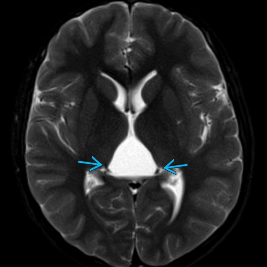
**Cyst of Velum Interpositum**
*Axial T2WI FS MR shows a cyst between the thalami and splaying the internal cerebral veins . Cysts of the cavum velum interpositum arise superior to the quadrigeminal cistern, but very large cysts may extend into or efface the quadrigeminal cistern.*

**Arachnoid Cyst**
*Sagittal T1 C+ MR shows a cyst dorsal to the normally enhancing pineal gland . There is mass effect on the superior vermis . This arachnoid cyst follows CSF on all sequences.*

**Neurocysticercosis**
*Axial T1 C+ FS MR shows leptomeningeal enhancement along the right suprasellar cistern extending along the right middle cerebral artery . Multiple cysts with enhancing rim extend along the right ambient cistern to the quadrigeminal cistern . Racemose neurocysticercosis presents with multiple extraaxial cysts.*

**Medial Atrial Diverticulum**
*Axial FLAIR MR shows medial bowing of the medial margin of the left atria of a dilated lateral ventricle, effacing the quadrigeminal plate cistern . The medial atrial wall is a weak point for pulsion diverticula from massive hydrocephalus.*

**Lipoma**
*Axial T2WI FS MR shows loss of signal in a quadrigeminal cistern mass . There was hyperintense T1 signal on T1WI, consistent with a lipoma. Lipomas are maldifferentiation, and persistence of the meninx primitiva and typically occur midline intracranially.*

**Epidermoid Cyst**
*Axial DWI b = 1000 MR shows a mass with uniform decreased diffusion arising in the quadrigeminal plate cistern. This lesion has a cystic appearance on T1 and T2 sequences (not shown) and was an epidermoid cyst at surgery.*
and compressing the quadrigeminal cistern .](images/app.statdx.com_image_thumbnail_57a3fd00-ac5b-45d9-b6e7-7026b903b949_annotated_true_size_900_quality_90_3723baad.jpg)
**Cyst of Velum Interpositum**
*Sagittal T1WI MR reveals a well-defined cavum velum interpositum , isointense with CSF, displacing the internal cerebral veins inferiorly and compressing the quadrigeminal cistern .*

**Arachnoid Cyst**
*Sagittal T1WI MR shows an arachnoid cyst compressing the vermis inferiorly , widening the quadrigeminal cistern , and extending into velum interpositum .*

**Neurocysticercosis**
*Sagittal STIR MR reveals multiple hyperintense cysts in the quadrigeminal cistern and basal subarachnoid spaces .*

**Lipoma**
*Sagittal T2WI MR demonstrates a fat intensity lipoma within the quadrigeminal cistern displacing the vermis, which suppressed with T1 fat suppression (not shown).*

**Epidermoid Cyst**
*Axial T1WI MR demonstrates a lobulated CSF-like epidermoid within the quadrigeminal cistern.*

**Vein of Galen Malformation**
*Sagittal T1WI MR demonstrates a prominent flow void within the quadrigeminal cistern from a vein of Galen malformation as well as associated fistulae .*

**Leptomeningeal Metastases**
*Coronal T1 C+ FS MR shows diffuse leptomeningeal enhancement involving the cerebellar folia , right temporal lobe , and structures surrounding the quadrigeminal plate cistern . This patient had a history of lung cancer.*

**Cyst of Velum Interpositum**
*Axial T2WI FS MR shows a cyst between the thalami and splaying the internal cerebral veins . Cysts of the cavum velum interpositum arise superior to the quadrigeminal cistern, but very large cysts may extend into or efface the quadrigeminal cistern.*

**Arachnoid Cyst**
*Sagittal T1 C+ MR shows a cyst dorsal to the normally enhancing pineal gland . There is mass effect on the superior vermis . This arachnoid cyst follows CSF on all sequences.*

**Neurocysticercosis**
*Axial T1 C+ FS MR shows leptomeningeal enhancement along the right suprasellar cistern extending along the right middle cerebral artery . Multiple cysts with enhancing rim extend along the right ambient cistern to the quadrigeminal cistern . Racemose neurocysticercosis presents with multiple extraaxial cysts.*

**Medial Atrial Diverticulum**
*Axial FLAIR MR shows medial bowing of the medial margin of the left atria of a dilated lateral ventricle, effacing the quadrigeminal plate cistern . The medial atrial wall is a weak point for pulsion diverticula from massive hydrocephalus.*

**Lipoma**
*Axial T2WI FS MR shows loss of signal in a quadrigeminal cistern mass . There was hyperintense T1 signal on T1WI, consistent with a lipoma. Lipomas are maldifferentiation, and persistence of the meninx primitiva and typically occur midline intracranially.*

**Epidermoid Cyst**
*Axial DWI b = 1000 MR shows a mass with uniform decreased diffusion arising in the quadrigeminal plate cistern. This lesion has a cystic appearance on T1 and T2 sequences (not shown) and was an epidermoid cyst at surgery.*