---
title: "Sensorineural Hearing Loss in Adult"
docid: "08d468da-fbc3-44f8-8212-6480e0a152c4"
authors:
- key: "eef2f839-5706-47b9-89c3-60d8315b2b3a"
value: "Nicholas A. Koontz, MD"
breadcrumbs:
-
name: "Head and Neck"
slug: "head-and-neck"
treeNodeId: "ed24ed8c-5d57-4629-879b-447b82d2973d"
-
name: "Differential Diagnosis"
slug: "differential-diagnosis"
treeNodeId: "40d68862-8975-4dde-ac2b-ebc43ab0fb5c"
-
name: "CPA-IAC and Posterior Fossa"
slug: "cpa-iac-and-posterior-fossa"
treeNodeId: "c590eedb-4a3b-4158-a04f-ad880564c992"
-
name: "Clinically Based Differentials"
slug: "clinically-based-differentials"
treeNodeId: "55dd15ac-e67d-48dd-8134-f52884dab28b"
-
name: "Sensorineural Hearing Loss in Adult"
slug: "sensorineural-hearing-loss-in-adult"
treeNodeId: null
category: "Head and Neck"
documentVersionId: "2268573f-6f13-4d60-b402-841d07de264c"
imageCount: 32
lastUpdated: "07/24/18"
pageDescription: "Sensorineural Hearing Loss in Adult"
pageKeywords: "Head and Neck, Differential Diagnosis, CPA-IAC and Posterior Fossa, Clinically Based Differentials, Sensorineural Hearing Loss in Adult"
pageTitle: "Sensorineural Hearing Loss in Adult | STATdx"
enhancedTitle: "Sensorineural Hearing Loss in Adult"
type: "DDX"
references: true
breadcrumbs:
- "Head and Neck"
- "Differential Diagnosis"
- "CPA-IAC and Posterior Fossa"
- "Clinically Based Differentials"
- "Sensorineural Hearing Loss in Adult"
---
# ESSENTIAL INFORMATION
- ## Key Differential Diagnosis Issues
- Many diagnoses cause sensorineural hearing loss (SNHL)
- Relative statistical incidence of major differential diagnoses
- **Vestibular schwannoma**: **90%** of lesions causing SNHL
- CPA meningioma, epidermoid cyst, aneurysm: 5% of all lesions causing SNHL
- All other diagnoses in SNHL differential diagnosis list: 5%
- Best imaging tool
- MR best for SNHL patients
- High-resolution/volumetric T2: Best for surgical anatomy, nerve of origin, & fundal CSF cap in setting of vestibular schwannoma
- T1 C+ T1 fat-saturated: Helps make labyrinthitis, vestibular neuritis, Ramsay Hunt syndrome diagnoses
- Increasing role of pre- & postcontrast 3D FLAIR in assessing sudden (onset < 72 hours) SNHL
- Differentiates vascular (methemoglobin → bright inner ear signal on T1 C- & 3D FLAIR C-) from inflammatory (proteinaceous exudate → bright inner ear signal only on 3D FLAIR C-) etiologies
- Inner ear enhancement on 3D FLAIR C+ identifies blood-labyrinth barrier breakdown
- ## Helpful Clues for Common Diagnoses
- **Vestibular Schwannoma**
- Morphology
- "Ice cream on cone" mass aligned with CPA-IAC
- Imaging findings
- T1 C+ MR: Enhancing lesion ± intramural cysts
- High-resolution/volumetric T2: Fundal cap size, relationship of tumor to cochlear nerve canal, & (if small) nerve of origin
- GRE/SWI: ± microhemorrhages with blooming artifact
- **Meningioma in CPA**
- Morphology
- Dural-based mass often asymmetric to porus acusticus
- Imaging findings
- T1 C+ MR: Enhancing mass ± dural tails ± CSF-vascular cleft between mass & brainstem
- **Epidermoid Cyst in CPA**
- Morphology
- Insinuating with brainstem margin
- Imaging findings
- T1 C+ MR: Nonenhancing mass may be difficult to see
- DWI: Reduced diffusivity makes diagnosis
- **Aneurysm in CPA**
- Morphology
- Ovoid or fusiform CPA mass; rarely in IAC
- Imaging findings
- T1 & T1 C+ MR: Complex signal mass from wall calcification, clot, & flow
- MRA, CTA, or catheter angiography confirmatory
- **T-Bone****Fracture**
- T-bone CT essential; imaging findings
- Transverse, longitudinal, or complex fracture crosses inner ear structures ± pneumolabyrinth
- ## Helpful Clues for Less Common Diagnoses
- **Cochlear Otosclerosis**
- Pathophysiology: Etiology unknown; osteodystrophy of otic capsule
- Imaging findings
- Bone CT: Radiolucent foci in bony labyrinth
- T1 C+ MR: Multiple enhancing foci in bony labyrinth
- **Metastases in CPA-IAC**
- Imaging findings
- T1 C+ MR: Multiple enhancing lesions involving flocculus, choroid plexus, pia-arachnoid, or dura
- **Facial Nerve Schwannoma in CPA-IAC**
- Imaging findings
- Bone CT: Labyrinthine segment facial nerve canal enlarged = labyrinthine tail
- T1 C+ MR: Enhancing tubular mass affects CPA-IAC & labyrinthine segment of facial nerve
- **Lipoma in CPA-IAC**
- Imaging findings
- NECT: Fatty lesion of CPA, IAC ± inner ear
- T1 MR: High-signal lesion as above; fat saturation suppresses fat signal
- **Large Endolymphatic Sac Anomaly (IP-2)**
- Most common lesion found in children with bilateral congenital SNHL
- Imaging findings
- Bone CT: Large bony vestibular aqueduct ± mild cochlear malformation
- Axial CT: ≥ 1 mm at midpoint, ≥ 2 mm at operculum
- MR: Large endolymphatic sac & duct ± incomplete cochlear partitioning &/or deficient modiolus
- **Intralabyrinthine Schwannoma**
- Name based on anatomic location: Intracochlear, intralabyrinthine, vestibulocochlear, transmodiolar, transmacular, & transotic types
- Imaging findings
- T1 C+ MR: Intralabyrinthine enhancing lesion
- High-resolution T2 MR: Focal filling defect within high-signal intralabyrinthine fluid
- **Labyrinthitis**
- Imaging findings
- T1 C+ MR: Diffuse (less commonly focal) enhancement of labyrinth
- Facial or vestibulocochlear nerves may also enhance
- **Vestibulocochlear****Neuritis**
- Imaging findings: T1 C+ MR: Linear enhancement in CPA-IAC cisterns
- **Paget Disease in T-Bone**
- Clinical: Patient > 50 years of age
- Imaging findings
- Bone CT: Expansile bony lesion with cotton-wool appearance; may involve otic capsule
- T1 C+ MR: Heterogeneous enhancement within expanded T-bone, skull base, & calvarium
- **Fibrous Dysplasia****in****T-Bone**
- Clinical: Patient < 30 years of age
- Imaging findings
- Bone CT: Expansile lesion with ground-glass/sclerotic & cystic components; spares otic capsule
- MR: Expansile lesion with heterogeneous signal
- T1 C+ MR: Heterogeneous avid enhancement mixed with areas of minimal to no enhancement
- ## Helpful Clues for Rare Diagnoses
- **Endolymphatic Sac Tumor**
- Tumor centered in endolymphatic duct or sac area of posterior T-bone
- Imaging findings
- Bone CT: Spiculated or coarse calcifications within tumor matrix with thin posterior marginal calcification
- T1 MR: Multifocal high-signal tumor foci (blood products in tumor matrix)
- **Sarcoidosis in CPA**
- Laboratory: CSF lymphocystosis; increased blood angiotensin-converting enzyme (ACE)
- Morphology: En plaque, nodular, or linear masses
- Imaging findings
- T1 C+ MR: Multifocal dural-based enhancing lesions
- **Superficial Siderosis****in****CPA-IAC**
- Clinical: Bilateral SNHL with ataxia
- Imaging findings
- GRE/SWI MR: Blooming dark signal (hemosiderin) along surface of cerebellum & cranial nerves
- MR may also be used to identify site of chronic bleeding: Surgical site, aneurysm, tumor, or arteriovenous malformation, including spin
- **IAC/Temporal Bone Facial Nerve Venous Malformation ("Hemangioma")**
- Imaging findings
- CT: Punctate calcification in IAC lesion
- T1 C+ MR: Enhancing lesion in IAC with focal low-signal areas (calcifications)
- **Ramsay Hunt Syndrome**
- Clinical: External ear vesicles ± CNVII or CNVIII neuropathy
- Imaging findings: T1 C+ MR: Linear enhancing foci in IAC
- **Susac Syndrome (Retinocochleocerebral Vasculopathy)**
- Clinical: Classic triad of branch retinal artery occlusions, SNHL (often with vestibular symptoms), & encephalopathy (including headaches)
- Imaging findings
- T2/FLAIR MR: Multifocal white matter (WM) lesions, often round & involving mid callosal region
- T1 C+ MR: Variable enhancement of WM lesions; ± leptomeningeal enhancement
## References
# Selected References
1. [Coffey N et al: Imaging findings in sensorineural hearing loss: a pictorial essay. Can Assoc Radiol J. 68(2):106-115, 2017](http://www.ncbi.nlm.nih.gov/pubmed/?term=27209216%5Bpmid%5D)
1. [Conte G et al: MR imaging in sudden sensorineural hearing loss. Time to Talk. AJNR Am J Neuroradiol. ePub, 2017](http://www.ncbi.nlm.nih.gov/pubmed/?term=28546251%5Bpmid%5D)
1. [Lee JI et al: Prognostic Value of Labyrinthine 3D-FLAIR Abnormalities in Idiopathic Sudden Sensorineural Hearing Loss. AJNR Am J Neuroradiol. 37(12):2317-2322, 2016](http://www.ncbi.nlm.nih.gov/pubmed/?term=27516239%5Bpmid%5D)
1. [Naganawa S et al: Heavily T₂-Weighted 3D-FLAIR Improves the Detection of Cochlear Lymph Fluid Signal Abnormalities in Patients with Sudden Sensorineural Hearing Loss. Magn Reson Med Sci. 15(2):203-11, 2016](http://www.ncbi.nlm.nih.gov/pubmed/?term=26597430%5Bpmid%5D)
1. [Pakdaman MN et al: Blood-Labyrinth Barrier Permeability in Menière Disease and Idiopathic Sudden Sensorineural Hearing Loss: Findings on Delayed Postcontrast 3D-FLAIR MRI. AJNR Am J Neuroradiol. ePub, 2016](http://www.ncbi.nlm.nih.gov/pubmed/?term=27256854%5Bpmid%5D)
1. [Cerqueira AC et al: Superficial siderosis of the central nervous system: an unusual cause of sensorineural hearing loss. Arq Neuropsiquiatr. 68(3):469-71, 2010](http://www.ncbi.nlm.nih.gov/pubmed/?term=20602058%5Bpmid%5D)
1. [Goyault G et al: Leptomeningeal carcinomatosis and sensorineural hearing loss: correlation of labyrinthine enhancement patterns with symptoms. J Neuroradiol. 36(2):98-101, 2009](http://www.ncbi.nlm.nih.gov/pubmed/?term=19144408%5Bpmid%5D)
1. [Thamburaj K et al: Intratumoral microhemorrhages on T2*-weighted gradient-echo imaging helps differentiate vestibular schwannoma from meningioma. AJNR Am J Neuroradiol. 29(3):552-7, 2008](http://www.ncbi.nlm.nih.gov/pubmed/?term=18079187%5Bpmid%5D)
1. [Daniels RL et al: Causes of unilateral sensorineural hearing loss screened by high-resolution fast spin echo magnetic resonance imaging: review of 1,070 consecutive cases. Am J Otol. 21(2):173-80, 2000](http://www.ncbi.nlm.nih.gov/pubmed/?term=10733180%5Bpmid%5D)
1. [Davidson HC et al: MR evaluation of vestibulocochlear anomalies associated with large endolymphatic duct and sac. AJNR Am J Neuroradiol. 20(8):1435-41, 1999](http://www.ncbi.nlm.nih.gov/pubmed/?term=10512225%5Bpmid%5D)
1. [Swartz JD: Sensorineural hearing deficit: a systematic approach based on imaging findings. Radiographics. 16(3):561-74, 1996](http://www.ncbi.nlm.nih.gov/pubmed/?term=8897624%5Bpmid%5D)
## Images
### Selected Images
 with avid, heterogeneous enhancement. Note tumor growth along the location of the vestibular nerve within the posterior IAC
  .](images/app.statdx.com_image_thumbnail_d3735149-87ba-4946-a6be-f0b1f08d305f_annotated_true_size_900_quality_90_530bc584132808c88933cbc23f234521e5322f13.jpg)
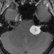
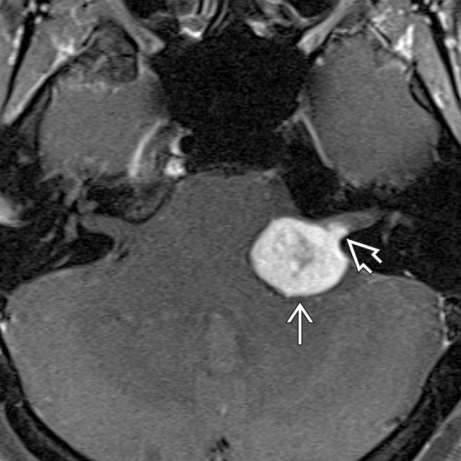
**Vestibular Schwannoma**
*Axial T1 C+ FS MR shows the typical appearance of a CPA-IAC vestibular schwannoma with avid, heterogeneous enhancement. Note tumor growth along the location of the vestibular nerve within the posterior IAC .*
**Vestibular Schwannoma**
*Axial T1 C+ FS MR shows the typical appearance of a CPA-IAC vestibular schwannoma with avid, heterogeneous enhancement. Note tumor growth along the location of the vestibular nerve within the posterior IAC .*
**Vestibular Schwannoma**
*Axial T1 C+ FS MR shows the typical appearance of a CPA-IAC vestibular schwannoma with avid, heterogeneous enhancement. Note tumor growth along the location of the vestibular nerve within the posterior IAC .*

**Vestibular Schwannoma**
*Axial T1 C+ FS MR shows the typical appearance of a CPA-IAC vestibular schwannoma with avid, heterogeneous enhancement. Note tumor growth along the location of the vestibular nerve within the posterior IAC .*
 from microhemorrhage, a feature that can help distinguish CPA-IAC schwannoma from meningioma.](images/app.statdx.com_image_thumbnail_0bd14ef9-fa2b-49f4-a274-290f0a016831_annotated_true_size_900_quality_90_8295787439dc0641cc294b36bcc813f2b7179fbb.jpg)
**Vestibular Schwannoma**
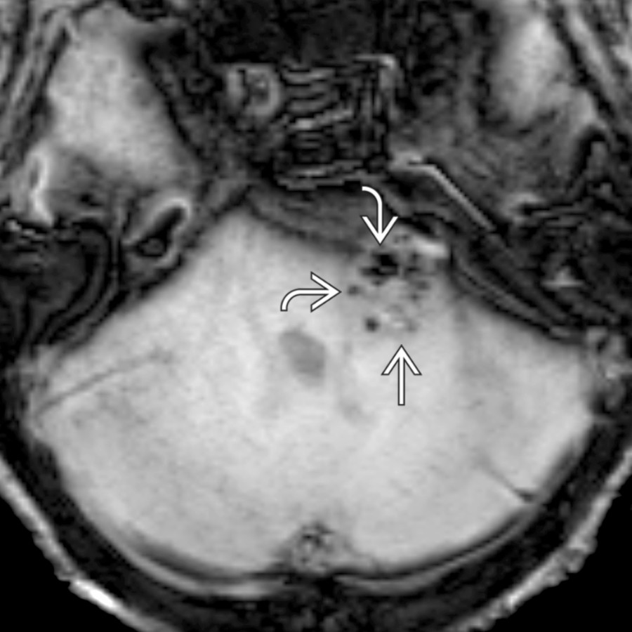
*Axial SWI MR in a CPA-IAC vestibular schwannoma shows punctate foci of gradient susceptibility from microhemorrhage, a feature that can help distinguish CPA-IAC schwannoma from meningioma.*
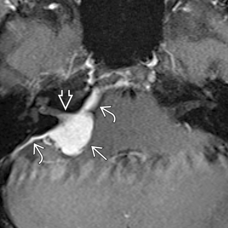
**Meningioma in CPA**
*Axial T1 C+ FS MR shows the typical appearance of a CPA meningioma , which is avidly enhancing and extends into the IAC . Note the characteristic dural tails , which help differentiate meningioma from schwannoma.*
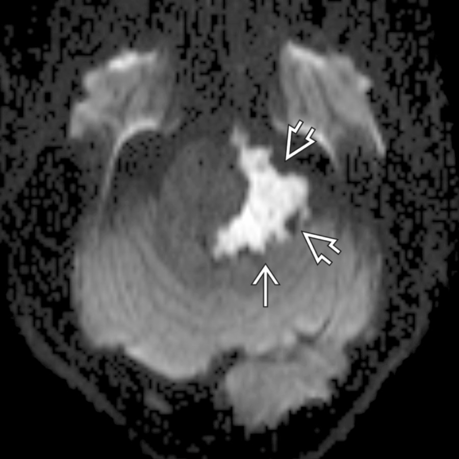
**Epidermoid Cyst in CPA**
*Axial DTI trace image through the posterior fossa shows a typical epidermoid cyst with reduced diffusivity. Note the scalloped, insinuating margins , which frequently encase cranial nerves and vessels and can make surgical resection fraught with peril.*
**Vestibular Schwannoma**
*Axial SWI MR in a CPA-IAC vestibular schwannoma shows punctate foci of gradient susceptibility from microhemorrhage, a feature that can help distinguish CPA-IAC schwannoma from meningioma.*

**Meningioma in CPA**
*Axial T1 C+ FS MR shows the typical appearance of a CPA meningioma , which is avidly enhancing and extends into the IAC . Note the characteristic dural tails , which help differentiate meningioma from schwannoma.*

**Epidermoid Cyst in CPA**
*Axial DTI trace image through the posterior fossa shows a typical epidermoid cyst with reduced diffusivity. Note the scalloped, insinuating margins , which frequently encase cranial nerves and vessels and can make surgical resection fraught with peril.*
 posterolaterally.](images/app.statdx.com_image_thumbnail_0a837932-9add-4268-8d4b-971743940ea0_annotated_true_size_900_quality_90_95859beecf4cdc34a5d2e7593427c4e9d7713a8b.jpg)
**Aneurysm in CPA**
*Axial T2WI MR shows a CPA vertebral artery aneurysm as an ovoid mass with complex wall signal, which bows the vestibulocochlear nerve posterolaterally.*
**T-Bone Fracture**
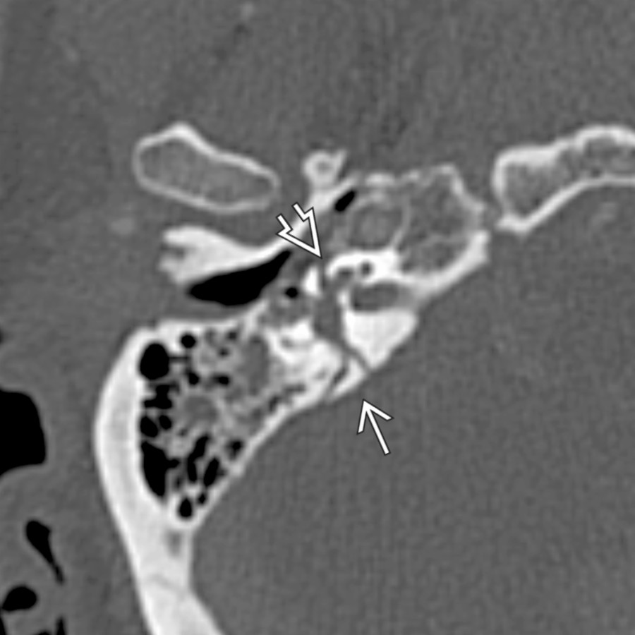
*Axial bone CT shows a complex, transversely oriented T-bone fracture that involves the otic capsule and disrupts the cochlea . The mastoid air cells and middle ear are opacified with blood, corresponding with hemotympanum on otoscopy.*
**Cochlear Otosclerosis**
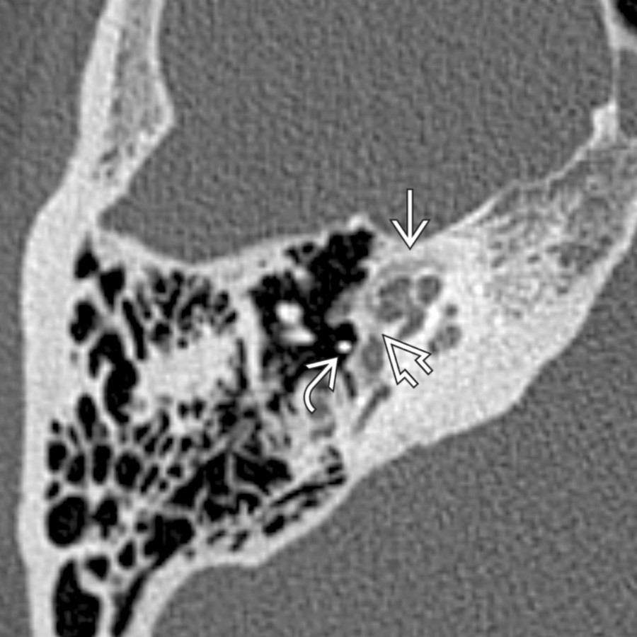
*Axial bone CT shows cochlear otosclerosis yielding SNHL. Note confluent otic capsule lucency adjacent to the cochlea. Patients commonly have fenestral involvement with lucency near the fissula ante fenestram , contributing a conductive component to hearing loss. Note the prior partial ossicular replacement prosthesis .*
**Metastases in CPA-IAC**
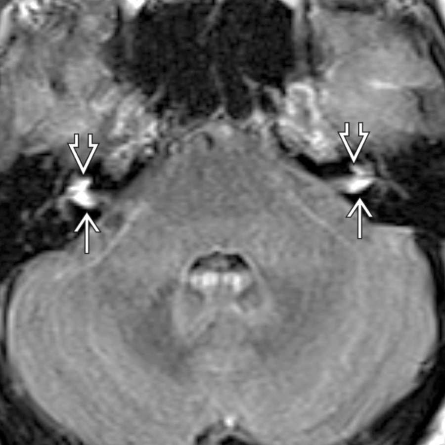
*Axial FLAIR C+ MR shows leptomeningeal metastases from melanoma with enhancement at the fundus of bilateral IACs extending into the basal turn of the cochlea bilaterally.*

**Facial Nerve Schwannoma in CPA-IAC**
*Axial T1 C+ SPGR MR shows a facial nerve schwannoma involving the left CPA-IAC. In this case, the labyrinthine tail of enhancement extends to the asymmetrically enhancing geniculate ganglion region , following the expected course of the facial nerve.*

**Lipoma in CPA-IAC**
*Coronal T1 MR of a left CPA-IAC lipoma shows a T1-bright lobular mass extending into the IAC . The presence of chemical shift artifact at the tumor-CSF (i.e., water) margin confirms the presence of fat within this mass.*
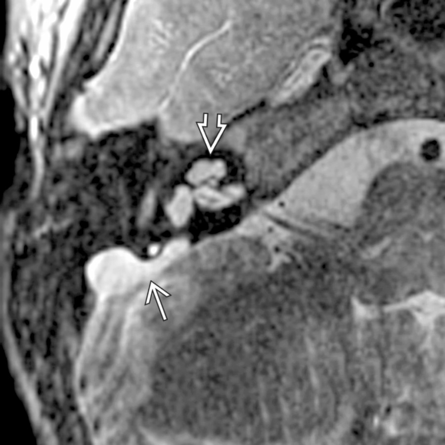
**Large Endolymphatic Sac Anomaly (IP-2)**
*Axial T2WI FS MR shows a large endolymphatic sac along the posterior wall of the T-bone associated with a malformed cochlea (modiolar deficiency, incomplete apical septation, and bulbous apical turn) .*
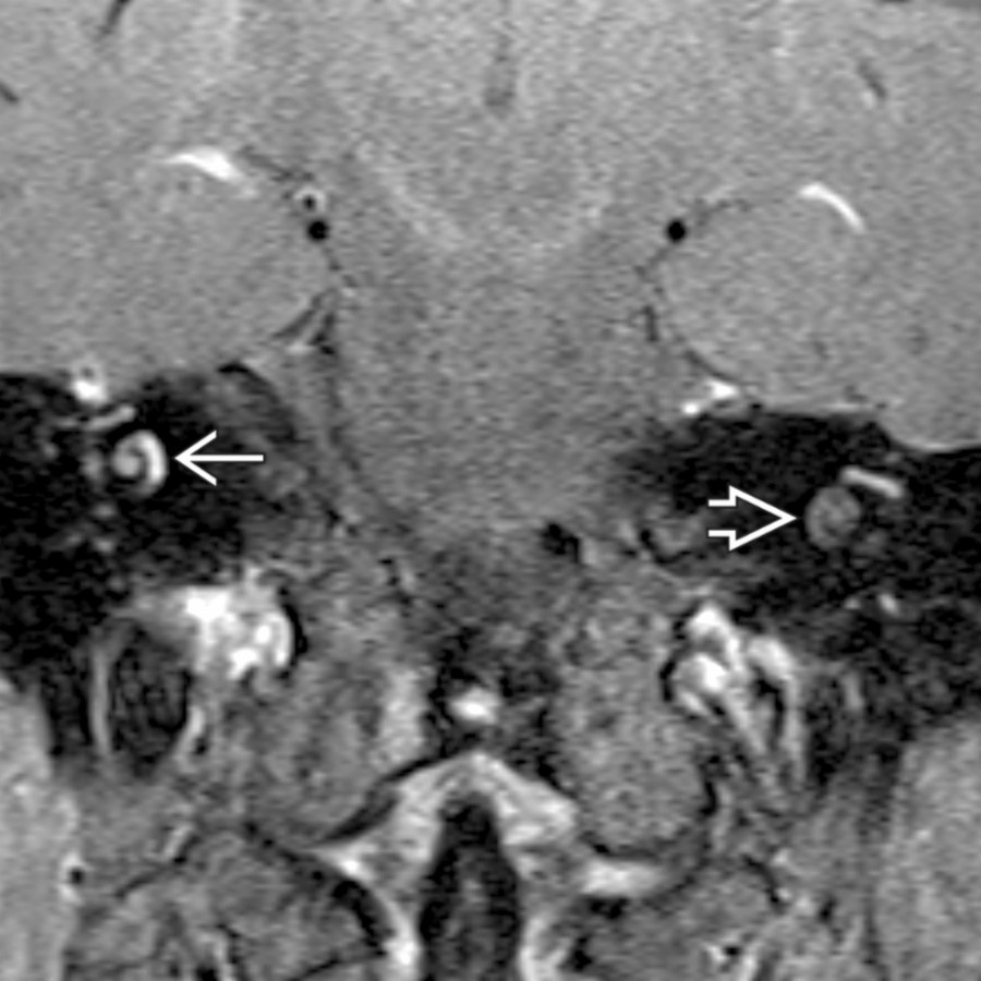
**Intralabyrinthine Schwannoma**
*Coronal T1 C+ FS MR of an intracochlear schwannoma shows abnormal enhancement in the middle turn of the right cochlea . Note the normal postcontrast appearance of the contralateral cochlea for comparison .*
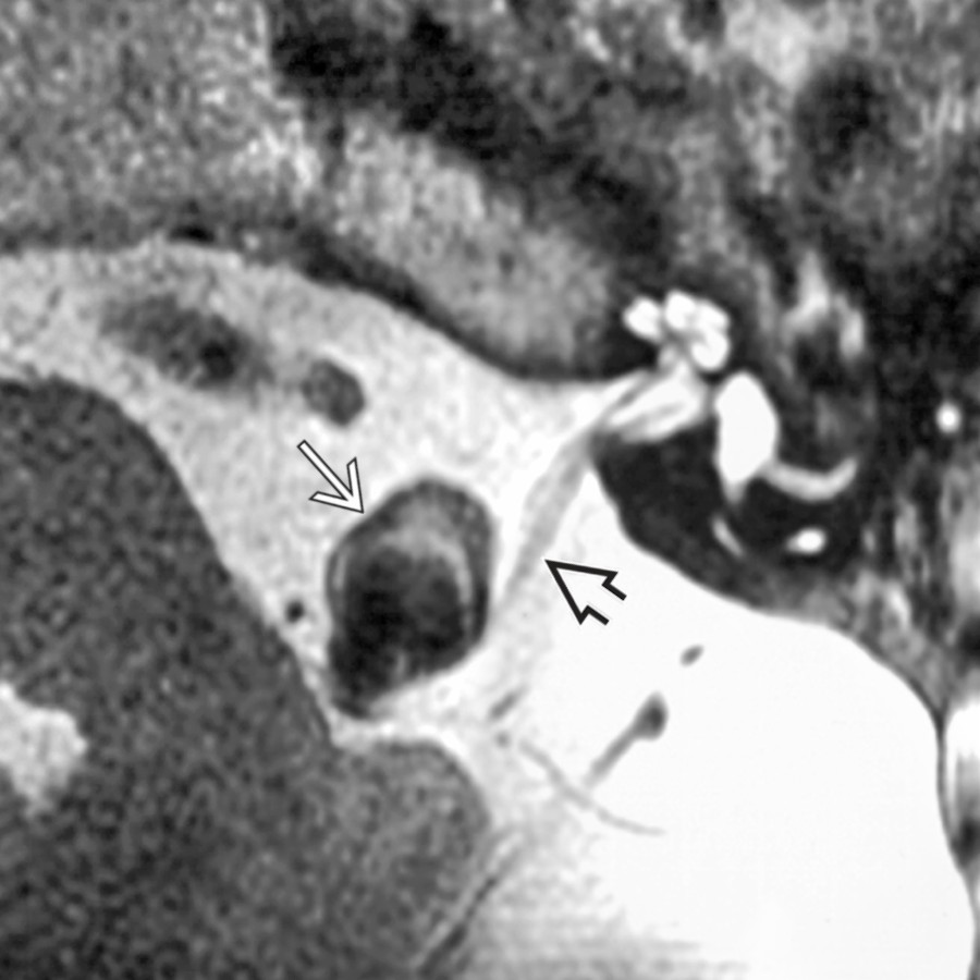
**Aneurysm in CPA**
*Axial T2WI MR shows a CPA vertebral artery aneurysm as an ovoid mass with complex wall signal, which bows the vestibulocochlear nerve posterolaterally.*

**T-Bone Fracture**
*Axial bone CT shows a complex, transversely oriented T-bone fracture that involves the otic capsule and disrupts the cochlea . The mastoid air cells and middle ear are opacified with blood, corresponding with hemotympanum on otoscopy.*

**Cochlear Otosclerosis**
*Axial bone CT shows cochlear otosclerosis yielding SNHL. Note confluent otic capsule lucency adjacent to the cochlea. Patients commonly have fenestral involvement with lucency near the fissula ante fenestram , contributing a conductive component to hearing loss. Note the prior partial ossicular replacement prosthesis .*

**Metastases in CPA-IAC**
*Axial FLAIR C+ MR shows leptomeningeal metastases from melanoma with enhancement at the fundus of bilateral IACs extending into the basal turn of the cochlea bilaterally.*

**Facial Nerve Schwannoma in CPA-IAC**
*Axial T1 C+ SPGR MR shows a facial nerve schwannoma involving the left CPA-IAC. In this case, the labyrinthine tail of enhancement extends to the asymmetrically enhancing geniculate ganglion region , following the expected course of the facial nerve.*

**Lipoma in CPA-IAC**
*Coronal T1 MR of a left CPA-IAC lipoma shows a T1-bright lobular mass extending into the IAC . The presence of chemical shift artifact at the tumor-CSF (i.e., water) margin confirms the presence of fat within this mass.*

**Large Endolymphatic Sac Anomaly (IP-2)**
*Axial T2WI FS MR shows a large endolymphatic sac along the posterior wall of the T-bone associated with a malformed cochlea (modiolar deficiency, incomplete apical septation, and bulbous apical turn) .*

**Intralabyrinthine Schwannoma**
*Coronal T1 C+ FS MR of an intracochlear schwannoma shows abnormal enhancement in the middle turn of the right cochlea . Note the normal postcontrast appearance of the contralateral cochlea for comparison .*
 due to marked inflammation.](images/app.statdx.com_image_thumbnail_339d6bcb-3739-4e06-8be4-a945b3e4d948_annotated_true_size_900_quality_90_2dcc16a8e0232f44a5755936e65bd2492e198879.jpg)
**Labyrinthitis**
*Axial T1 C+ FS MR in a patient with acute otomastoiditis complicated by labyrinthitis shows enhancement in the cochlea . There is additional vestibulocochlear neuritis with linear enhancement at the IAC fundus . Note enhancing mastoid/middle ear disease and dural enhancement due to marked inflammation.*
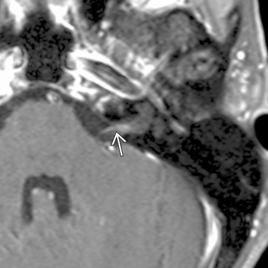
**Vestibulocochlear Neuritis**
*Axial T1 C+ FS MR shows vestibulocochlear neuritis in a patient with rapid onset of sensorineural hearing loss. Note the area of linear enhancement in the proximal IAC .*
### Additional Images
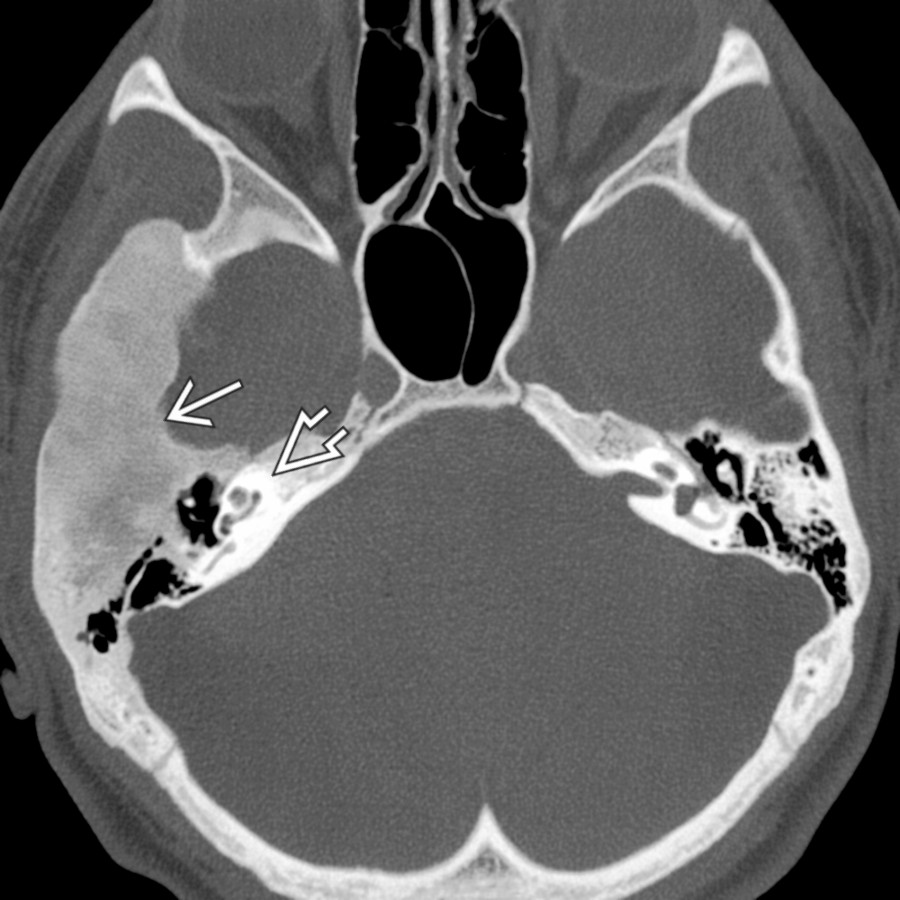
**Fibrous Dysplasia in T-Bone**
*Axial bone CT shows expansile ground-glass density focus of fibrous dysplasia affecting the squamous, tympanic, & mastoid portions of the T-bone. Note relative sparing of the otic capsule .*

**Endolymphatic Sac Tumor**
*Axial T1WI MR reveals a tumor along the posterior wall of the T-bone with high-signal foci that is highly suggestive of an endolymphatic sac tumor.*
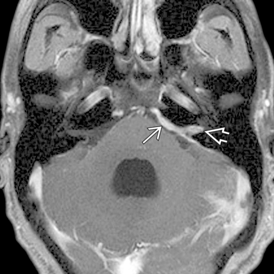
**Sarcoidosis in CPA**
*Axial T1 C+ FS MR demonstrates thick enhancing dural-based sarcoidosis in the left CPA and IAC mimicking en plaque meningioma.*
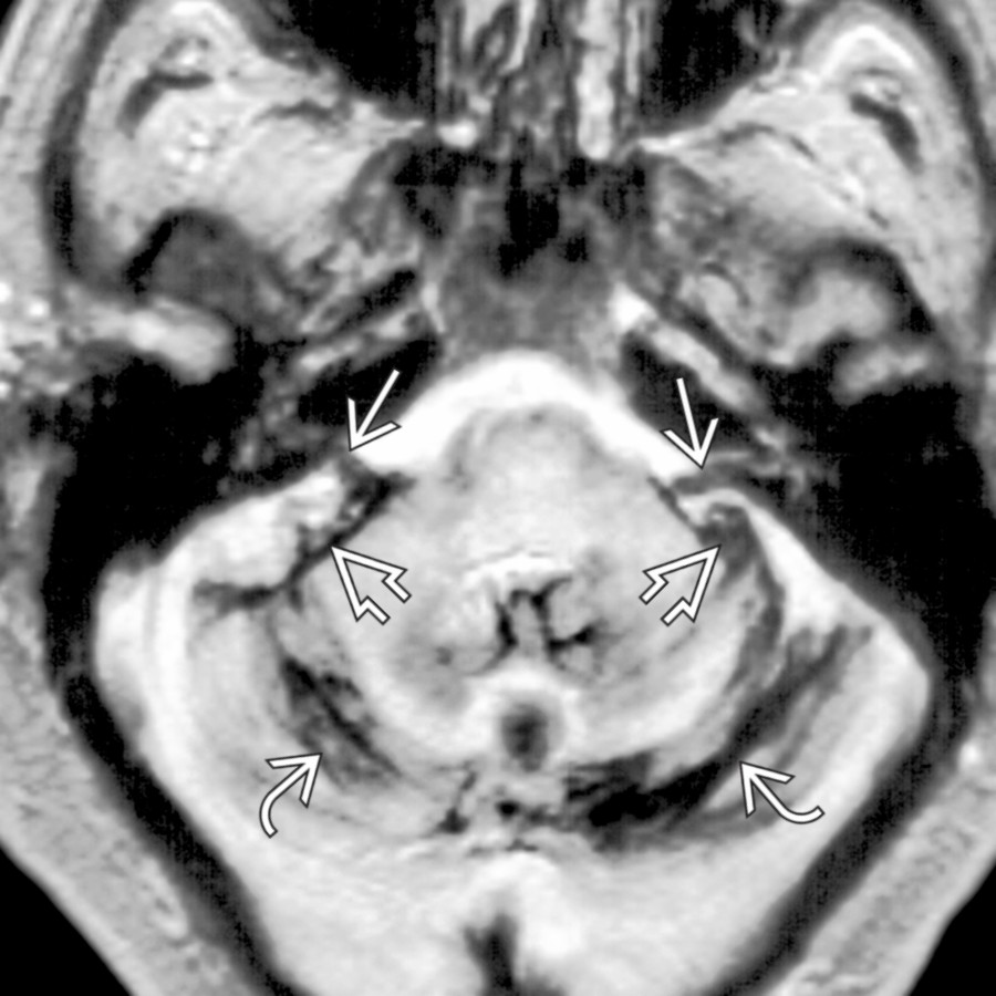
**Superficial Siderosis in CPA-IAC**
*Axial T2* GRE MR shows superficial siderosis on the surface of the posterior fossa structures (linear low signal), including cerebellar folia , flocculi , and vestibulocochlear nerves .*

**IAC/Temporal Bone Facial Nerve Venous Malformation ("Hemangioma")**
*Axial bone CT reveals punctate calcifications in the IAC suggesting the diagnosis of IAC hemangioma. Enhanced MR showed an enhancing mass in this location.*

**Ramsay Hunt Syndrome**
*Axial T1 C+ MR shows area of crescentic enhancement in the IAC fundus associated with tympanic segment of the facial nerve asymmetric enhancement . EAC vesicles were present, characteristic of Ramsey Hunt syndrome.*
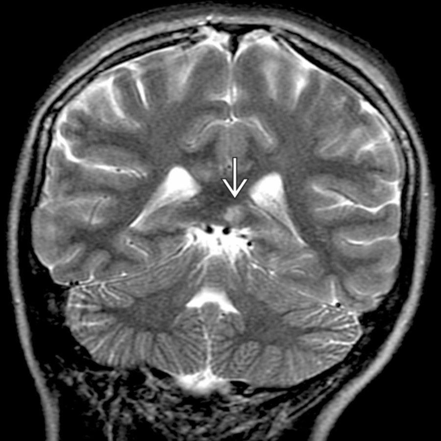
**Susac Syndrome (Retinocochleocerebral Vasculopathy)**
*Coronal T2 FS MR in a patient with retinocochleocerebral vasculopathy (Susac syndrome) shows a characteristic round, hyperintense lesion in the splenium of the corpus callosum .*
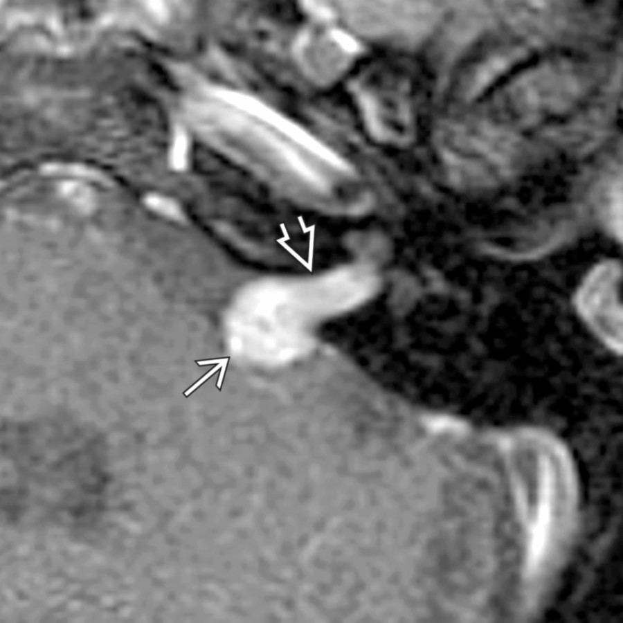
**Vestibular Schwannoma**
*Axial T1 C+ FS MR shows typical mid sized CPA-IAC vestibular schwannoma. "Ice cream" (CPA component) "on cone" (IAC component) morphology is highly suggestive of this diagnosis.*
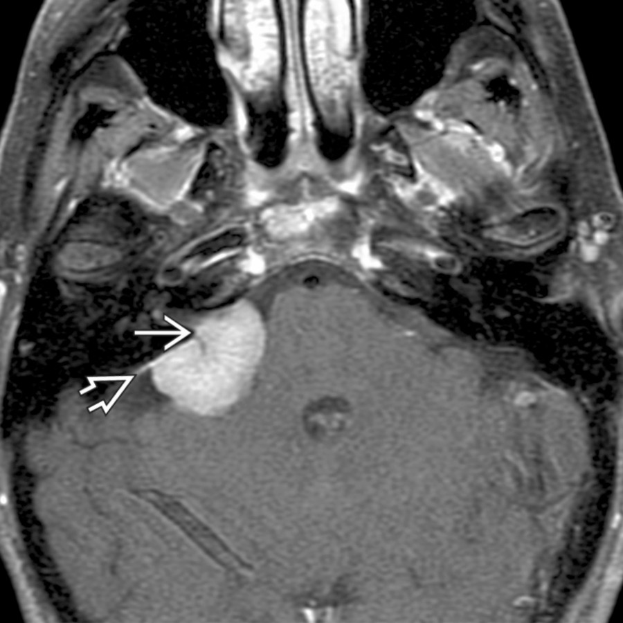
**Meningioma in CPA**
*Axial T1 C+ FS MR shows the sessile morphology of meningioma as it "sits" on the posterior T-bone wall. Note the characteristic dural tail and dural artery feeding the tumor center.*
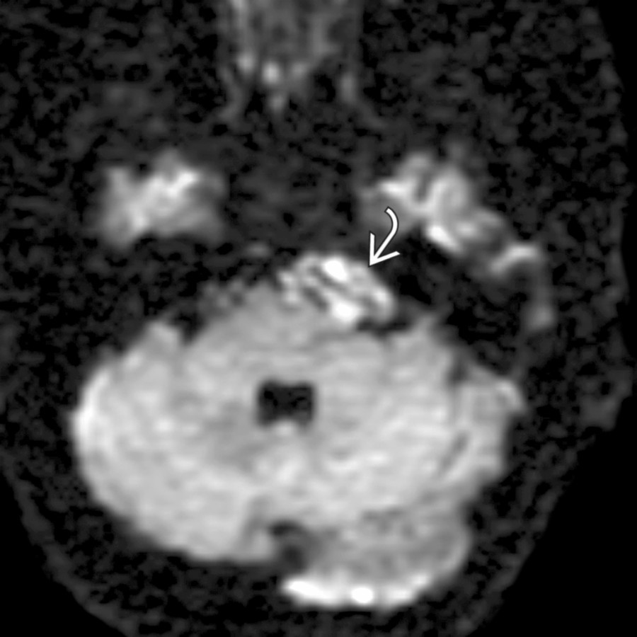
**Epidermoid Cyst in CPA**
*Axial DWI MR shows a CPA epidermoid identified by its reduced diffusivity . CPA epidermoids often are not directly over the porus acusticus, as in this case.*

**T-Bone Fracture**
*Axial bone CT shows an oblique T-bone fracture traversing the oval window and extending to the IAC area . Notice the air bubble (pneumolabyrinth) along the anterior margin of the vestibule .*

**Cochlear Otosclerosis**
*Axial bone CT shows severe otosclerosis as radiolucent foci along the medial middle ear wall (fenestral otosclerosis) and within the bony labyrinth (cochlear otosclerosis).*
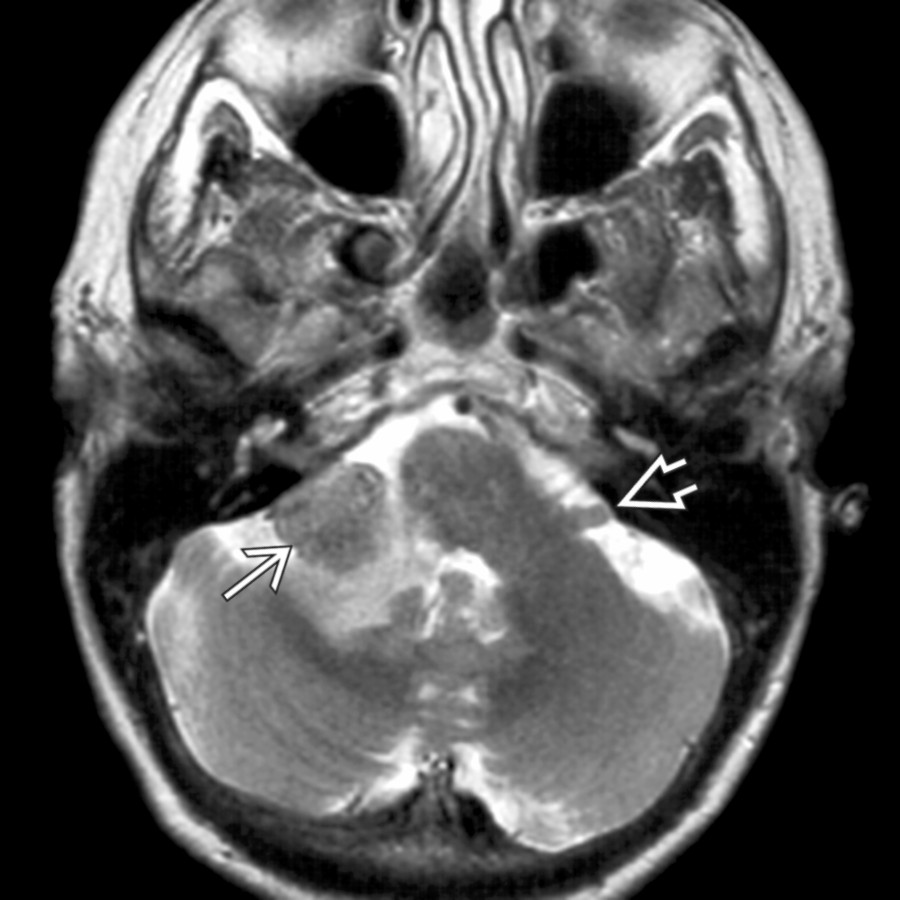
**Metastases in CPA-IAC**
*Axial T2WI MR demonstrates a right floccular metastasis with associated high-signal cerebellar and brachium pontis edema. A normal left flocculus is also seen.*
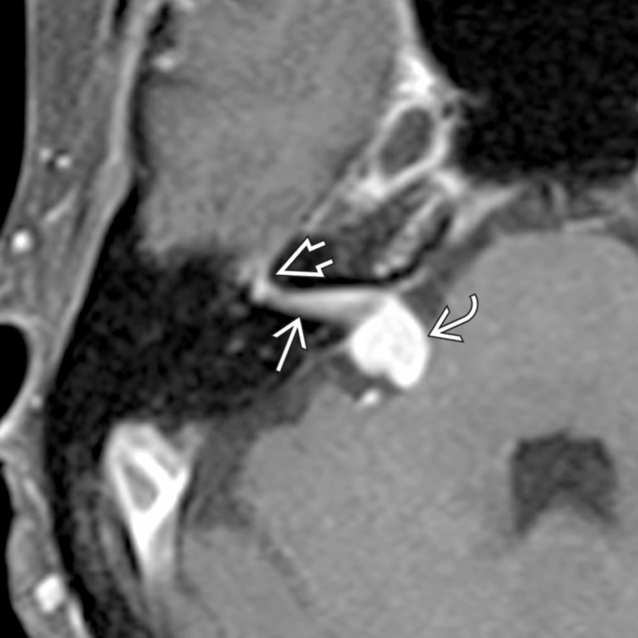
**Facial Nerve Schwannoma in CPA-IAC**
*Axial T1 C+ FS MR reveals an enhancing facial nerve schwannoma traversing the CPA and IAC into the facial nerve labyrinthine segment . This labyrinthine tail is characteristic.*
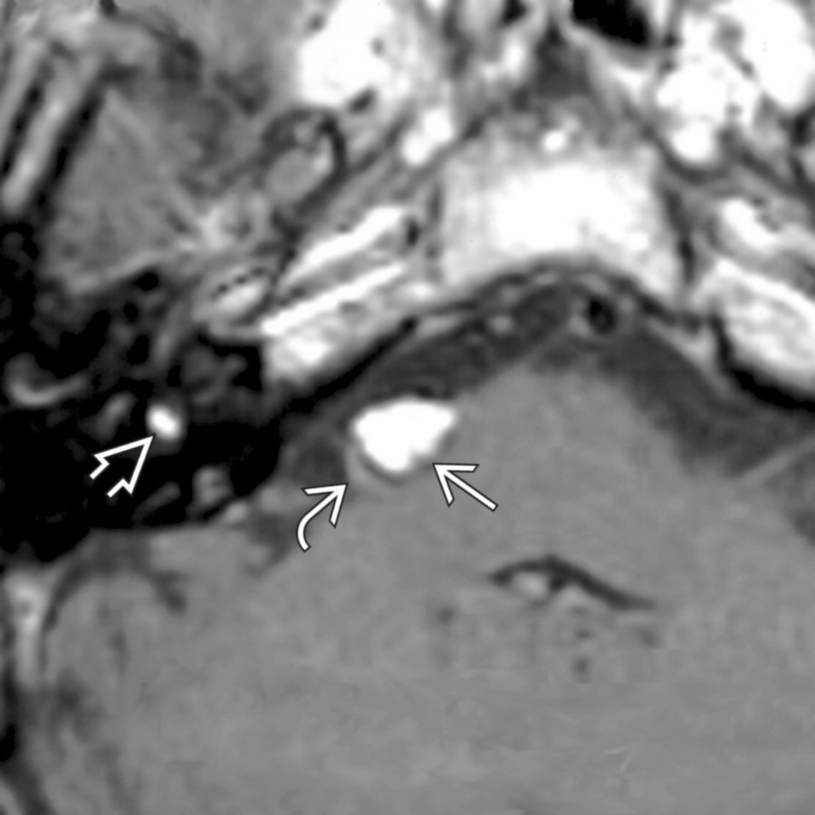
**Lipoma in CPA-IAC**
*Axial T1 MR demonstrates a CPA and intravestibular lipoma. Notice that the CPA lipoma effaces the most proximal vestibulocochlear nerve bundle . These lesions are left alone.*
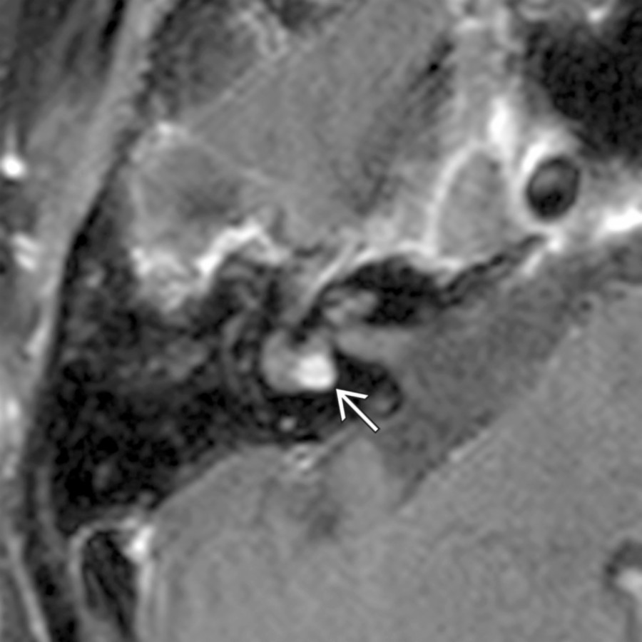
**Intralabyrinthine Schwannoma**
*Axial T1 C+ FS MR demonstrates a nodule of enhancing tissue in the vestibule of the inner ear secondary to intralabyrinthine schwannoma. CT of the T-bone was normal.*
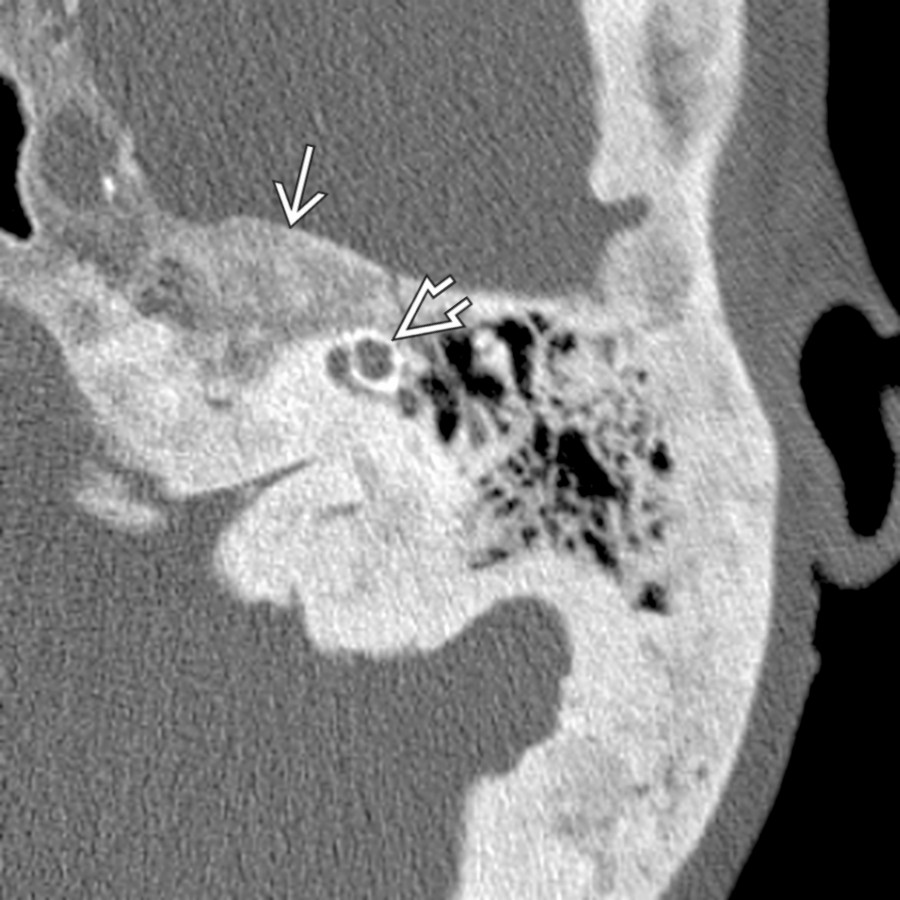
**Paget Disease in T-Bone**
*Axial bone CT shows late-phase Paget disease with diffuse bony expansion with areas of demineralization . Notice that the bony labyrinth along the anterior cochlear surface is involved .*

**Labyrinthitis**
*Axial T1 C+ FS MR shows enhancement of the middle ear , inner ear membranous labyrinth , and IAC in this pediatric patient with acute actinomycosis.*
due to marked inflammation.](images/app.statdx.com_image_thumbnail_339d6bcb-3739-4e06-8be4-a945b3e4d948_annotated_true_size_900_quality_90_2dcc16a8e0232f44a5755936e65bd2492e198879.jpg)
**Labyrinthitis**
*Axial T1 C+ FS MR in a patient with acute otomastoiditis complicated by labyrinthitis shows enhancement in the cochlea . There is additional vestibulocochlear neuritis with linear enhancement at the IAC fundus . Note enhancing mastoid/middle ear disease and dural enhancement due to marked inflammation.*

**Vestibulocochlear Neuritis**
*Axial T1 C+ FS MR shows vestibulocochlear neuritis in a patient with rapid onset of sensorineural hearing loss. Note the area of linear enhancement in the proximal IAC .*
### Additional Images

**Fibrous Dysplasia in T-Bone**
*Axial bone CT shows expansile ground-glass density focus of fibrous dysplasia affecting the squamous, tympanic, & mastoid portions of the T-bone. Note relative sparing of the otic capsule .*

**Endolymphatic Sac Tumor**
*Axial T1WI MR reveals a tumor along the posterior wall of the T-bone with high-signal foci that is highly suggestive of an endolymphatic sac tumor.*

**Sarcoidosis in CPA**
*Axial T1 C+ FS MR demonstrates thick enhancing dural-based sarcoidosis in the left CPA and IAC mimicking en plaque meningioma.*

**Superficial Siderosis in CPA-IAC**
*Axial T2* GRE MR shows superficial siderosis on the surface of the posterior fossa structures (linear low signal), including cerebellar folia , flocculi , and vestibulocochlear nerves .*

**IAC/Temporal Bone Facial Nerve Venous Malformation ("Hemangioma")**
*Axial bone CT reveals punctate calcifications in the IAC suggesting the diagnosis of IAC hemangioma. Enhanced MR showed an enhancing mass in this location.*

**Ramsay Hunt Syndrome**
*Axial T1 C+ MR shows area of crescentic enhancement in the IAC fundus associated with tympanic segment of the facial nerve asymmetric enhancement . EAC vesicles were present, characteristic of Ramsey Hunt syndrome.*

**Susac Syndrome (Retinocochleocerebral Vasculopathy)**
*Coronal T2 FS MR in a patient with retinocochleocerebral vasculopathy (Susac syndrome) shows a characteristic round, hyperintense lesion in the splenium of the corpus callosum .*

**Vestibular Schwannoma**
*Axial T1 C+ FS MR shows typical mid sized CPA-IAC vestibular schwannoma. "Ice cream" (CPA component) "on cone" (IAC component) morphology is highly suggestive of this diagnosis.*

**Meningioma in CPA**
*Axial T1 C+ FS MR shows the sessile morphology of meningioma as it "sits" on the posterior T-bone wall. Note the characteristic dural tail and dural artery feeding the tumor center.*

**Epidermoid Cyst in CPA**
*Axial DWI MR shows a CPA epidermoid identified by its reduced diffusivity . CPA epidermoids often are not directly over the porus acusticus, as in this case.*

**T-Bone Fracture**
*Axial bone CT shows an oblique T-bone fracture traversing the oval window and extending to the IAC area . Notice the air bubble (pneumolabyrinth) along the anterior margin of the vestibule .*

**Cochlear Otosclerosis**
*Axial bone CT shows severe otosclerosis as radiolucent foci along the medial middle ear wall (fenestral otosclerosis) and within the bony labyrinth (cochlear otosclerosis).*

**Metastases in CPA-IAC**
*Axial T2WI MR demonstrates a right floccular metastasis with associated high-signal cerebellar and brachium pontis edema. A normal left flocculus is also seen.*

**Facial Nerve Schwannoma in CPA-IAC**
*Axial T1 C+ FS MR reveals an enhancing facial nerve schwannoma traversing the CPA and IAC into the facial nerve labyrinthine segment . This labyrinthine tail is characteristic.*

**Lipoma in CPA-IAC**
*Axial T1 MR demonstrates a CPA and intravestibular lipoma. Notice that the CPA lipoma effaces the most proximal vestibulocochlear nerve bundle . These lesions are left alone.*

**Intralabyrinthine Schwannoma**
*Axial T1 C+ FS MR demonstrates a nodule of enhancing tissue in the vestibule of the inner ear secondary to intralabyrinthine schwannoma. CT of the T-bone was normal.*

**Paget Disease in T-Bone**
*Axial bone CT shows late-phase Paget disease with diffuse bony expansion with areas of demineralization . Notice that the bony labyrinth along the anterior cochlear surface is involved .*

**Labyrinthitis**
*Axial T1 C+ FS MR shows enhancement of the middle ear , inner ear membranous labyrinth , and IAC in this pediatric patient with acute actinomycosis.*