---
title: "Skull Base Chondrosarcoma"
docid: "d52746d2-9761-43a6-8f47-a2ccc01d612d"
authors:
- key: "94f835c8-fa13-4e8a-995b-53048e6b0605"
value: "Philip R. Chapman, MD"
breadcrumbs:
-
name: "Head and Neck"
slug: "head-and-neck"
treeNodeId: "ed24ed8c-5d57-4629-879b-447b82d2973d"
-
name: "Diagnosis"
slug: "diagnosis"
treeNodeId: "19b6b986-97d0-40e7-b317-00f0c5cd8fa2"
-
name: "Skull Base Lesions"
slug: "skull-base-lesions"
treeNodeId: "e2f573c9-3c74-435d-86f7-fe1d6c157f65"
-
name: "Diffuse or Multifocal Skull Base Disease"
slug: "diffuse-or-multifocal-skull-base-d-"
treeNodeId: "b7ce2ad6-77dc-4c50-a083-5fd04739fa1e"
-
name: "Skull Base Chondrosarcoma"
slug: "skull-base-chondrosarcoma"
treeNodeId: null
category: "Head and Neck"
cmeTopicId: "2e3b4196-d650-4dad-9cfb-638a0a54444e"
documentVersionId: "b1cf1027-9e19-466e-976d-ea1d55114892"
imageCount: 16
lastUpdated: "08/10/21"
pageDescription: "Skull Base Chondrosarcoma"
pageKeywords: "Head and Neck, Diagnosis, Skull Base Lesions, Diffuse or Multifocal Skull Base Disease, Skull Base Chondrosarcoma"
pageTitle: "Skull Base Chondrosarcoma | STATdx"
enhancedTitle: "Skull Base Chondrosarcoma"
type: "DX"
references: true
breadcrumbs:
- "Head and Neck"
- "Diagnosis"
- "Skull Base Lesions"
- "Diffuse or Multifocal Skull Base Disease"
- "Skull Base Chondrosarcoma"
---
# KEY FACTS
- ## Terminology
- Skull base chondrosarcoma (CSa-SB): Chondroid malignancy of skull base
- ## Imaging
- Typical location **off-midline**, centered on**petrooccipital fissure**
- CT
- Characteristic **chondroid tumor matrix calcification** **in** **50%**
- Arc or ring-like calcifications
- Sharp, narrow, nonsclerotic transition zone to adjacent normal bone
- MR
- High T2 signal with scattered hypointense foci (calcifications)
- Heterogeneously enhancing
- Whorls of enhancing lines within tumor matrix often seen
- ## Top Differential Diagnoses
- Chordoma
- Skull base metastasis
- Plasmacytoma
- Nasopharyngeal carcinoma (invasive)
- Meningioma
- Benign petrous apex lesions
- ## Clinical Issues
- Typically, middle-aged patient with insidious onset of headache and cranial nerve palsies (especially CNVI)
- ## Diagnostic Checklist
- Is lesion **off-midline** (CSa) or in midline (chordoma)?
- Do calcifications represent **arc-whorl intralesional calcifications** (CSa) or fragmented destroyed bone (chordoma)?
- Consider MR angiography or CTA for preoperative characterization of vessel involvement
# TERMINOLOGY
- ## Abbreviations
- Skull base chondrosarcoma (CSa-SB)
- ## Definitions
- CSa-SB: Chondroid malignancy of skull base
# IMAGING
- ## General Features
- ### Best diagnostic clue
- Solitary enhancing osteolytic soft tissue mass, centered at petrooccipital fissure (POF) ± chondroid matrix
- ### Location
- Off-midline at **POF** (2/3)
- Anterior basisphenoid (1/3)
- Rarely multiple
- ### Size
- Variable, usually > 3 cm at time of diagnosis
- ### Morphology
- Well-circumscribed, lobulated margins
- Lobulated growth in 3 dimensions results in cauliflower shape
- ## CT Findings
- ### NECT
- Soft tissue component is relatively dense
- ### CECT
- Variable, heterogeneous enhancement
- ### Bone CT
- Expansile mass at POF producing erosive or destructive bone changes in clivus and petrous apex
- ~ 50% will have radiographically classic chondroid matrix with **"rings and arcs" calcification**
- Sharp, narrow, nonsclerotic transition zone to adjacent normal bone
- ## MR Findings
- ### T1WI
- Low to intermediate signal intensity relative to gray matter
- ↓ signal foci within tumor may suggest underlying coarse matrix mineralization or fibrocartilaginous elements
- ### T2WI
- Variable, usually **high signal**
- CSa-SB may be homogeneously hyperintense or simply contain localized zones of hyperintensity
- Degree of T2 hyperintensity is inversely related to degree of calcified matrix
- Steady-state free precession T2 images may be low signal (even if bright on T2 spin echo)
- Hypointense foci (calcifications) less conspicuous than on CT
- ### PD/intermediate
- High signal
- ### T1WI C+
- Heterogeneous enhancement
- Usually has nodular or focal enhancement within tumor
- Whorls of enhancing lines within tumor matrix often seen
- ## Angiographic Findings
- Avascular or hypovascular mass
- Internal carotid artery displacement ± encasement
- ## Imaging Recommendations
- ### Best imaging tool
- Combination of multiplanar, gadolinium-enhanced MR and high-resolution bone CT
- ### Protocol advice
- High-resolution axial bone CT for evaluation of chondroid matrix and pattern of bone destruction
- MR of skull base to include T2WI and multiplanar T1WI, followed by T1WI C+ FS in same planes for direct comparison
- MRA and MRV, or CTA helpful to assess vascular involvement preoperatively
- Carotid injury risk higher for endoscopic resection of CSa-SB
- Preoperative angiography with test occlusion if vessel sacrifice necessary
# DIFFERENTIAL DIAGNOSIS
- [Chordoma](/document/chordoma/84148164-fa26-479a-b615-6e932a4a280e)
- Destructive clival lesion; bone fragments within matrix
- Midline > lateral location
- Low T1 and markedly high T2 MR signal; enhancing mass
- Chondroid chordomas more aggressive and worse prognosis
- May be impossible to distinguish chordoma from chondrosarcoma on routine imaging
- [Skull Base Metastasis](/document/skull-base-metastasis/54838a9e-d712-4a48-8928-a1ca925d6aea)
- Bone CT: Destructive mass that can be anywhere in skull base
- MR: Often multiple enhancing, invasive lesions
- Typically low to intermediate T2 signal
- Known primary tumor
- [Plasmacytoma & Multiple Myeloma](/document/skull-base-multiple-myeloma/f6984d5f-5d95-4f22-b91b-ea6b5d0a3a23)
- Usually more midline, within clivus
- **T2 signal is low** to intermediate
- > 50% have concurrent multiple myeloma
- Multiple lesions of skull base or calvarium generally excludes CSa-SB
- [Nasopharyngeal Carcinoma](/document/nasopharyngeal-carcinoma/1e193b95-6d8b-4605-b83e-3de8d22982b4)
- Primary mass in nasopharyngeal mucosal space
- Tumor invades superiorly to clivus, foramen lacerum, and POF
- [Meningioma](/document/skull-base-meningioma/e264a03f-f5c1-42f3-8725-155c648c3e45)
- Calcification in meningioma can mimic chondroid matrix
- Hyperostosis possible; not typically destructive in absence of invasion
- Low to intermediate T2 MR signal; enhancing with dural tails
- Skull base meningioma occurring just above POF with bone invasion could closely resemble chondrosarcoma
- ## Non-Hodgkin Lymphoma
- Lymphoproliferative neoplasm with focal or multifocal, osseous &/or extraosseous involvement of skull base
- Low to intermediate T2 MR signal, may restrict on DWI
- ## Nonneoplastic Lesion of Petrous Apex
- Includes benign, expansile lesions: Cholesteatoma, mucocele, cholesterol granuloma
- Should be nonenhancing, smoothly marginated without calcified matrix
- High signal from asymmetric petrous apex marrow can mimic enhancing tumor
- Petrous apicitis could demonstrate inflammatory enhancement of petrous apex with permeative bone changes
- Expect acute clinical syndrome with fever, leukocytosis, Gradenigo syndrome
- ## Chondromyxoid Fibroma
- Rare, expansile, noninfiltrating skull base mass
- Areas of ground-glass density may be seen
- Appearance may overlap with CSa-SB
# PATHOLOGY
- ## General Features
- ### Etiology
- Arises from remnants of embryonal cartilage, endochondral bone, or from primitive mesenchymal cells in meninges
- May arise from metaplasia of meningeal fibroblasts
- ### Genetics
- May complicate Ollier disease and Maffucci syndrome
- ## Staging, Grading, & Classification
- Classification
- Conventional CSa: Hyaline (7%), myxoid (30%), or mixed (63%)
- Clear cell
- Mesenchymal
- Dedifferentiated
- Grading from low to high grade
- Based on degree of cellularity, pleomorphism, mitoses, and multinucleated cells
- ## Gross Pathologic & Surgical Features
- Smooth, lobulated mass "welling up" from POF
- Cut surface shows gray-white, glistening parenchyma
- ## Microscopic Features
- Hypercellular tumor composed of chondrocytes with hyperchromatic, pleomorphic nuclei and prominent nucleoli
- Binucleate or multinucleate cells are rule
- Hyaline matrix may calcify in "ringlets"
- Intercellular matrix is solid in hyaline type compared to mucinous/gelatinous matrix in myxoid or mixed types
- Histology may overlap with or be confused with that of chordoma
- Histology particularly confusing in chondroid chordoma, myxoid CSa
- Differentiation facilitated by immunohistochemical staining
# CLINICAL ISSUES
- ## Presentation
- ### Most common signs/symptoms
- **Abducens (CNVI) palsy** due to proximity of Dorello canal
- **Headache**
- Mean duration of symptoms at diagnosis = 27 months
- ### Other signs/symptoms
- Other cranial nerve palsies (CNIII, V, VII, VIII)
- ### Clinical profile
- Middle-aged patient with insidious onset of headaches and cranial nerve palsies
- ## Demographics
- ### Age
- Range: 10-80 years
- Mean: 40 years
- ### Epidemiology
- 6% of all skull base tumors
- 75% of all cranial CSa occur in skull base
- ## Natural History & Prognosis
- Prognosis depends on extent at diagnosis, histologic grade, and completeness of surgical resection
- Disease-specific 10-year survival rates of 99% recently reported
- Most central CSa-SBs are well to moderately differentiated
- High-grade CSa metastasizes to bones and lung more frequently
- Conventional CSa: Indolent growth pattern
- Most are slow growing, locally invasive, but rarely metastasize
- Mesenchymal and dedifferentiated forms: Aggressive behavior; poor prognosis
- ## Treatment
- Aggressive resection associated with significant morbidity and low likelihood of complete resection
- Basal subfrontal approach used for tumor that invades clivus and extends anteriorly into sphenoid and ethmoid sinuses
- Subtemporal and preauricular infratemporal approach used when CSa extends laterally beyond petrous internal carotid artery
- Combined radical resection and postoperative, high-dose, fractionated precision conformal radiation therapy most often utilized
- Charged particle radiation therapy (protons or carbon ions) alone or combined with subtotal resection
# DIAGNOSTIC CHECKLIST
- ## Consider
- Is lesion in off-midline (CSa) vs. midline (chordoma)?
- Do calcifications represent arc-whorl intralesional calcifications (CSa) or fragmented destroyed bone (chordoma)?
- Does patient have known primary neoplasm (metastasis), myeloma (plasmacytoma), or nasopharyngeal mass (nasopharyngeal carcinoma)?
- ## Image Interpretation Pearls
- Classic appearance: Heterogeneously enhancing tumor located at **POF** with hyperintense signal on T2 MR
- CT shows chondroid mineralization and bone destruction
- When no tumor matrix found, difficult to tell from CSa plasmacytoma, focal metastasis, or chondromyxoid fibroma
c47564a6-1df8-40c1-97cb-e02a5fd09f92
## References
# Selected References
1. [Edem I et al: Advances in the management of primary bone sarcomas of the skull base. J Neurooncol. 150(3):393-403, 2020](http://www.ncbi.nlm.nih.gov/pubmed/?term=32306199%5Bpmid%5D)
1. [Kano H et al: Radiosurgery for chordoma and chondrosarcoma. Prog Neurol Surg. 34:207-14, 2019](http://www.ncbi.nlm.nih.gov/pubmed/?term=31096236%5Bpmid%5D)
1. [Purgina B et al: Distinctive head and neck bone and soft tissue neoplasms. Surg Pathol Clin. 10(1):223-79, 2017](http://www.ncbi.nlm.nih.gov/pubmed/?term=28153134%5Bpmid%5D)
1. [Awad M et al: Skull base chondrosarcoma. J Clin Neurosci. 24:1-5, 2016](http://www.ncbi.nlm.nih.gov/pubmed/?term=26724847%5Bpmid%5D)
1. [Van Gompel JJ et al: Chordoma and chondrosarcoma. Otolaryngol Clin North Am. 48(3):501-14, 2015](http://www.ncbi.nlm.nih.gov/pubmed/?term=25863568%5Bpmid%5D)
1. [Bag AK et al: Neuroimaging: intrinsic lesions of the central skull base region. Semin Ultrasound CT MR. 34(5):412-35, 2013](http://www.ncbi.nlm.nih.gov/pubmed/?term=24216451%5Bpmid%5D)
1. [Bloch O et al: Skull base chondrosarcoma: evidence-based treatment paradigms. Neurosurg Clin N Am. 24(1):89-96, 2013](http://www.ncbi.nlm.nih.gov/pubmed/?term=23174360%5Bpmid%5D)
1. [Gardner PA et al: Carotid artery injury during endoscopic endonasal skull base surgery: incidence and outcomes. Neurosurgery. 73(2 Suppl Operative):ons261-9; discussion ons269-70, 2013](http://www.ncbi.nlm.nih.gov/pubmed/?term=23695646%5Bpmid%5D)
1. [Sbaihat A et al: Skull base chondrosarcomas: surgical treatment and results. Ann Otol Rhinol Laryngol. 122(12):763-70, 2013](http://www.ncbi.nlm.nih.gov/pubmed/?term=24592579%5Bpmid%5D)
1. [Amichetti M et al: A systematic review of proton therapy in the treatment of chondrosarcoma of the skull base. Neurosurg Rev. 33(2):155-65, 2010](http://www.ncbi.nlm.nih.gov/pubmed/?term=19921291%5Bpmid%5D)
1. [Bloch OG et al: Cranial chondrosarcoma and recurrence. Skull Base. 20(3):149-56, 2010](http://www.ncbi.nlm.nih.gov/pubmed/?term=21318031%5Bpmid%5D)
1. [Ares C et al: Effectiveness and safety of spot scanning proton radiation therapy for chordomas and chondrosarcomas of the skull base: first long-term report. Int J Radiat Oncol Biol Phys. 75(4):1111-8, 2009](http://www.ncbi.nlm.nih.gov/pubmed/?term=19386442%5Bpmid%5D)
1. [Borges A: Imaging of the central skull base. Neuroimaging Clin N Am. 19(4):669-96, 2009](http://www.ncbi.nlm.nih.gov/pubmed/?term=19959012%5Bpmid%5D)
1. [Gallia GL et al: Skull base chondrosarcoma presenting with hemorrhage. Can J Neurol Sci. 36(6):774-5, 2009](http://www.ncbi.nlm.nih.gov/pubmed/?term=19960759%5Bpmid%5D)
1. [Güneş M et al: Intracranial chondrosarcoma: a case report and review of the literature. Minim Invasive Neurosurg. 52(5-6):238-41, 2009](http://www.ncbi.nlm.nih.gov/pubmed/?term=20077365%5Bpmid%5D)
1. [Hong P et al: Chondrosarcoma of the head and neck: report of 11 cases and literature review. J Otolaryngol Head Neck Surg. 38(2):279-85, 2009](http://www.ncbi.nlm.nih.gov/pubmed/?term=19442379%5Bpmid%5D)
1. [Samii A et al: Surgical treatment of skull base chondrosarcomas. Neurosurg Rev. 32(1):67-75; discussion 75, 2009](http://www.ncbi.nlm.nih.gov/pubmed/?term=18818961%5Bpmid%5D)
1. [Abdelmalek M et al: Recurrent chondrosarcoma of the right skull base in a patient with Maffucci syndrome. Am J Clin Dermatol. 9(1):61-5, 2008](http://www.ncbi.nlm.nih.gov/pubmed/?term=18092846%5Bpmid%5D)
1. [Cho YH et al: Chordomas and chondrosarcomas of the skull base: comparative analysis of clinical results in 30 patients. Neurosurg Rev. 31(1):35-43; discussion 43, 2008](http://www.ncbi.nlm.nih.gov/pubmed/?term=17922295%5Bpmid%5D)
1. [Ferrer Pérez AI et al: Skull base chondrosarcoma: a case treated by the CyberKnife system. Clin Transl Oncol. 10(10):670-2, 2008](http://www.ncbi.nlm.nih.gov/pubmed/?term=18940749%5Bpmid%5D)
1. [Gelderblom H et al: The clinical approach towards chondrosarcoma. Oncologist. 2008 Mar;13(3):320-9. Review. Erratum in: Oncologist. 13(5):618, 2008](http://www.ncbi.nlm.nih.gov/pubmed/?term=18378543%5Bpmid%5D)
1. [Nguyen QN et al: Emerging role of proton beam radiation therapy for chordoma and chondrosarcoma of the skull base. Curr Oncol Rep. 10(4):338-43, 2008](http://www.ncbi.nlm.nih.gov/pubmed/?term=18778560%5Bpmid%5D)
1. [Sanna M et al: Chondrosarcomas of the jugular foramen. Laryngoscope. 118(10):1719-28, 2008](http://www.ncbi.nlm.nih.gov/pubmed/?term=18622309%5Bpmid%5D)
1. [Seizeur R et al: Chondrosarcoma of the skull base in Maffucci's syndrome. Br J Neurosurg. 22(6):778-80, 2008](http://www.ncbi.nlm.nih.gov/pubmed/?term=19085363%5Bpmid%5D)
1. [Zhang Q et al: Endoscopic endonasal surgery for clival chordoma and chondrosarcoma. ORL J Otorhinolaryngol Relat Spec. 70(2):124-9, 2008](http://www.ncbi.nlm.nih.gov/pubmed/?term=18408411%5Bpmid%5D)
1. [Almefty K et al: Chordoma and chondrosarcoma: similar, but quite different, skull base tumors. Cancer. 110(11):2457-67, 2007](http://www.ncbi.nlm.nih.gov/pubmed/?term=17894390%5Bpmid%5D)
1. [Dini LI et al: Maffucci's syndrome complicated by intracranial chondrosarcoma: two new illustrative cases. Arq Neuropsiquiatr. 65(3B):816-21, 2007](http://www.ncbi.nlm.nih.gov/pubmed/?term=17952287%5Bpmid%5D)
1. [Lustig LR et al: Chondrosarcomas of the skull base and temporal bone. J Laryngol Otol. 121(8):725-35, 2007](http://www.ncbi.nlm.nih.gov/pubmed/?term=17319989%5Bpmid%5D)
1. [Schulz-Ertner D et al: Carbon ion radiotherapy of skull base chondrosarcomas. Int J Radiat Oncol Biol Phys. 67(1):171-7, 2007](http://www.ncbi.nlm.nih.gov/pubmed/?term=17056193%5Bpmid%5D)
1. [Baehring JM et al: Chondrosarcoma of the skull base. J Neurooncol. 76(1):49, 2006](http://www.ncbi.nlm.nih.gov/pubmed/?term=16402277%5Bpmid%5D)
1. [Brackmann DE et al: Chondrosarcoma of the skull base: long-term follow-up. Otol Neurotol. 27(7):981-91, 2006](http://www.ncbi.nlm.nih.gov/pubmed/?term=17006349%5Bpmid%5D)
1. [Frank G et al: The endoscopic transnasal transsphenoidal approach for the treatment of cranial base chordomas and chondrosarcomas. Neurosurgery. 59(1 Suppl 1):ONS50-7; discussion ONS50-7, 2006](http://www.ncbi.nlm.nih.gov/pubmed/?term=16888551%5Bpmid%5D)
1. [Harada K et al: Chondrosarcoma of the clivus presenting with intratumoral hemorrhage: case report. J Neurooncol. 77(2):221-3, 2006](http://www.ncbi.nlm.nih.gov/pubmed/?term=16333684%5Bpmid%5D)
1. [Tzortzidis F et al: Patient outcome at long-term follow-up after aggressive microsurgical resection of cranial base chondrosarcomas. Neurosurgery. 58(6):1090-8; discussion 1090-8, 2006](http://www.ncbi.nlm.nih.gov/pubmed/?term=16723888%5Bpmid%5D)
1. [Krishnan S et al: Radiosurgery for cranial base chordomas and chondrosarcomas. Neurosurgery. 56(4):777-84; discussion 777-84, 2005](http://www.ncbi.nlm.nih.gov/pubmed/?term=15792516%5Bpmid%5D)
1. [Oghalai JS et al: Skull base chondrosarcoma originating from the petroclival junction. Otol Neurotol. 26(5):1052-60, 2005](http://www.ncbi.nlm.nih.gov/pubmed/?term=16151358%5Bpmid%5D)
1. [Noël G et al: Chondrosarcomas of the base of the skull in Ollier's disease or Maffucci's syndrome--three case reports and review of the literature. Acta Oncol. 43(8):705-10, 2004](http://www.ncbi.nlm.nih.gov/pubmed/?term=15764214%5Bpmid%5D)
1. [Raghu M et al: Chondrosarcomas of the temporal bone: presentation and management. J Laryngol Otol. 118(7):551-5, 2004](http://www.ncbi.nlm.nih.gov/pubmed/?term=15318964%5Bpmid%5D)
1. [Neff B et al: Chondrosarcoma of the skull base. Laryngoscope. 112(1):134-9, 2002](http://www.ncbi.nlm.nih.gov/pubmed/?term=11802051%5Bpmid%5D)
1. [Schmidinger A et al: Natural history of chondroid skull base lesions--case report and review. Neuroradiology. 44(3):268-71, 2002](http://www.ncbi.nlm.nih.gov/pubmed/?term=11942386%5Bpmid%5D)
1. [Crockard HA et al: A multidisciplinary team approach to skull base chondrosarcomas. J Neurosurg. 95(2):184-9, 2001](http://www.ncbi.nlm.nih.gov/pubmed/?term=11780886%5Bpmid%5D)
1. [Richardson MS: Pathology of skull base tumors. Otolaryngol Clin North Am. 34(6):1025-42, vii, 2001](http://www.ncbi.nlm.nih.gov/pubmed/?term=11728930%5Bpmid%5D)
1. [Hug EB et al: Proton radiation therapy for chordomas and chondrosarcomas of the skull base. J Neurosurg. 91(3):432-9, 1999](http://www.ncbi.nlm.nih.gov/pubmed/?term=10470818%5Bpmid%5D)
1. [Rosenberg AE et al: Chondrosarcoma of the base of the skull: a clinicopathologic study of 200 cases with emphasis on its distinction from chordoma. Am J Surg Pathol. 23(11):1370-8, 1999](http://www.ncbi.nlm.nih.gov/pubmed/?term=10555005%5Bpmid%5D)
1. [Korten AG et al: Intracranial chondrosarcoma: review of the literature and report of 15 cases. J Neurol Neurosurg Psychiatry. 65(1):88-92, 1998](http://www.ncbi.nlm.nih.gov/pubmed/?term=9667567%5Bpmid%5D)
1. [Keel SB et al: Chondromyxoid fibroma of the skull base: a tumor which may be confused with chordoma and chondrosarcoma. A report of three cases and review of the literature. Am J Surg Pathol. 21(5):577-82, 1997](http://www.ncbi.nlm.nih.gov/pubmed/?term=9158683%5Bpmid%5D)
1. [Ramina R et al: Maffucci's syndrome associated with a cranial base chondrosarcoma: case report and literature review. Neurosurgery. 41(1):269-72, 1997](http://www.ncbi.nlm.nih.gov/pubmed/?term=9218317%5Bpmid%5D)
1. [Rapidis AD et al: Chondrosarcomas of the skull base: review of the literature and report of two cases. J Craniomaxillofac Surg. 25(6):322-7, 1997](http://www.ncbi.nlm.nih.gov/pubmed/?term=9504309%5Bpmid%5D)
1. [Geirnaerdt MJ et al: Calcified meningioma of the skull base simulating chondrosarcoma. Eur J Radiol. 21(2):148-51, 1995](http://www.ncbi.nlm.nih.gov/pubmed/?term=8850513%5Bpmid%5D)
1. [Weber AL et al: Cartilaginous tumors and chordomas of the cranial base. Otolaryngol Clin North Am. 28(3):453-71, 1995](http://www.ncbi.nlm.nih.gov/pubmed/?term=7675464%5Bpmid%5D)
1. [Brown E et al: Chondrosarcoma of the skull base. Neuroimaging Clin N Am. 4(3):529-41, 1994](http://www.ncbi.nlm.nih.gov/pubmed/?term=7952954%5Bpmid%5D)
1. [Stapleton SR et al: Chondrosarcoma of the skull base: a series of eight cases. Neurosurgery. 32(3):348-55; discussion 355-6, 1993](http://www.ncbi.nlm.nih.gov/pubmed/?term=8455759%5Bpmid%5D)
1. [Meyers SP et al: Chondrosarcomas of the skull base: MR imaging features. Radiology. 184(1):103-8, 1992](http://www.ncbi.nlm.nih.gov/pubmed/?term=1609064%5Bpmid%5D)
1. [Kveton JF et al: Chondrosarcoma of the skull base. Otolaryngol Head Neck Surg. 94(1):23-32, 1986](http://www.ncbi.nlm.nih.gov/pubmed/?term=3081852%5Bpmid%5D)
## Images
### Selected Images
 . Note the normal right petrooccipital fissure
  . Chondroid calcifications, depicted in yellow, are present within the lesion
  .](images/app.statdx.com_image_thumbnail_114d5426-d4b3-4f63-b60b-6ff5d23a92af_annotated_true_size_900_quality_90_98594d53.jpg)
*Axial graphic depicts the classic location of a chondrosarcoma of the skull base centered in the left petrooccipital fissure . Note the normal right petrooccipital fissure . Chondroid calcifications, depicted in yellow, are present within the lesion .*

*Axial graphic depicts the classic location of a chondrosarcoma of the skull base centered in the left petrooccipital fissure . Note the normal right petrooccipital fissure . Chondroid calcifications, depicted in yellow, are present within the lesion .*

*Axial graphic depicts the classic location of a chondrosarcoma of the skull base centered in the left petrooccipital fissure . Note the normal right petrooccipital fissure . Chondroid calcifications, depicted in yellow, are present within the lesion .*

*Axial graphic depicts the classic location of a chondrosarcoma of the skull base centered in the left petrooccipital fissure . Note the normal right petrooccipital fissure . Chondroid calcifications, depicted in yellow, are present within the lesion .*
 .](images/app.statdx.com_image_thumbnail_78819c6a-7888-4d7b-bad6-f07ebe05e023_annotated_true_size_900_quality_90_e929202a.jpg)
*Axial T2 MR reveals a large, high-signal chondrosarcoma of the left petrooccipital fissure. Note that the vertical segment of the petrous internal carotid artery is compressed . Note the normal right petrooccipital fissure .*
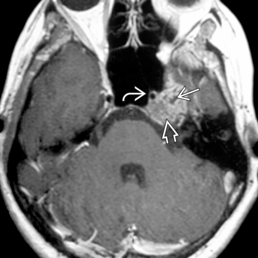
*Axial T1 C+ MR shows mottled enhancement within a chondrosarcoma centered at the left petrooccipital fissure. Calcified matrix is seen as a focal low signal intensity area within the otherwise enhancing tumor. The left internal carotid artery is patent .*

*Axial bone CT demonstrates typical chondroid calcification in a left petrooccipital fissure chondrosarcoma. In this case, no significant destruction of the adjacent petrous apex is appreciated.*

*Axial bone CT shows rounded and arc-like calcified foci in this large chondrosarcoma centered at the petrooccipital fissure. Up to 50% of chondrosarcomas demonstrate matrix calcification. Note slight narrowing of the left vidian canal .*
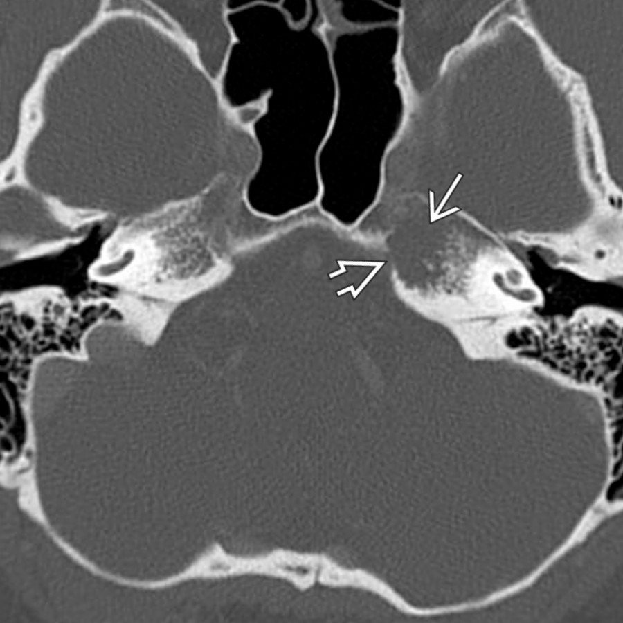
*Axial bone window CT shows subtle bone destruction with cortical erosion in this small left petrous apex chondrosarcoma. No calcified matrix is seen. MR showed a corresponding T2-hyperintense and enhancing mass in this location.*
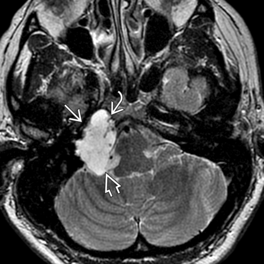
*Axial T2 MR shows a hyperintense chondrosarcoma involving the right petrous apex and extending into the right cerebellopontine angle cistern (CPA) . The petrous carotid artery is displaced anteriorly .*

*Axial T1 C+ FS MR demonstrates avid enhancement in this chondrosarcoma. Note petrous apex and CPA cistern involvement. Most skull base chondrosarcomas emanate from the petrooccipital fissure. However, when they become large, it may be hard to see the point of origin.*

*Axial T2 MR reveals a large, high-signal chondrosarcoma of the left petrooccipital fissure. Note that the vertical segment of the petrous internal carotid artery is compressed . Note the normal right petrooccipital fissure .*

*Axial T1 C+ MR shows mottled enhancement within a chondrosarcoma centered at the left petrooccipital fissure. Calcified matrix is seen as a focal low signal intensity area within the otherwise enhancing tumor. The left internal carotid artery is patent .*

*Axial bone CT demonstrates typical chondroid calcification in a left petrooccipital fissure chondrosarcoma. In this case, no significant destruction of the adjacent petrous apex is appreciated.*

*Axial bone CT shows rounded and arc-like calcified foci in this large chondrosarcoma centered at the petrooccipital fissure. Up to 50% of chondrosarcomas demonstrate matrix calcification. Note slight narrowing of the left vidian canal .*

*Axial bone window CT shows subtle bone destruction with cortical erosion in this small left petrous apex chondrosarcoma. No calcified matrix is seen. MR showed a corresponding T2-hyperintense and enhancing mass in this location.*

*Axial T2 MR shows a hyperintense chondrosarcoma involving the right petrous apex and extending into the right cerebellopontine angle cistern (CPA) . The petrous carotid artery is displaced anteriorly .*

*Axial T1 C+ FS MR demonstrates avid enhancement in this chondrosarcoma. Note petrous apex and CPA cistern involvement. Most skull base chondrosarcomas emanate from the petrooccipital fissure. However, when they become large, it may be hard to see the point of origin.*
 and basilar
  arterial displacement by the mass.](images/app.statdx.com_image_thumbnail_4e3e510c-875c-47aa-b0c6-6093f0422f42_annotated_true_size_900_quality_90_cc36cb69.jpg)
*Axial T2 MR shows a hyperintense right parasellar chondrosarcoma that extends into the prepontine cistern and sphenoid sinus . Note internal carotid and basilar arterial displacement by the mass.*

*Axial T1 C+ FS MR shows moderate enhancement within a right parasellar chondrosarcoma . The patient presented with intractable headache, epistaxis, and diplopia. Tumor invades prepontine cistern and sphenoid sinus , partly encases the right ICA , and displaces the basilar artery .*
### Additional Images

*Axial bone CT of the skull base shows a chondrosarcoma centered over the left petrooccipital fissure. Notice the absence of chondroid calcifications within the tumor matrix in this case.*

*Axial bone CT in a patient with a petro-occipital chondrosarcoma shows coarse chondroid calcifications within the tumor matrix .*

*Axial T2 FS MR shows intermediate, heterogeneous signal in the cephalad margin of a petrooccipital fissure chondrosarcoma . Curvilinear and punctate hypointensities in the lesion are due to chondroid calcifications.*

*Axial T2 MR shows a hyperintense small early left petrous apex chondrosarcoma . Although one might consider trapped fluid in a pneumatized petrous apex based on the T2 image, associated enhancement and bone destruction on CT suggested a more aggressive process. The cisternal 6th cranial nerves are faintly seen, and their proximity to the petrous temporal bone illustrates why chondrosarcomas here frequently present with 6th nerve involvement.*

*Axial CECT demonstrates a left cavernous sinus/paracavernous myxoid chondrosarcoma . Orbital apex involvement is present . Note absence of chondroid calcifications.*

*Axial T2 FS MR in the same patient reveals a homogeneously hyperintense left cavernous and paracavernous myxoid chondrosarcoma . Notice that the cavernous internal carotid artery is nearly surrounded by tumor.*
arterial displacement by the mass.](images/app.statdx.com_image_thumbnail_4e3e510c-875c-47aa-b0c6-6093f0422f42_annotated_true_size_900_quality_90_cc36cb69.jpg)
*Axial T2 MR shows a hyperintense right parasellar chondrosarcoma that extends into the prepontine cistern and sphenoid sinus . Note internal carotid and basilar arterial displacement by the mass.*

*Axial T1 C+ FS MR shows moderate enhancement within a right parasellar chondrosarcoma . The patient presented with intractable headache, epistaxis, and diplopia. Tumor invades prepontine cistern and sphenoid sinus , partly encases the right ICA , and displaces the basilar artery .*
### Additional Images

*Axial bone CT of the skull base shows a chondrosarcoma centered over the left petrooccipital fissure. Notice the absence of chondroid calcifications within the tumor matrix in this case.*

*Axial bone CT in a patient with a petro-occipital chondrosarcoma shows coarse chondroid calcifications within the tumor matrix .*

*Axial T2 FS MR shows intermediate, heterogeneous signal in the cephalad margin of a petrooccipital fissure chondrosarcoma . Curvilinear and punctate hypointensities in the lesion are due to chondroid calcifications.*

*Axial T2 MR shows a hyperintense small early left petrous apex chondrosarcoma . Although one might consider trapped fluid in a pneumatized petrous apex based on the T2 image, associated enhancement and bone destruction on CT suggested a more aggressive process. The cisternal 6th cranial nerves are faintly seen, and their proximity to the petrous temporal bone illustrates why chondrosarcomas here frequently present with 6th nerve involvement.*

*Axial CECT demonstrates a left cavernous sinus/paracavernous myxoid chondrosarcoma . Orbital apex involvement is present . Note absence of chondroid calcifications.*

*Axial T2 FS MR in the same patient reveals a homogeneously hyperintense left cavernous and paracavernous myxoid chondrosarcoma . Notice that the cavernous internal carotid artery is nearly surrounded by tumor.*