---
title: "Small Ventricles"
docid: "2f99bc62-163e-41aa-b190-0da8a4de6d11"
authors:
- key: "d19354f3-7ff2-495a-ad3f-064122e45602"
value: "Bernadette L. Koch, MD"
- key: "f184750a-90b4-47a7-907b-23b05d70357a"
value: "Chang Yueh Ho, MD"
breadcrumbs:
-
name: "Brain"
slug: "brain"
treeNodeId: "6d8829f1-14d7-45af-8675-255189aa526a"
-
name: "Differential Diagnosis"
slug: "differential-diagnosis"
treeNodeId: "a7fdd139-664e-4bb8-8d18-400e4733ff60"
-
name: "Ventricles, Periventricular Regions"
slug: "ventricles-periventricular-regions"
treeNodeId: "353c434a-a6fc-4ef1-8786-d30a1988a4dc"
-
name: "Generic Imaging Patterns"
slug: "generic-imaging-patterns"
treeNodeId: "969c31a2-ef56-4fc3-9125-05857cf9aac3"
-
name: "Small Ventricles"
slug: "small-ventricles"
treeNodeId: null
category: "Brain"
documentVersionId: "5b4a0ab0-ffd6-4209-a606-7d38fb6ec442"
imageCount: 27
lastUpdated: "01/26/23"
pageDescription: "Small Ventricles"
pageKeywords: "Brain, Differential Diagnosis, Ventricles, Periventricular Regions, Generic Imaging Patterns, Small Ventricles"
pageTitle: "Small Ventricles | STATdx"
enhancedTitle: "Small Ventricles"
type: "DDX"
references: true
breadcrumbs:
- "Brain"
- "Differential Diagnosis"
- "Ventricles, Periventricular Regions"
- "Generic Imaging Patterns"
- "Small Ventricles"
---
# ESSENTIAL INFORMATION
- ## Key Differential Diagnosis Issues
- Clinical features usually help define cause
- ## Helpful Clues for Common Diagnoses
- **Normal Variant (Young Brain)**
- Small ventricles in children & young adults may be normal
- Look for visualization of sulci at vertex & sylvian fissures to exclude diffuse cerebral edema
- **CSF Shunts & Complications**
- **Slit ventricle syndrome**: Symptoms of ↑ ICP with collapsed shunted ventricles
- Noncompliant ventricles, stiff brain, or overshunting
- Anti-siphon devices (gravitational shunt valves) to prevent overshunting
- **Posttraumatic Brain Swelling**
- Diffuse edema with sulcal & ventricular effacement
- Frequently with associated diffuse axonal injury
- Focal edema from contusions may cause ventricular effacement & herniation
- **Herniation Syndromes, Intracranial**
- Ventricular effacement from herniation due to hematoma & tumors
- Increasing subfalcine herniation → ipsilateral ventricular effacement & contralateral ventricular dilatation
- **Adult Hypoxic-Ischemic Injury**
- Hypoxic-ischemic injury (HII) = global HII, global anoxic injury, & cerebral hypoperfusion injury or cerebral hypoperfusion → diffuse cytotoxic edema
- CT: Diffuse cerebral edema + small ventricles & sulci
- ↓ gray-white differentiation, hypodense basal ganglia, bright cerebellar reversal sign
- MR: Diffuse cortical ↑ T2 signal & ↓ diffusion
- ± pattern of central gray ↓ diffusion
- **Neonatal Hypoxic-Ischemic Injury**
- Hypoxic-ischemic encephalopathy from perinatal hypoxia or hypoperfusion
- Deep or central pattern: Deep gray ± brainstem injury & loss of cortical ribbon on T2WI
- Most likely to cause small ventricles
- Peripheral pattern = injury to watershed zones, less likely to cause diffuse cerebral swelling
- ## Helpful Clues for Less Common Diagnoses
- **Hyperglycemia**
- Diabetic ketoacidosis (DKA): Diffuse cerebral edema
- ± osmotic demyelination
- **Cerebral Infection**
- Gray & white matter T2 hyperintensity & edema
- Mild restriction on DWI common in viral encephalitis
- **Intracranial Hypotension**
- "Slumping" midbrain, acquired tonsillar herniation, diffusely enhancing dura, veins/dural sinuses distended
- Interpeduncular cistern lower than dorsum sella
- Best evaluated on sagittal images
- **Idiopathic Intracranial Hypertension**
- Dilated optic nerve sheaths, posterior globe flattening, empty sella, slit-like ventricles rare
- ## Helpful Clues for Rare Diagnoses
- **Inborn Errors of Metabolism (Acute Presentation)**
- Acute exacerbation + diffuse edema & brain injury → mass effect on ventricles
## References
# Selected References
1. [Panagopoulos D et al: Shunt over-drainage, slit ventricle syndrome, programmable valves and anti-siphon devices. a narrative review of a multifactorial and intractable problem. J Integr Neurosci. 21(3):84, 2022](http://www.ncbi.nlm.nih.gov/pubmed/?term=35633165%5Bpmid%5D)
1. [Bond KM et al: Spontaneous intracranial hypotension: atypical radiologic ppearances, imaging mimickers, and clinical look-alikes. AJNR Am J Neuroradiol. 41(8):1339-47, 2020](http://www.ncbi.nlm.nih.gov/pubmed/?term=32646948%5Bpmid%5D)
1. [Kranz PG et al: Spontaneous intracranial hypotension: pathogenesis, diagnosis, and treatment. Neuroimaging Clin N Am. 29(4):581-94, 2019](http://www.ncbi.nlm.nih.gov/pubmed/?term=31677732%5Bpmid%5D)
1. [Rekate HL: Shunt-related headaches: the slit ventricle syndromes. Childs Nerv Syst. 24(4):423-30, 2008](http://www.ncbi.nlm.nih.gov/pubmed/?term=18259760%5Bpmid%5D)
## Images
### Selected Images
 . Slit ventricle syndrome must have clinical symptoms of ↑ intracranial pressure with slit appearance of the lateral ventricles due to shunt.](0728b805-3262-47ee-8be6-1021849a885b)
**CSF Shunts & Complications**
*Axial NECT shows completely collapsed lateral ventricles with a shunt catheter . Slit ventricle syndrome must have clinical symptoms of ↑ intracranial pressure with slit appearance of the lateral ventricles due to shunt.*
**Posttraumatic Brain Swelling**
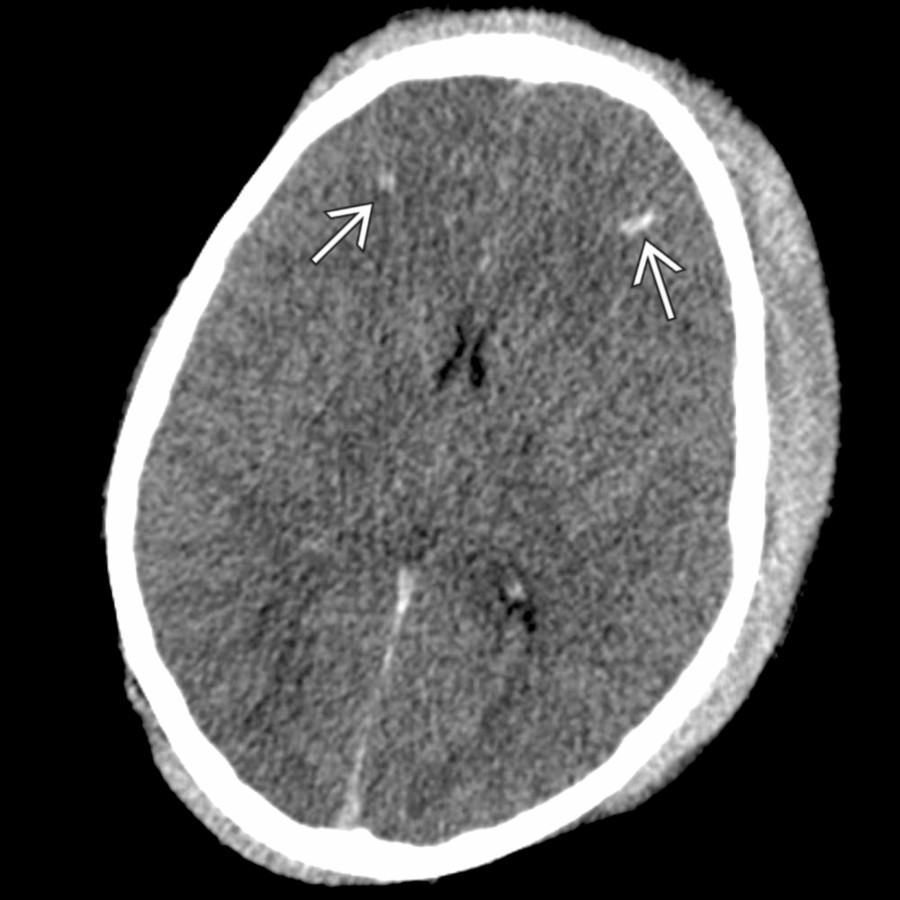
*Axial CT in a 9-year-old injured by a horse shows bilateral scalp hematomas, effacement of sulci, loss of gray-white matter differentiation, and small lateral ventricles. Also note small foci of hemorrhagic shear-type injury at the gray-white junction in the frontal lobes.*

**CSF Shunts & Complications**
*Axial NECT shows completely collapsed lateral ventricles with a shunt catheter . Slit ventricle syndrome must have clinical symptoms of ↑ intracranial pressure with slit appearance of the lateral ventricles due to shunt.*

**Posttraumatic Brain Swelling**
*Axial CT in a 9-year-old injured by a horse shows bilateral scalp hematomas, effacement of sulci, loss of gray-white matter differentiation, and small lateral ventricles. Also note small foci of hemorrhagic shear-type injury at the gray-white junction in the frontal lobes.*
 from entrapment.](images/app.statdx.com_image_thumbnail_59079b5e-7d2d-4336-a5ad-aa23a5785112_annotated_true_size_900_quality_90_39d39744_20251018T165215Z.jpg)
**Herniation Syndromes, Intracranial**
*Axial NECT shows complete loss of the left middle cerebral artery gray-white distinction with cytotoxic edema and left-to-right subfalcine herniation . Note the compression of the ipsilateral left lateral ventricle and enlargement of the right lateral ventricle from entrapment.*

**Adult Hypoxic-Ischemic Injury**
*Axial NECT shows diffuse effacement of ventricles and sulci from diffuse cerebral edema. There is focal symmetric hypodensity of the globi pallidi in this patient with a history of cardiogenic shock.*

**Neonatal Hypoxic-Ischemic Injury**
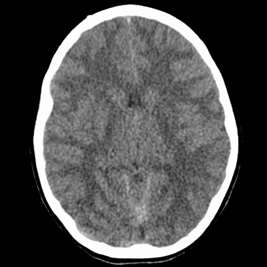
*Axial PD MR in a 2-day-old with severe deep/central pattern of hypoxic-ischemic injury demonstrates small lateral ventricles and cerebral sulci, as well as symmetric, hyperintense signal in the bilateral globus pallidus, putamen, and thalamus .*
**Hyperglycemia**
*Axial NECT in a 12-year-old with altered mental status and diabetic ketoacidosis shows near-complete effacement of the lateral ventricles, perimesencephalic cisterns, and cerebral sulci related to diffuse cerebral edema.*
**Cerebral Infection**
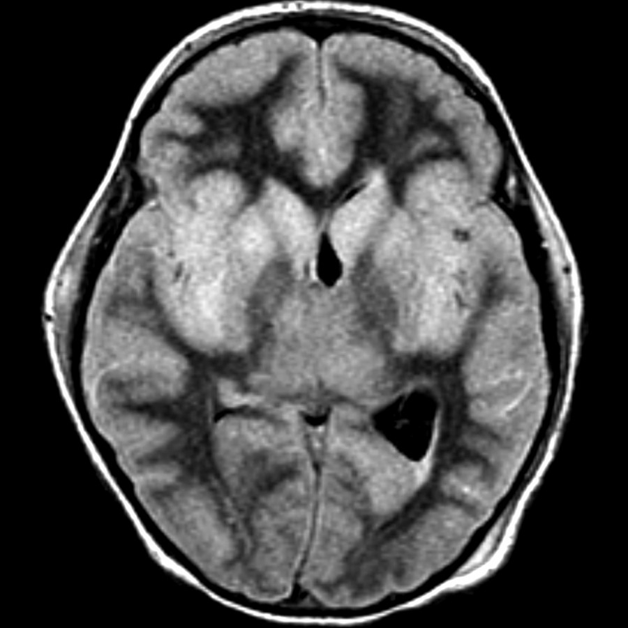
*Axial FLAIR MR shows diffuse gray matter swelling and hyperintensity with resulting effacement of the cerebral sulci and right lateral ventricle. This patient eventually succumbed to rabies encephalitis.*
**Herniation Syndromes, Intracranial**
*Axial NECT shows complete loss of the left middle cerebral artery gray-white distinction with cytotoxic edema and left-to-right subfalcine herniation . Note the compression of the ipsilateral left lateral ventricle and enlargement of the right lateral ventricle from entrapment.*

**Adult Hypoxic-Ischemic Injury**
*Axial NECT shows diffuse effacement of ventricles and sulci from diffuse cerebral edema. There is focal symmetric hypodensity of the globi pallidi in this patient with a history of cardiogenic shock.*

**Neonatal Hypoxic-Ischemic Injury**
*Axial PD MR in a 2-day-old with severe deep/central pattern of hypoxic-ischemic injury demonstrates small lateral ventricles and cerebral sulci, as well as symmetric, hyperintense signal in the bilateral globus pallidus, putamen, and thalamus .*

**Hyperglycemia**
*Axial NECT in a 12-year-old with altered mental status and diabetic ketoacidosis shows near-complete effacement of the lateral ventricles, perimesencephalic cisterns, and cerebral sulci related to diffuse cerebral edema.*

**Cerebral Infection**
*Axial FLAIR MR shows diffuse gray matter swelling and hyperintensity with resulting effacement of the cerebral sulci and right lateral ventricle. This patient eventually succumbed to rabies encephalitis.*
 , which has moved below the level of the dorsum sella, related to "brainstem sagging."](6e228d55-4868-4c43-95d8-556d5988bb6d)
**Intracranial Hypotension**
*Sagittal T1 MR shows downward displacement of the cerebellar tonsils and brainstem. There is effacement of the suprasellar cistern . Note effacement of the interpeduncular cistern , which has moved below the level of the dorsum sella, related to "brainstem sagging."*
### Additional Images

**CSF Shunts & Complications**
*Axial NECT shows small ventricles and indeterminate shunt position . Symptomatic ventricular collapse is known as slit-like ventricle syndrome and suggests overshunting.*
**Posttraumatic Brain Swelling**
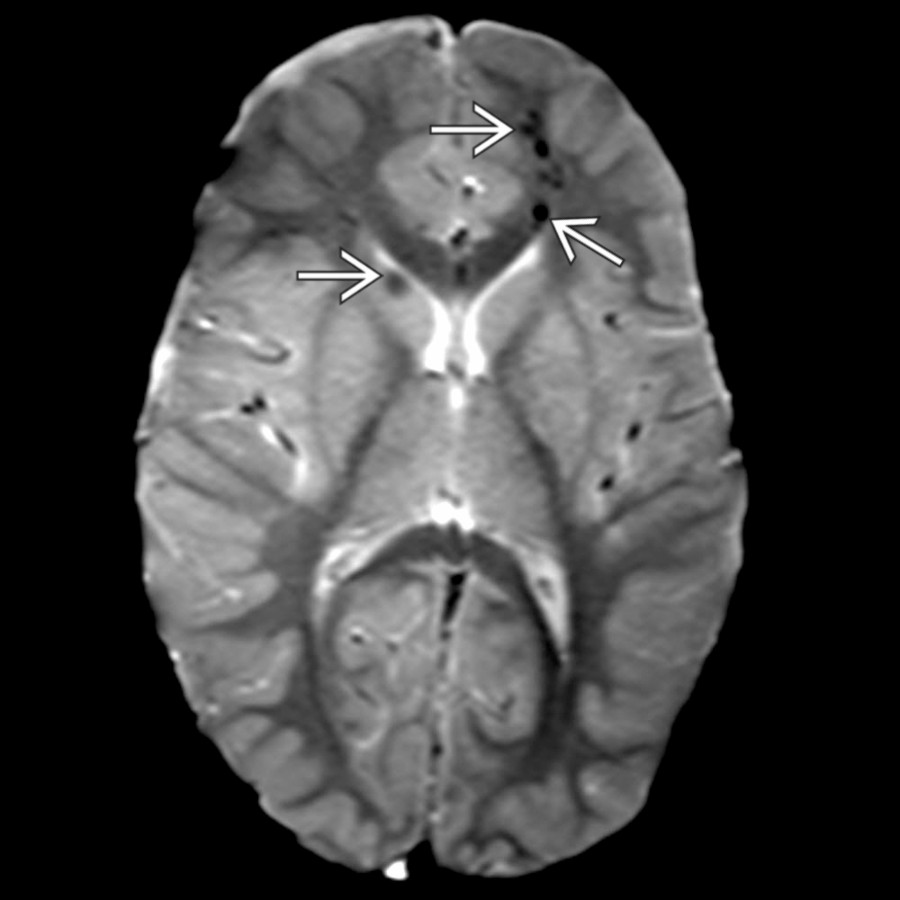
*Axial GRE MR in an 4-year-old pedestrian struck by a motor vehicle shows scalp swelling, small ventricles and sulci, and multifocal areas of hemorrhagic shear type injury .*

**Posttraumatic Brain Swelling**
*Axial NECT shows hyperdense foci of diffuse axonal injury , which is commonly associated with traumatic cerebral edema. Note sulcal and ventricular effacement . Loss of gray-white differentiation is common.*
**Posttraumatic Brain Swelling**
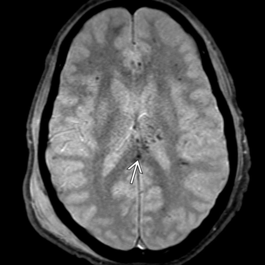
*Axial GRE MR shows multiple punctate areas of susceptibility in the subcortical frontal white matter but also in the splenium of the corpus callosum from diffuse axonal injury. Less well appreciated on this sequence are small lateral ventricles and cerebral edema.*

**Herniation Syndromes, Intracranial**
*Axial NECT shows low-density subacute infarcts in the cerebellar hemispheres. Basal cisterns are effaced , as is the 4th ventricle in this patient with transtentorial herniation. Herniation syndromes typically result from trauma, ischemia, or mass.*
**Herniation Syndromes, Intracranial**
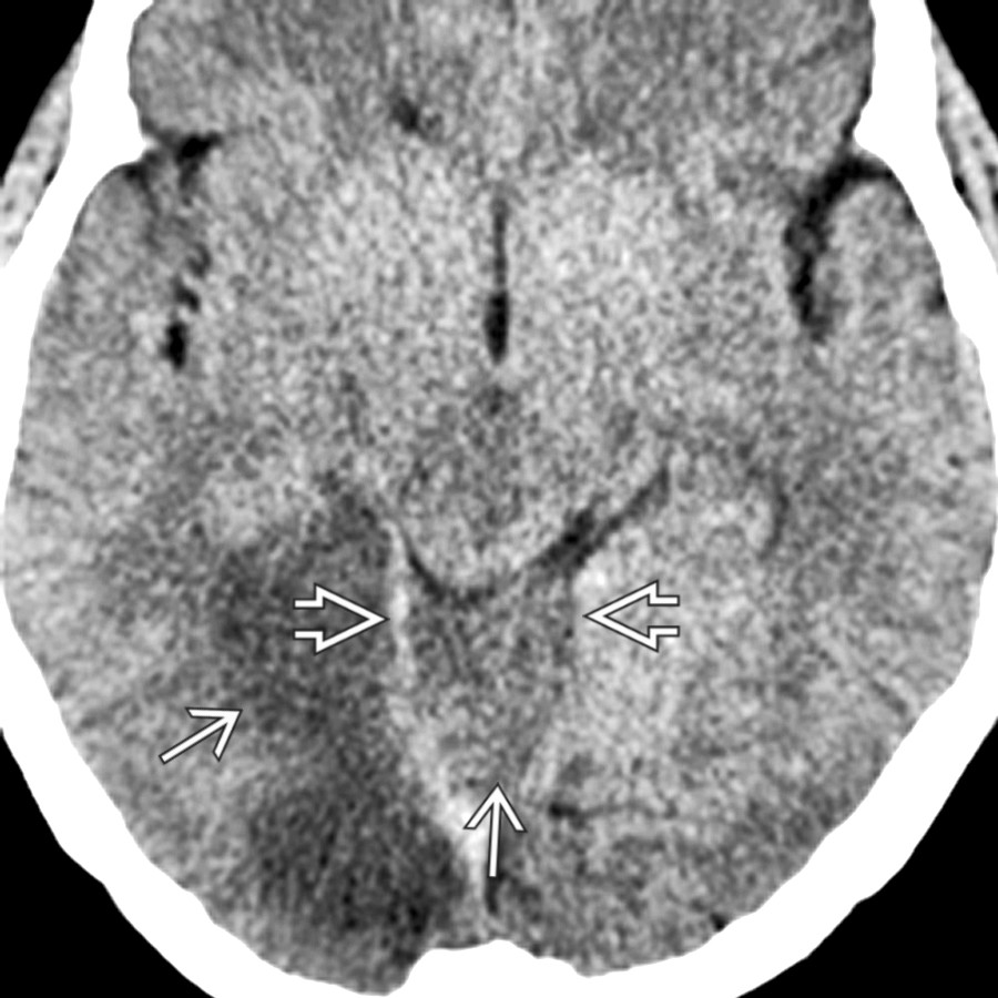
*Axial NECT shows small ventricles and right occipital and vermian low density due to infarct. Basal cisterns are effaced. Vermis is herniated upward through the tentorial incisura , displacing midbrain anteriorly and superiorly.*
**Adult Hypoxic-Ischemic Injury**
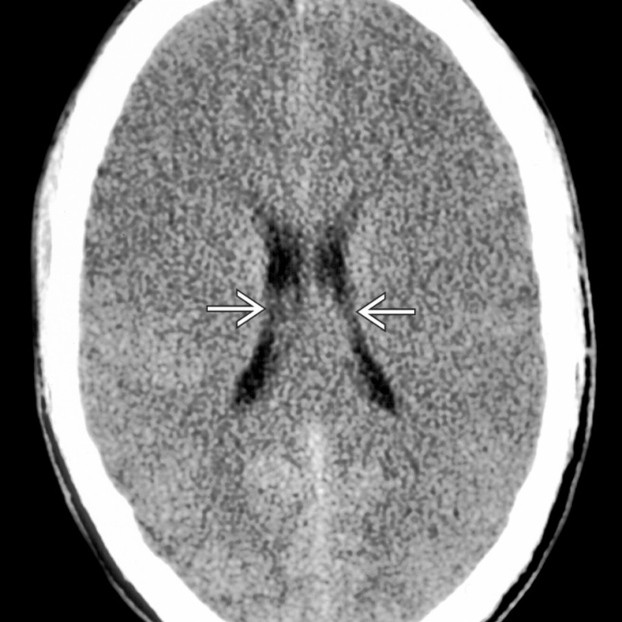
*Axial NECT shows a typical case of impending brain death with diffuse cerebral edema. Note diffuse sulcal effacement, small ventricles , and decreased gray-white matter differentiation within the cerebral hemispheres.*

**Intracranial Hypotension**
*Sagittal T1 MR shows downward displacement of the cerebellar tonsils and brainstem. There is effacement of the suprasellar cistern . Note effacement of the interpeduncular cistern , which has moved below the level of the dorsum sella, related to "brainstem sagging."*
### Additional Images

**CSF Shunts & Complications**
*Axial NECT shows small ventricles and indeterminate shunt position . Symptomatic ventricular collapse is known as slit-like ventricle syndrome and suggests overshunting.*

**Posttraumatic Brain Swelling**
*Axial GRE MR in an 4-year-old pedestrian struck by a motor vehicle shows scalp swelling, small ventricles and sulci, and multifocal areas of hemorrhagic shear type injury .*

**Posttraumatic Brain Swelling**
*Axial NECT shows hyperdense foci of diffuse axonal injury , which is commonly associated with traumatic cerebral edema. Note sulcal and ventricular effacement . Loss of gray-white differentiation is common.*

**Posttraumatic Brain Swelling**
*Axial GRE MR shows multiple punctate areas of susceptibility in the subcortical frontal white matter but also in the splenium of the corpus callosum from diffuse axonal injury. Less well appreciated on this sequence are small lateral ventricles and cerebral edema.*

**Herniation Syndromes, Intracranial**
*Axial NECT shows low-density subacute infarcts in the cerebellar hemispheres. Basal cisterns are effaced , as is the 4th ventricle in this patient with transtentorial herniation. Herniation syndromes typically result from trauma, ischemia, or mass.*

**Herniation Syndromes, Intracranial**
*Axial NECT shows small ventricles and right occipital and vermian low density due to infarct. Basal cisterns are effaced. Vermis is herniated upward through the tentorial incisura , displacing midbrain anteriorly and superiorly.*

**Adult Hypoxic-Ischemic Injury**
*Axial NECT shows a typical case of impending brain death with diffuse cerebral edema. Note diffuse sulcal effacement, small ventricles , and decreased gray-white matter differentiation within the cerebral hemispheres.*
 .](images/app.statdx.com_image_thumbnail_9f391b27-1dc1-4c50-b042-f1697cebd49c_annotated_true_size_900_quality_90_7f7b09b9_20251018T165216Z.jpg)
**Adult Hypoxic-Ischemic Injury**
*Axial PD FSE MR shows enlarged, bilateral, hyperintense deep nuclei and small ventricles from mass effect in hypoxic-ischemic encephalopathy. Cortical hyperintensity is less prominent than on DWI (not shown) except for more advanced bilateral occipital involvement .*

**Cerebral Infection**
*Axial FLAIR MR shows near-confluent T2 hyperintensity in the deep white matter and small ventricles related to mild mass effect from encephalitis.*

**Cerebral Infection**
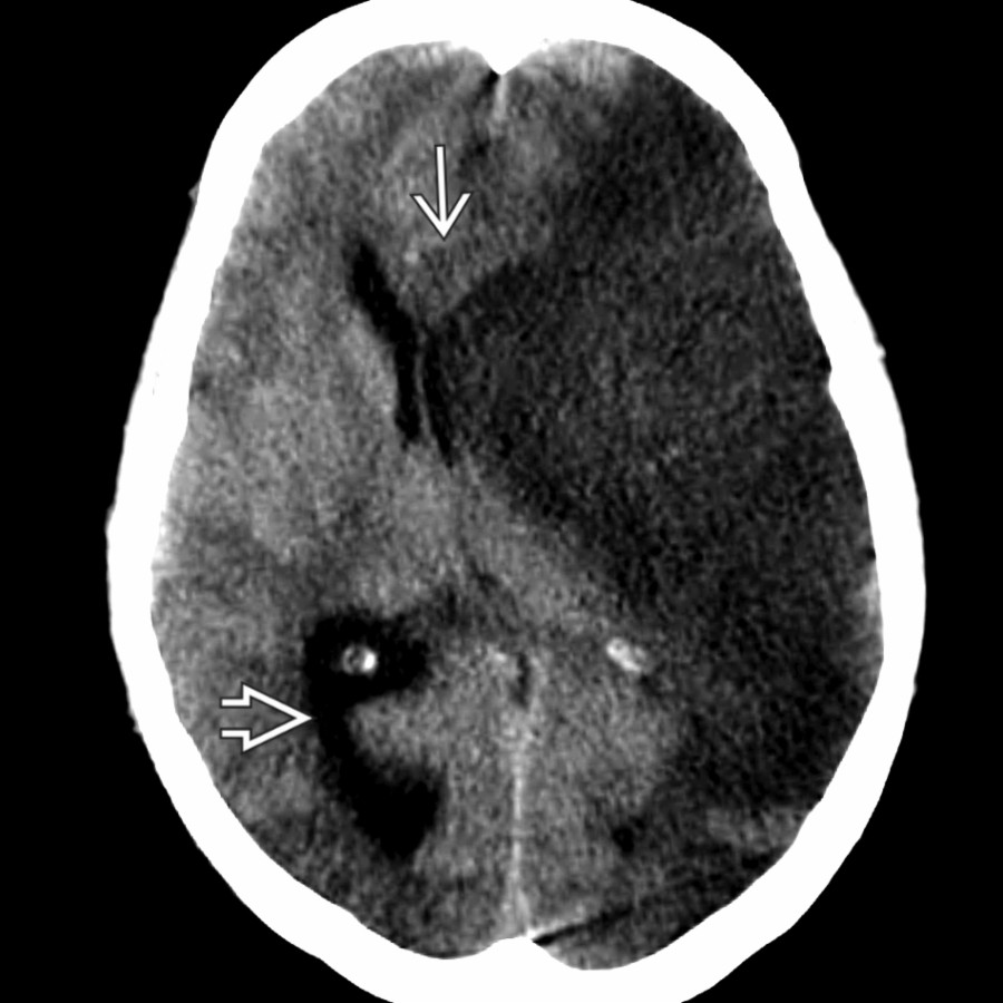
*Axial CECT shows thick, enhancing frontal leptomeninges with adjacent frontal lobe hypodensity from edema and mass effect on the frontal horns. This was pyogenic meningitis with adjacent cerebritis.*
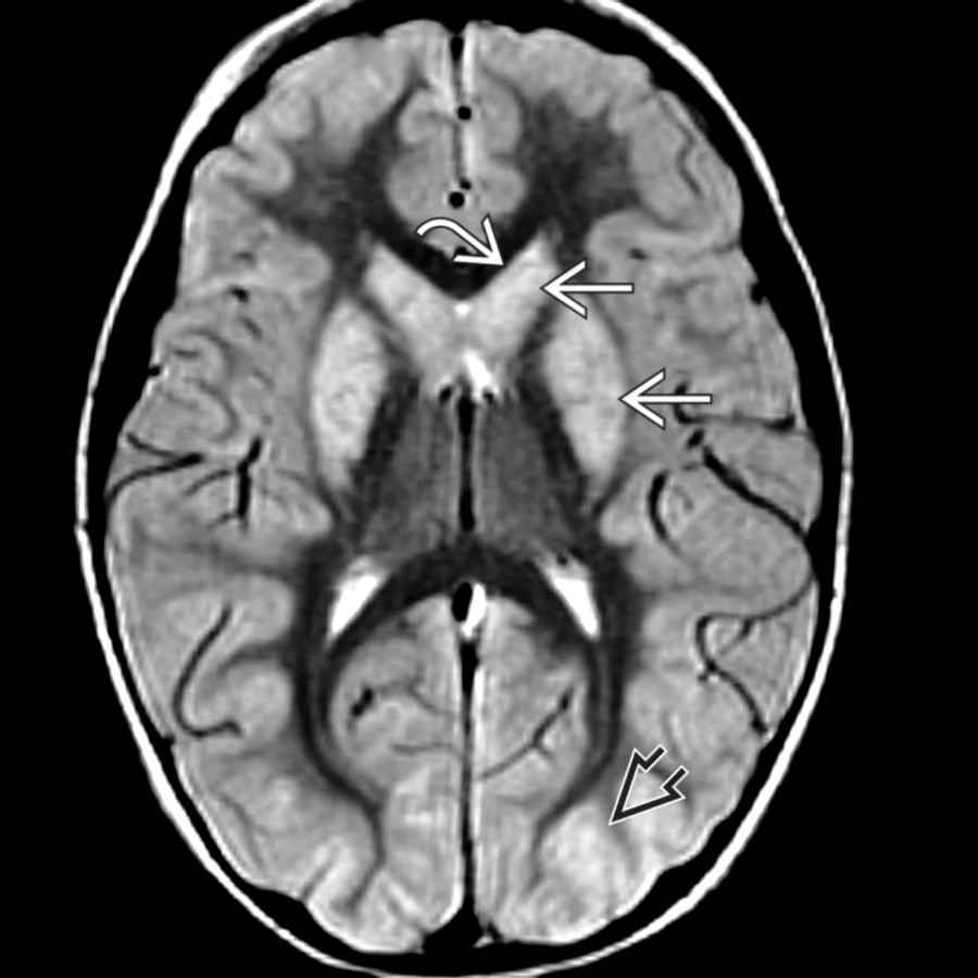
**Adult Hypoxic-Ischemic Injury**
*Axial PD FSE MR shows enlarged, bilateral, hyperintense deep nuclei and small ventricles from mass effect in hypoxic-ischemic encephalopathy. Cortical hyperintensity is less prominent than on DWI (not shown) except for more advanced bilateral occipital involvement .*

**Cerebral Infection**
*Axial FLAIR MR shows near-confluent T2 hyperintensity in the deep white matter and small ventricles related to mild mass effect from encephalitis.*

**Cerebral Infection**
*Axial CECT shows thick, enhancing frontal leptomeninges with adjacent frontal lobe hypodensity from edema and mass effect on the frontal horns. This was pyogenic meningitis with adjacent cerebritis.*
 due to diffuse brain edema, although when pial exudates obstruct CSF flow, hydrocephalus is more common.](d0e3444e-a542-4be2-8ff1-366016be79a4)
**Cerebral Infection**
*Axial T1 C+ FS MR shows subtle enhancement of the pial surface of the brain . Note associated ependymitis and choroid plexitis , which may complicate meningitis. Ventricles are small due to diffuse brain edema, although when pial exudates obstruct CSF flow, hydrocephalus is more common.*

**Cerebral Infection**
*Coronal T1 C+ MR shows meningitis complicated by local cerebritis, as evidenced in this patient with focal mass effect, small ventricles, and ill-defined enhancement .*

**Idiopathic Intracranial Hypertension**
*Sagittal T1 MR shows an empty sella and small ventricles in a patient with idiopathic intracranial hypertension or "pseudotumor cerebri."*

**Idiopathic Intracranial Hypertension**
*Coronal T1 MR in a young woman with headaches and papilledema shows very small lateral ventricles . Superficial sulci also look somewhat less prominent than normal. Pituitary gland is normal for a young, menstruating female.*

**Idiopathic Intracranial Hypertension**
*Axial T2 MR shows increased fluid in the optic nerve sheaths and mild flattening of the globes at the optic nerve heads . Note the partly empty sella . Findings suggest idiopathic intracranial hypertension that can be confirmed with CSF opening pressures.*

**Intracranial Hypotension**
*Sagittal T1 MR shows a "slumping midbrain," where the midbrain is displaced below the dorsum sellae . Ventricles are commonly small . The cerebellar tonsils are typically low-lying or herniated. Diffuse dural enhancement is characteristic. Subdural collections are uncommon (15%).*

**Intracranial Hypotension**
*Axial T1 C+ MR shows symmetric, small ventricles and smooth, diffuse, linear pachymeningeal thickening and enhancement .*

**Inborn Errors of Metabolism (Acute Presentation)**
*Axial T2 MR shows diffuse, acute brain swelling in maple syrup urine disease and small ventricles due to subacute edema of deep white matter , thalami , and internal capsules.*

**Intracranial Hypotension**
*Axial T1 C+ MR shows diffuse dural enhancement and leptomeningeal enhancement from venous distention. There are small lateral ventricles. Intracranial hypotension is commonly from CSF leak through a dural defect or from LP.*
due to diffuse brain edema, although when pial exudates obstruct CSF flow, hydrocephalus is more common.](d0e3444e-a542-4be2-8ff1-366016be79a4)
**Cerebral Infection**
*Axial T1 C+ FS MR shows subtle enhancement of the pial surface of the brain . Note associated ependymitis and choroid plexitis , which may complicate meningitis. Ventricles are small due to diffuse brain edema, although when pial exudates obstruct CSF flow, hydrocephalus is more common.*

**Cerebral Infection**
*Coronal T1 C+ MR shows meningitis complicated by local cerebritis, as evidenced in this patient with focal mass effect, small ventricles, and ill-defined enhancement .*

**Idiopathic Intracranial Hypertension**
*Sagittal T1 MR shows an empty sella and small ventricles in a patient with idiopathic intracranial hypertension or "pseudotumor cerebri."*

**Idiopathic Intracranial Hypertension**
*Coronal T1 MR in a young woman with headaches and papilledema shows very small lateral ventricles . Superficial sulci also look somewhat less prominent than normal. Pituitary gland is normal for a young, menstruating female.*

**Idiopathic Intracranial Hypertension**
*Axial T2 MR shows increased fluid in the optic nerve sheaths and mild flattening of the globes at the optic nerve heads . Note the partly empty sella . Findings suggest idiopathic intracranial hypertension that can be confirmed with CSF opening pressures.*

**Intracranial Hypotension**
*Sagittal T1 MR shows a "slumping midbrain," where the midbrain is displaced below the dorsum sellae . Ventricles are commonly small . The cerebellar tonsils are typically low-lying or herniated. Diffuse dural enhancement is characteristic. Subdural collections are uncommon (15%).*

**Intracranial Hypotension**
*Axial T1 C+ MR shows symmetric, small ventricles and smooth, diffuse, linear pachymeningeal thickening and enhancement .*

**Inborn Errors of Metabolism (Acute Presentation)**
*Axial T2 MR shows diffuse, acute brain swelling in maple syrup urine disease and small ventricles due to subacute edema of deep white matter , thalami , and internal capsules.*

**Intracranial Hypotension**
*Axial T1 C+ MR shows diffuse dural enhancement and leptomeningeal enhancement from venous distention. There are small lateral ventricles. Intracranial hypotension is commonly from CSF leak through a dural defect or from LP.*