---
title: "Spigelian Hernia"
docid: "3bbee7e5-dcd2-423c-a079-ce530cdb08c0"
authors:
- key: "c1df94ab-4a9f-44c4-add7-1f174fb9ac45"
value: "Siva P. Raman, MD"
breadcrumbs:
-
name: "Gastrointestinal"
slug: "gastrointestinal"
treeNodeId: "b52263f7-5978-4a22-a17d-7260e0033943"
-
name: "Diagnosis"
slug: "diagnosis"
treeNodeId: "5a7c51af-b1c6-4629-8f0e-d99e6fe57a98"
-
name: "Peritoneum, Mesentery, and Abdominal Wall"
slug: "peritoneum-mesentery-and-abdominal-"
treeNodeId: "a3fb9f00-f894-4b38-9e01-2f78406cf547"
-
name: "External Hernias"
slug: "external-hernias"
treeNodeId: "71ab3f79-4332-463c-9f60-d3dd2902d974"
-
name: "Spigelian Hernia"
slug: "spigelian-hernia"
treeNodeId: null
category: "Gastrointestinal"
documentVersionId: "7720c8c2-19fd-4aa0-a7dc-c23026d61a8a"
imageCount: 13
lastUpdated: "03/12/25"
pageDescription: "Spigelian Hernia"
pageKeywords: "Gastrointestinal, Diagnosis, Peritoneum, Mesentery, and Abdominal Wall, External Hernias, Spigelian Hernia"
pageTitle: "Spigelian Hernia | STATdx"
enhancedTitle: "Spigelian Hernia"
type: "DX"
references: true
ddx: true
cases: 2
breadcrumbs:
- "Gastrointestinal"
- "Diagnosis"
- "Peritoneum, Mesentery, and Abdominal Wall"
- "External Hernias"
- "Spigelian Hernia"
---
# KEY FACTS
- ## Terminology
- Hernia through defect in aponeurosis of internal oblique and transverse abdominal muscles
- ## Imaging
- Hernia occurs along lateral border of rectus abdominis muscles, inferior/lateral to umbilicus, at level of arcuate line
- Occurs at or below arcuate line due to lack of posterior rectus sheath at this level
- Lies deep to external oblique aponeurosis and muscle
- 90% within **Spigelian belt**, 6-cm transverse band above line joining anterior superior iliac spines
- Most often contains portions of greater omentum, small bowel, or colon
- Rarely can involve appendix, bladder, and other abdominal/pelvic structures
- Defect size is usually small (usually < 2 cm), resulting in narrow hernia neck and high risk of strangulation
- ## Top Differential Diagnoses
- Other abdominal wall hernias
- Subcutaneous or intramuscular lipoma
- Subcutaneous masses, fluid collections, or hematoma
- ## Pathology
- Probably multifactorial etiology, including congenital weakness of Spigelian fascia
- Usually congenital in children and acquired in adults
- Prior history of abdominal surgery and obesity are biggest risk factors in adults
- Other risk factors include multiple pregnancies, rapid weight loss, COPD, and trauma
- ## Clinical Issues
- Rare hernia that accounts for 1-2% of anterior abdominal wall hernias
- Difficult to diagnose clinically due to deep anatomic location, especially in obese patients
- Most common symptoms are pain and palpable bulge most apparent when standing
- Surgical treatment indicated in virtually all patients due to high risk of strangulation and incarceration
# TERMINOLOGY
- ## Abbreviations
- Spigelian hernia (SH)
- ## Synonyms
- Lateral ventral hernia; anterolateral hernia; hernia through conjoint tendon
- ## Definitions
- Hernia through defect in aponeurosis of internal oblique and transverse abdominal muscles
- Spigelian aponeurosis: Aponeurosis of internal oblique and transverse abdominal muscles
# IMAGING
- ## General Features
- ### Best diagnostic clue
- Hernia located lateral to rectus muscle and caudal/lateral to umbilicus, which is covered by intact external oblique muscle
- ### Location
- Hernia sac extends through defect in aponeurosis of transverse abdominal and internal oblique muscles
- Typically at or below level of arcuate line = semicircular line
- Occur at or below arcuate line due to lack of posterior rectus sheath at this level
- Lies deep to external oblique aponeurosis
- External oblique aponeurosis usually remains intact (hernial sac is intermuscular)
- Occurs along lateral border of rectus abdominis muscles, inferior and lateral to umbilicus, at level of arcuate line
- 90% of SHs are within **Spigelian belt of Spangen**, 6-cm transverse band above line joining both anterior superior iliac spines
- 2 primary types
- **Interstitial or interparietal SH**: Located below major oblique muscle among muscle layers of abdominal wall
- By far most common (98% of cases)
- **Subcutaneous SH**: Hernia sac crosses major oblique aponeurosis and becomes superficial (crosses beyond muscle layer of wall)
- Very rare
- May have slight left-sided predominance, although exact reason is unknown
- ### Morphology
- Defect size range: 1- to 7.5-cm diameter (most defects small and < 2 cm in size)
- Most often contains portions of greater omentum, small bowel, or colon
- Rarely can involve appendix, bladder, and other abdominal/pelvic structures
- ## CT Findings
- Hernia defect in aponeurosis of transverse abdominal and internal oblique muscles
- Lateral to rectus sheath, caudal to umbilicus, and deep to external oblique muscle and fascia
- Herniation of omentum ± bowel loops
- ## Imaging Recommendations
- ### Best imaging tool
- CECT
- ## Ultrasonographic Findings
- Begin at lateral margin of rectus abdominis in transverse plane at umbilicus
- As transducer is moved inferiorly, inferior epigastric artery can be identified
- SH can be visualized along linea semilunaris
- Cough and Valsalva maneuver during exam may help increase conspicuity of hernia
# DIFFERENTIAL DIAGNOSIS
- [Ventral Hernia](/document/ventral-hernia/ab08cd87-4342-4825-948b-d02fc178078f)
- Majority occur through midline aponeurosis
- Incisional hernia through off-midline incision can mimic SH
- No intact external oblique muscle or fascia
- [Umbilical Hernia](/document/umbilical-hernia/ff51faeb-9832-4e87-a6cc-ca45673d754f)
- Bowel, fat, or ascites protruding through umbilical defect in midline
- SHs occur inferior and lateral to umbilicus
- [Hernia Through Laparoscopy Port](/document/ventral-hernia/ab08cd87-4342-4825-948b-d02fc178078f)
- Usually smaller defect and tend to be located medial or lateral to spigelian site
- [Rectus Sheath Hematoma](/document/retroperitoneal-hemorrhage/56d5188d-85d3-49ff-af89-2b9d08608931)
- Cylindrical, heterogeneous mass within sheath without true fascial defect or hernia
- Easily differentiated from hernia on CT
- ## Subcutaneous or Intramuscular Lipoma
- Should be easily distinguished from hernia on CT
# PATHOLOGY
- ## General Features
- ### Etiology
- Probably multifactorial etiology, including congenital weakness of Spigelian fascia
- Abdominal wall at site of SH intrinsically weak due to lack of posterior sheath behind rectus muscle
- Usually congenital defect in children and infants
- Usually acquired defects in adults
- Prior history of abdominal surgery and obesity are biggest risk factors in adults for development of SH
- Other risk factors include multiple pregnancies, rapid weight loss, COPD, and trauma
- ### Associated abnormalities
- Undescended testis (17%) in children; ipsilateral
- Other anterior wall defects (e.g., omphalocele, bladder extrophy, prune belly)
- Coexisting ventral, inguinal, umbilical hernia
- ## Gross Pathologic & Surgical Features
- Interparietal or interstitial herniation (i.e., hernias cross transversus abdominis and internal oblique muscles but are behind external oblique aponeurosis)
- Contents: Omentum ± short segment of small or large bowel
# CLINICAL ISSUES
- ## Presentation
- Can be asymptomatic
- Most common symptoms are pain and palpable bulge or mass, usually most apparent when standing
- Symptoms of bowel obstruction due to high predisposition for incarceration
- Difficult to diagnose clinically due to deep anatomic location, especially in obese patients
- ## Demographics
- ### Age
- Patients between 40-70 years of age
- ### Sex
- Children: M:F = 2:1
- Essentially equal among adults, although possibly very minimal female predominance
- ### Epidemiology
- Accounts for 1-2% of anterior abdominal hernias
- Bilateral hernia: 15% incidence in children
- ## Natural History & Prognosis
- Omentum within SH may infarct and cause symptoms
- Hernia defect tends to be tight with narrow diameter of hernia neck (usually < 2 cm), making strangulation very common (24% of patients at presentation)
- ## Treatment
- Surgical treatment recommended in virtually all patients due to high risk of strangulation and incarceration
- Surgical treatment can be performed using either open or laparoscopic technique, although laparoscopic treatment has now become preferred option
- Surgical treatment usually performed with primary mesh repair or mesh reinforcement
- Recurrence rates after mesh repair are quite low
# DIAGNOSTIC CHECKLIST
- ## Consider
- Pre-/intraoperative US for accurate localization of SH, especially in obese patients
- Extensive intraoperative dissection, distortion of tissue planes, and morbidity risks may be avoided
- ## Image Interpretation Pearls
- Incisional hernias lateral to rectus muscle can mimic SH
- Incisional hernias have no intact external oblique muscle or aponeurosis
f4a8e939-fd6a-4921-859e-0b6f62cfc0f4
## References
# Selected References
1. [Shrestha P et al: Spigelian hernia: a case report. JNMA J Nepal Med Assoc. 62(270):145-7, 2024](http://www.ncbi.nlm.nih.gov/pubmed/?term=38409978%5Bpmid%5D)
1. [Hanzalova I et al: Spigelian hernia: current approaches to surgical treatment-a review. Hernia. 26(6):1427-33, 2022](http://www.ncbi.nlm.nih.gov/pubmed/?term=34665343%5Bpmid%5D)
1. [Azar SF et al: MDCT imaging in Spigelian hernia, clinical, and surgical implications. Clin Imaging. 74:131-8, 2021](http://www.ncbi.nlm.nih.gov/pubmed/?term=33493970%5Bpmid%5D)
1. [Lavin A et al: Incarcerated Spigelian hernias: a rare cause of a high-grade small bowel obstruction. Cureus. 12(3):e7397, 2020](http://www.ncbi.nlm.nih.gov/pubmed/?term=32337123%5Bpmid%5D)
1. [Webber V et al: Contemporary thoughts on the management of Spigelian hernia. Hernia. 21(3):355-61, 2017](http://www.ncbi.nlm.nih.gov/pubmed/?term=28097450%5Bpmid%5D)
1. [Martin M et al: Spigelian hernia: CT findings and clinical relevance. Abdom Imaging. 38(2):260-4, 2013](http://www.ncbi.nlm.nih.gov/pubmed/?term=22476335%5Bpmid%5D)
1. [Mustaffa N et al: Education and imaging. Gastrointestinal: Spigelian hernia; an uncommon cause of longstanding intermittent abdominal pain. J Gastroenterol Hepatol. 28(1):202, 2013](http://www.ncbi.nlm.nih.gov/pubmed/?term=23278154%5Bpmid%5D)
1. [Sucandy I et al: Spigelian hernia, diagnosis, and minimally invasive repair: a case series of 11 patients. Am Surg. 79(8):E284-5, 2013](http://www.ncbi.nlm.nih.gov/pubmed/?term=23896238%5Bpmid%5D)
1. [Perrakis A et al: Spigel hernia: a single center experience in a rare hernia entity. Hernia. 16(4):439-44, 2012](http://www.ncbi.nlm.nih.gov/pubmed/?term=22644060%5Bpmid%5D)
1. [Salameh JR: Primary and unusual abdominal wall hernias. Surg Clin North Am. 88(1):45-60, viii, 2008](http://www.ncbi.nlm.nih.gov/pubmed/?term=18267161%5Bpmid%5D)
1. [Jamadar DA et al: Sonography of inguinal region hernias. AJR Am J Roentgenol. 187(1):185-90, 2006](http://www.ncbi.nlm.nih.gov/pubmed/?term=16794175%5Bpmid%5D)
1. [Losanoff JE et al: Spigelian hernia in a child: case report and review of the literature. Hernia. 6(4): 191-3, 2002](http://www.ncbi.nlm.nih.gov/pubmed/?term=12424600%5Bpmid%5D)
1. [Losanoff JE et al: Recurrent Spigelian hernia: a rare cause of colonic obstruction. Hernia. 5(2): 101-4, 2001](http://www.ncbi.nlm.nih.gov/pubmed/?term=11505645%5Bpmid%5D)
1. [Losanoff JE et al: Incarcerated Spigelian hernia in morbidly obese patients: the role of intraoperative ultrasonography for hernia localization. Obes Surg. 7(3): 211-4, 1997](http://www.ncbi.nlm.nih.gov/pubmed/?term=9730551%5Bpmid%5D)
1. [Harrison LA et al: Abdominal wall hernias: review of herniography and correlation with cross-sectional imaging. Radiographics. 15(2):315-32, 1995](http://www.ncbi.nlm.nih.gov/pubmed/?term=7761638%5Bpmid%5D)
## Differential diagnosis
### Abdominal Wall Mass
DDX:d51e2268-67b6-4a60-9222-f5a86f61ddec
### Acute Left Abdominal Pain
DDX:65c32297-ce9e-41dd-80b5-60fd7160f2a6
### Defect in Abdominal Wall (Hernia)
DDX:5af046fa-59ef-45b5-952b-acbcdee36196
## Cases
- {'cases': [{'authors': [{'key': '64470aa4-5429-4786-81fa-be44c86ca020', 'value': 'Kristine S Spinelli, MD'}], 'caseVersionId': 'c5463905-e3f8-4cd0-a8de-0aa0b61e8e55', 'description': 'Classic spigelian hernia on CECT.\n\nA loop of large intestine is seen herniated lateral to the rectus abdominus muscle (arrow, #1-3), consistent with a spigelian hernia.', 'history': None, 'imagePoolId': 'af273cd7-9a32-4a1b-b833-5dac98949716', 'name': 'Classic', 'teachingPoint': None, 'demographics': '65 Years old male'}, {'authors': [{'key': '3d84d682-9451-4b02-99b2-e34970a5b440', 'value': 'Michael P. Federle, MD, FACR'}], 'caseVersionId': 'cc436c73-ea4f-45ce-bd88-4db6ade70016', 'description': 'Axial CECT shows aortic and iliac aneurysms and an incidental spigelian hernia. The hernia (open arrow, #1) contains a segment of ascending colon, but there is no sign of bowel obstruction.', 'history': 'Elderly man being evaluated for an abdominal aortic aneurysm.', 'imagePoolId': '9571e5a8-af45-4e4b-8cd5-6a171815798c', 'name': 'Incidental finding', 'teachingPoint': None, 'demographics': '76 Years old male'}, {'authors': [{'key': '3d84d682-9451-4b02-99b2-e34970a5b440', 'value': 'Michael P. Federle, MD, FACR'}], 'caseVersionId': 'e7340c27-1d19-43a2-917f-e918df5add1c', 'description': 'CT shows a small bowel obstruction and a spigelian hernia.\n\nThe hernia (open arrow, #2) is evident, just lateral to the rectus muscle. A segment of small bowel herniates through the defect, resulting in partial obstruction. The hernia and obstruction were confirmed and corrected at surgery.', 'history': 'Middle-aged woman with distended abdomen and abdominal pain.', 'imagePoolId': 'e81acda9-0fe8-4001-8c86-e483141eeebd', 'name': 'With partial bowel obstruction', 'teachingPoint': None, 'demographics': '62 Years old female'}, {'authors': [{'key': '3d84d682-9451-4b02-99b2-e34970a5b440', 'value': 'Michael P. Federle, MD, FACR'}], 'caseVersionId': 'ea9291ee-372a-4332-b115-a57f974b92d1', 'description': 'CT shows herniation of descending colon through spigelian aponeurosis.\n\nA frontal radiograph (#1) shows the distorted and displaced descending colon (arrow). Note the defect (open arrow, #4) in the abdominal wall lateral to the rectus muscle, through which a portion of the descending colon herniates. Note that the external oblique muscle (curved arrow, #2) and its aponeurosis remain intact, making the hernia sac an intramuscular process, which is classic for a spigelian hernia.', 'history': None, 'imagePoolId': '54664b68-275e-40eb-90aa-a8a8a0fe1aff', 'name': 'Classic, with colon', 'teachingPoint': None, 'demographics': '52 Years old male'}], 'caseType': 'typical', 'name': 'TYPICAL'}
- {'cases': [{'authors': [{'key': '3d84d682-9451-4b02-99b2-e34970a5b440', 'value': 'Michael P. Federle, MD, FACR'}], 'caseVersionId': '8fe62ec3-1a09-40ef-b14a-99463fe13e8c', 'description': 'CT shows distended, ischemic bowel, obstructed in a spigelian hernia.\n\nFree air (curved arrow, #1) indicated bowel perforation. The small intestine is dilated and pneumatosis (arrow, #2-4) indicates bowel ischemia. The point of obstruction was a spigelian hernia (open arrow, #4).\n\nComment: Infarcted bowel was resected at surgery, but the patient expired soon after surgery.', 'history': 'Elderly woman with distended abdomen and pain.', 'imagePoolId': '5290ab4d-9f98-4d51-b5b2-1ce3ef0f25ac', 'name': 'With bowel obstruction and ischemia', 'teachingPoint': None, 'demographics': '92 Years old female'}, {'authors': [{'key': '3d84d682-9451-4b02-99b2-e34970a5b440', 'value': 'Michael P. Federle, MD, FACR'}], 'caseVersionId': 'c4d2db95-3bb0-4f9c-9388-b9d0a3ee2490', 'description': 'Axial CECT shows a spigelian hernia, with herniation of omental fat through a defect in the aponeurosis of the transverse abdominal and internal oblique muscles, covered by an intact aponeurosis of the external oblique muscle. Also present within the hernia is a thick-walled tubular structure (arrows, #1-4) (inflamed appendix), seen arising from the tip of the cecum.', 'history': 'Right lower quadrant pain & fever.', 'imagePoolId': '9333dddf-fbad-4976-ba1b-8d74d01ca29b', 'name': 'Inflamed appendix within spigelian hernia', 'teachingPoint': None, 'demographics': '81 Years old male'}, {'authors': [{'key': '3d84d682-9451-4b02-99b2-e34970a5b440', 'value': 'Michael P. Federle, MD, FACR'}], 'caseVersionId': '0a940857-19ac-4e61-89d0-ca2c275d88ab', 'description': 'A series of axial (#1-7) and coronal reformatted (#8-14) CT sections shows an intact external oblique muscle and its aponeurosis (curved arrows, #2-5,10-13), though these are thin and stretched over a subtle fat density bulge in the left lower abdominal wall, caudal and lateral to the umbilicus (open arrows, #1,8). A defect in the aponeurosis of the internal oblique and transverse abdominal muscles (arrows, #6,12-13) is a Spigelian hernia, and the herniated omental fat accounts for the other signs and symptoms.\n\nComment: The intact external oblique muscle is a distinguishing feature not found in cases of incisional hernias, which may occur in the same location.', 'history': 'Pain sharply localized to the left lower quadrant on straining.', 'imagePoolId': '24dd810d-998e-4ae6-8a53-edbd3aaaa6a1', 'name': 'Small and subtle', 'teachingPoint': None, 'demographics': '37 Years old male'}], 'caseType': 'variant', 'name': 'VARIANT'}
## Images
### Selected Images
 arising just lateral to the rectus muscle. A segment of small bowel herniates through the defect, resulting in partial obstruction. The hernia and obstruction were confirmed and corrected at surgery.](images/app.statdx.com_image_thumbnail_94407542-524f-49c5-9f97-6434bb7818b8_annotated_true_size_900_quality_90_f4a59611bee398d7f32409432f07aa833cc5d194.jpg)
*Axial CECT demonstrates a Spigelian hernia arising just lateral to the rectus muscle. A segment of small bowel herniates through the defect, resulting in partial obstruction. The hernia and obstruction were confirmed and corrected at surgery.*
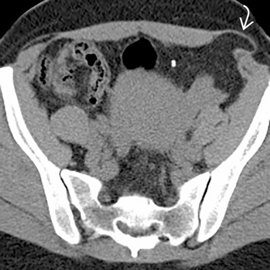
*Axial NECT demonstrates a small Spigelian hernia in the left lower quadrant, inferior and lateral to the umbilicus. Note the layer of the external oblique muscle overlying the hernia sac.*
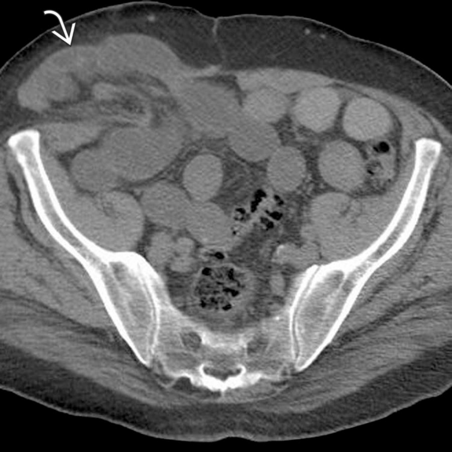
*Axial CECT demonstrates a Spigelian hernia arising just lateral to the rectus muscle. A segment of small bowel herniates through the defect, resulting in partial obstruction. The hernia and obstruction were confirmed and corrected at surgery.*

*Axial NECT demonstrates a small Spigelian hernia in the left lower quadrant, inferior and lateral to the umbilicus. Note the layer of the external oblique muscle overlying the hernia sac.*
 contains a segment of ascending colon, but there is no sign of bowel obstruction.](images/app.statdx.com_image_thumbnail_94280700-6734-48c7-9595-d780b4a5cdab_annotated_true_size_900_quality_90_0e7bab138ada79cd3fe82b25b2861878c69af69d.jpg)
*Axial NECT in a patient being imaged for iliac aneurysms demonstrates an incidental spigelian hernia. The hernia contains a segment of ascending colon, but there is no sign of bowel obstruction.*
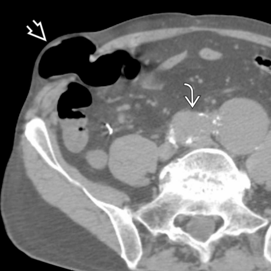
*Axial NECT in a patient being imaged for iliac aneurysms demonstrates an incidental spigelian hernia. The hernia contains a segment of ascending colon, but there is no sign of bowel obstruction.*
 . A segment of small bowel herniates through the defect, resulting in obstruction and dilatation of the upstream bowel .](images/app.statdx.com_image_thumbnail_e308a95d-5b1b-4bc5-b508-c51a2161620d_annotated_true_size_900_quality_90_a6b5d18fc83dc412062fa90d252757ca1aaa5ebd.jpg)
*Axial CECT shows a small bowel obstruction and a Spigelian hernia . A segment of small bowel herniates through the defect, resulting in obstruction and dilatation of the upstream bowel .*
### Additional Images
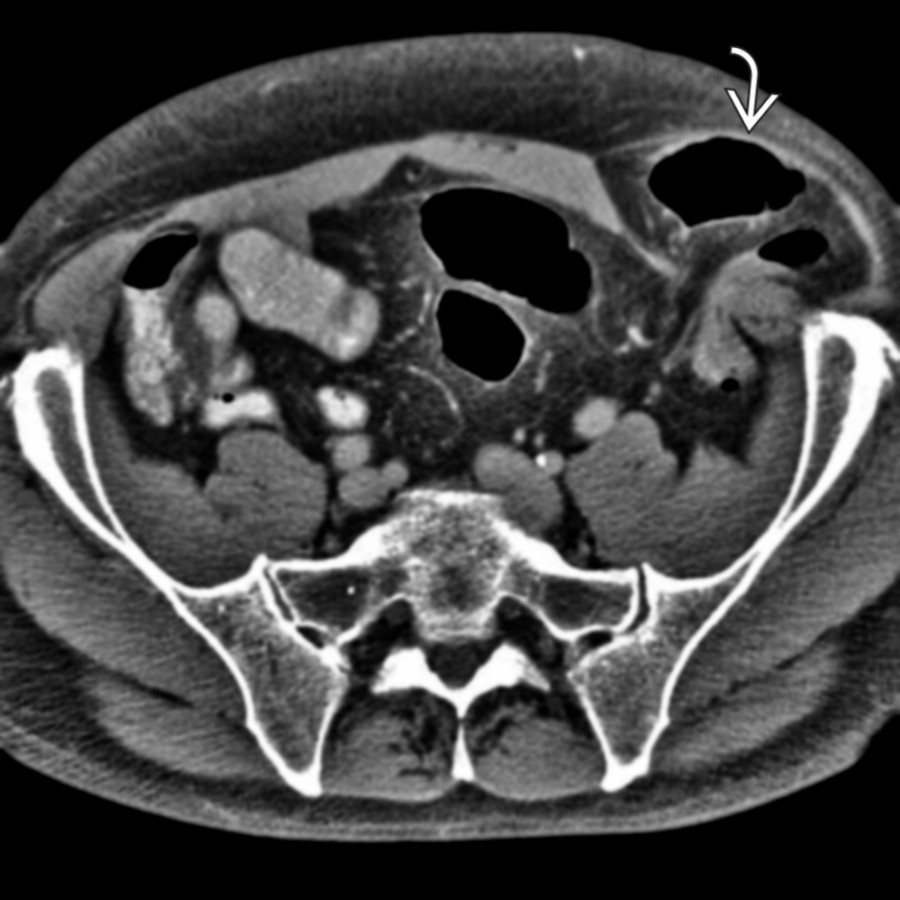
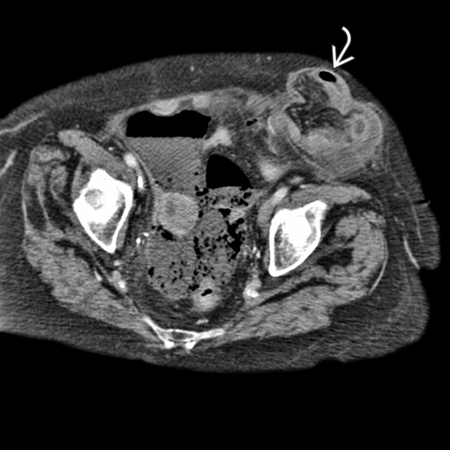
*Axial CECT shows a Spigelian hernia just lateral to the rectus muscle with an obstructed, herniated descending colon.*
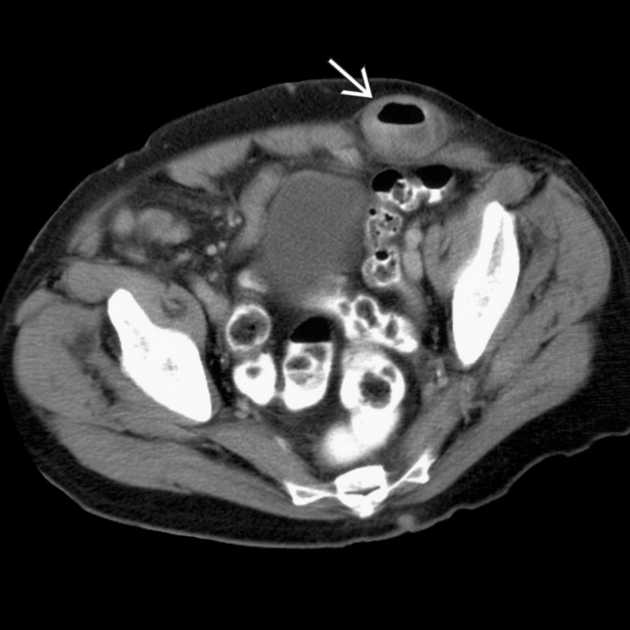
*Axial CECT shows a left Spigelian hernia .*
*Axial NECT shows a loop of large intestine herniating lateral to the rectus abdominis muscle, consistent with a Spigelian hernia.*
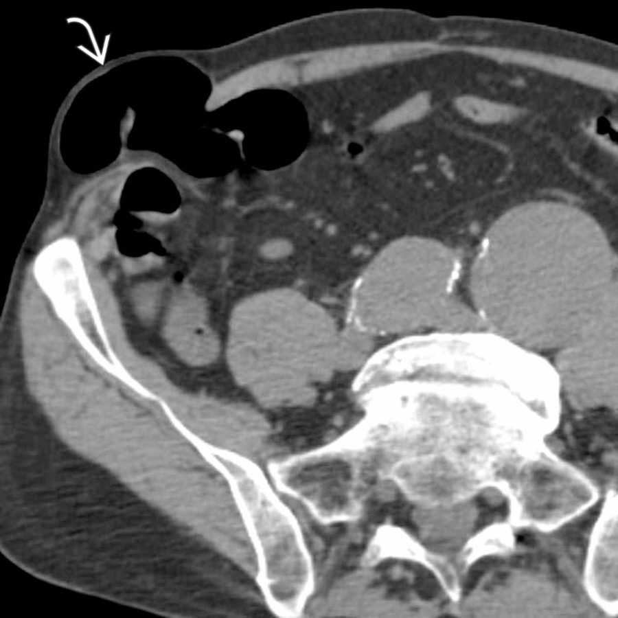
*Axial NECT shows aortic and iliac aneurysms and an incidental Spigelian hernia . The hernia contains a segment of ascending colon, but there is no sign of bowel obstruction.*

*Axial CECT shows a Spigelian hernia with herniation of omental fat through a defect in the aponeurosis of the transverse abdominal and internal oblique muscles, covered by an intact aponeurosis of the external oblique muscle. Also present within the hernia is a thick-walled tubular structure (inflamed appendix) seen arising from the tip of the cecum.*

*Axial CECT shows a Spigelian hernia with herniation of omental fat through a defect in the aponeurosis of the transverse abdominal and internal oblique muscles, covered by an intact aponeurosis of the external oblique muscle. Also present within the hernia is a thick-walled tubular structure (inflamed appendix) seen arising from the tip of the cecum.*

*Axial T2 MR demonstrates a Spigelian hernia in the left lower quadrant, inferior and lateral to the umbilicus. Note the layer of the external oblique muscle overlying the hernia sac.*

*Axial CECT in a patient with left lower quadrant pain demonstrates a Spigelian hernia containing a fat-containing lesion , which extends from the surface of the colon with adjacent fat stranding or inflammation. This represents epiploic appendagitis within a Spigelian hernia.*

*Axial CECT demonstrates a Spigelian hernia containing a fat-containing lesion , which extends from the surface of the colon with adjacent fat stranding or inflammation. This represents epiploic appendagitis within a Spigelian hernia.*
. A segment of small bowel herniates through the defect, resulting in obstruction and dilatation of the upstream bowel .](images/app.statdx.com_image_thumbnail_e308a95d-5b1b-4bc5-b508-c51a2161620d_annotated_true_size_900_quality_90_a6b5d18fc83dc412062fa90d252757ca1aaa5ebd.jpg)
*Axial CECT shows a small bowel obstruction and a Spigelian hernia . A segment of small bowel herniates through the defect, resulting in obstruction and dilatation of the upstream bowel .*
### Additional Images

*Axial CECT shows a Spigelian hernia just lateral to the rectus muscle with an obstructed, herniated descending colon.*

*Axial CECT shows a left Spigelian hernia .*

*Axial NECT shows a loop of large intestine herniating lateral to the rectus abdominis muscle, consistent with a Spigelian hernia.*

*Axial NECT shows aortic and iliac aneurysms and an incidental Spigelian hernia . The hernia contains a segment of ascending colon, but there is no sign of bowel obstruction.*

*Axial CECT shows a Spigelian hernia with herniation of omental fat through a defect in the aponeurosis of the transverse abdominal and internal oblique muscles, covered by an intact aponeurosis of the external oblique muscle. Also present within the hernia is a thick-walled tubular structure (inflamed appendix) seen arising from the tip of the cecum.*

*Axial CECT shows a Spigelian hernia with herniation of omental fat through a defect in the aponeurosis of the transverse abdominal and internal oblique muscles, covered by an intact aponeurosis of the external oblique muscle. Also present within the hernia is a thick-walled tubular structure (inflamed appendix) seen arising from the tip of the cecum.*

*Axial T2 MR demonstrates a Spigelian hernia in the left lower quadrant, inferior and lateral to the umbilicus. Note the layer of the external oblique muscle overlying the hernia sac.*

*Axial CECT in a patient with left lower quadrant pain demonstrates a Spigelian hernia containing a fat-containing lesion , which extends from the surface of the colon with adjacent fat stranding or inflammation. This represents epiploic appendagitis within a Spigelian hernia.*

*Axial CECT demonstrates a Spigelian hernia containing a fat-containing lesion , which extends from the surface of the colon with adjacent fat stranding or inflammation. This represents epiploic appendagitis within a Spigelian hernia.*