---
title: "Temporal Bone Facial Nerve Schwannoma"
docid: "cf2bcc82-4a1b-4989-adeb-f4e82116111b"
authors:
- key: "07a2c087-6202-49e7-870b-7aa162d18f06"
value: "Bronwyn E. Hamilton, MD"
- key: "33151213-01b2-4542-9105-342e006b3915"
value: "H. Ric Harnsberger, MD"
breadcrumbs:
-
name: "Head and Neck"
slug: "head-and-neck"
treeNodeId: "ed24ed8c-5d57-4629-879b-447b82d2973d"
-
name: "Diagnosis"
slug: "diagnosis"
treeNodeId: "19b6b986-97d0-40e7-b317-00f0c5cd8fa2"
-
name: "Temporal Bone"
slug: "temporal-bone"
treeNodeId: "9ad7d7b2-b2e4-4de2-be04-55ce607560c9"
-
name: "Intratemporal Facial Nerve"
slug: "intratemporal-facial-nerve"
treeNodeId: "35b77f60-796d-460f-8bac-4a187a150171"
-
name: "Benign and Malignant Tumors"
slug: "benign-and-malignant-tumors"
treeNodeId: "353e7622-0bb3-4dce-96d6-f5f889dad7cb"
-
name: "Temporal Bone Facial Nerve Schwannoma"
slug: "temporal-bone-facial-nerve-schwann-"
treeNodeId: null
category: "Head and Neck"
cmeTopicId: "e4d7bd7c-9fb3-4ba4-a909-8cd12d4f09e5"
documentVersionId: "925a57f2-14ad-4c18-93d5-9586c1b3d8a0"
imageCount: 18
lastUpdated: "08/18/21"
pageDescription: "Temporal Bone Facial Nerve Schwannoma"
pageKeywords: "Head and Neck, Diagnosis, Temporal Bone, Intratemporal Facial Nerve, Benign and Malignant Tumors, Temporal Bone Facial Nerve Schwannoma"
pageTitle: "Temporal Bone Facial Nerve Schwannoma | STATdx"
enhancedTitle: "Temporal Bone Facial Nerve Schwannoma"
type: "DX"
references: true
ddx: true
cases: 2
breadcrumbs:
- "Head and Neck"
- "Diagnosis"
- "Temporal Bone"
- "Intratemporal Facial Nerve"
- "Benign and Malignant Tumors"
- "Temporal Bone Facial Nerve Schwannoma"
---
# KEY FACTS
- ## Terminology
- Facial nerve schwannoma (FNS): Rare benign tumor of Schwann cells that invests intratemporal facial nerve (CNVII)
- ## Imaging
- Temporal bone CT: Tubular mass spanning multiple intratemporal CNVII segments with smooth enlargement of bony CNVII canal
- **> 90%** of FNS span ≥ 3 intratemporal CNVII segments
- T1 C+ MR: Homogeneously enhancing tubular mass ± intramural cysts
- Temporal bone CT appearance dictated by specific location
- **Geniculate fossa FNS**: Ovoid, smooth enlargement of geniculate fossa with projections into labyrinthine ± anterior tympanic segments of CNVII
- **Tympanic segment FNS**: Pedunculated FNS emanates from tympanic CNVII into middle ear
- **Mastoid segment FNS**: Either tubular with sharp margins or globular with irregular margins (breaks into mastoid air cells)
- **Greater superficial petrosal nerve (****GSPN) schwannoma**: Enlargement of GSPN canal; middle cranial fossa mass
- ## Top Differential Diagnoses
- Normal intratemporal facial nerve enhancement
- Bell palsy (herpetic facial paralysis)
- Intratemporal facial nerve venous malformation
- Intratemporal CNVII perineural malignancy
- ## Clinical Issues
- Symptoms: Hearing loss (70%), CNVII paresis (50%)
- Treatment options
- Conservative: Observation
- Surgical treatment: Complete removal is goal
- Radiotherapy: Nerve edema and hearing loss limit utility
# TERMINOLOGY
- ## Abbreviations
- Facial nerve schwannoma (FNS)
- ## Synonyms
- Facial neuroma, facial neurilemmoma
- ## Definitions
- FNS: Rare benign tumor of Schwann cells that invests intratemporal facial nerve (CNVII)
# IMAGING
- ## General Features
- ### Best diagnostic clue
- Temporal bone CT: Tubular mass spanning multiple intratemporal CNVII segments with smooth enlargement of bony CNVII canal
- T1 C+ MR: Homogeneously enhancing tubular mass ± intramural cysts
- ### Location
- Most common location: Geniculate ganglion
- **> 90%** of FNS span ≥ **3 intratemporal CNVII segments**
- ### Size
- Often long (multiple centimeters)
- Cross-sectional measurement usually < 1 cm
- ### Morphology
- Location dependent
- Geniculate fossa: Ovoid or triangular
- Greater superficial petrosal nerve (GSPN): Ovoid, projects into middle cranial fossa
- Tympanic CNVII: Lobulates into middle ear
- Mastoid CNVII: Irregular margin if breaks into surrounding air cells
- Parotid CNVII: Tubular or ovoid mass along CNVII intraparotid course
- **Tubular shape** along multiple CNVII segments
- ## CT Findings
- ### CECT
- No role for CECT in this diagnosis
- Use enhanced MR instead
- ### Bone CT
- General temporal bone CT appearances
- Tubular enlargement of CNVII canal
- Bony margins are smooth, benign-appearing
- Temporal bone CT appearance is dictated by specific location of FNS along CNVII
- **Geniculate fossa FNS**: Ovoid, smooth enlargement of geniculate fossa
- Tumor projects into labyrinthine ± anterior tympanic segments of CNVII
- **Tympanic segment FNS**: Pedunculated FNS emanates from tympanic segment of CNVII into middle ear cavity
- **Mastoid segment FNS**: Either tubular with sharp margins or globular with irregular margins
- Shape depends on whether FNS breaks into surrounding mastoid air cells
- **GSPN schwannoma**: Ovoid enlargement of GSPN canal anteromedial to geniculate fossa
- ## MR Findings
- ### T1WI
- Intermediate- to low-signal lesion
- ### T2WI
- High-signal lesion
- ### T1WI C+
- **Geniculate ganglion FNS**: Ovoid, enhancing mass in enlarged geniculate fossa
- Tumor tails project into labyrinthine ± anterior tympanic segments of CNVII
- **Tympanic segment FNS**: Pedunculates into middle ear cavity
- **Mastoid segment FNS**
- Either tubular with sharp margins or globular with irregular margins
- Depends on whether it breaks into surrounding mastoid air cells
- **GSPN schwannoma**
- Diagnosed when enhancing mass is seen in location of GSPN
- Just anteromedial to geniculate fossa
- Middle cranial fossa enhancing mass with connection to geniculate fossa
- May be difficult to establish extraaxial nature of this schwannoma
- ## Imaging Recommendations
- ### Best imaging tool
- Patient presents with hearing loss ± CNVII paresis
- Start with thin-section T1 C+ fat-saturated MR in axial and coronal plane through internal auditory canal (IAC) and temporal bone
- If intratemporal, tubular enhancing mass is diagnosed on MR, then temporal bone CT helps delineate nature of lesion based on bone changes
# DIFFERENTIAL DIAGNOSIS
- [Normal Intratemporal Facial Nerve Enhancement](/document/intratemporal-facial-nerve-enhance-/a3569ec5-a566-411d-877f-41ad832e3fd2)
- Clinical: Asymptomatic
- Temporal bone CT: Intratemporal CNVII canal is normal
- T1 C+ MR: Geniculate ganglion, anterior tympanic ± mastoid segments enhance normally
- Labyrinthine CNVII does not enhance normally
- [Bell Palsy (Herpetic Facial Paralysis)](/document/bell-palsy/0958e575-8f76-4d70-b806-0dbed9c62a67)
- Clinical: Sudden onset of peripheral CNVII paralysis
- Temporal bone CT: Normal intratemporal CNVII canal
- T1 C+ MR: Intratemporal + IAC fundal CNVII enhancement
- [Intratemporal Facial Nerve Venous Malformation](/document/temporal-bone-facial-nerve-venous--/dcd6a44e-cbe6-457c-9b03-598a2b874ece)
- Clinical: Sudden unilateral peripheral CNVII paralysis
- Temporal bone CT: Intratumoral honeycomb or bone spicules
- T1 C+ MR: Poorly circumscribed, geniculate fossa enhancing mass
- ## Intratemporal CNVII Perineural Malignancy
- Clinical: Known or recurrent parotid malignancy
- Temporal bone CT: Mastoid CNVII canal is enlarged but less than in FNS
- T1 C+ MR: Infiltrating parotid mass is present
# PATHOLOGY
- ## General Features
- ### Etiology
- Slowly growing, benign tumor from Schwann cells investing intratemporal CNVII
- ### Genetics
- If multiple schwannomas ± meningiomas, think neurofibromatosis type 2 (NF2)
- ### Associated abnormalities
- NF2: Bilateral vestibular schwannomas; other schwannoma and meningioma possible
- ## Gross Pathologic & Surgical Features
- Tan, ovoid-tubular, encapsulated mass
- Arises from outer nerve sheath layer of CNVII, expanding eccentrically away from nerve
- ## Microscopic Features
- Benign, encapsulated tumor made up of bundles of spindle-shaped Schwann cells forming whorled pattern
- Cellular architecture consists of densely cellular (Antoni A) areas ± loose, myxomatous (Antoni B) areas
- S100 protein stain: Strongly and diffusely positive in both nucleus and cytoplasm
- May display **intramural cystic changes**
# CLINICAL ISSUES
- ## Presentation
- ### Most common signs/symptoms
- Hearing loss present in ~ 70%
- Facial nerve symptoms present in ~ 50%
- CNVII weakness or paralysis > involuntary facial movements
- Bell palsy-like CNVII paralysis is rare
- Ear ± facial pain
- ### Other signs/symptoms
- Cerebellopontine angle (CPA)-IAC FNS: Sensorineural hearing loss, vertigo, and tinnitus
- Larger tympanic and mastoid segments FNS
- Avascular retrotympanic mass
- Conductive hearing loss
- ## Demographics
- ### Age
- Mean age at presentation: 50 years
- ### Epidemiology
- FNS is rare tumor (< 1% of intrapetrous tumors)
- Within temporal bone > > intraparotid > CPA-IAC
- ## Natural History & Prognosis
- Slow-growing benign tumor
- Eventually enlarges sufficiently to cause hearing loss and other cranial neuropathy
- Some tumors (< 10%) do not grow or become symptomatic
- Risk of facial weakness ↑ with intratemporal involvement
- Risk for facial weakness and hearing loss ↑ with more segments involved
- Risk for hearing loss ↑ with more proximal tumor
- ## Treatment
- **Conservative management**
- If CNVII paralysis is absent or mild when diagnosed, surgical cure can be worse than disease
- Incomplete recovery of full CNVII function may occur despite surgical restoration of CNVII continuity
- Follow until CNVII symptoms begin to develop
- Treatment used in older adult patients
- **Surgical treatment**
- Goal = complete FNS removal with preservation of hearing and CNVII function restoration
- Size-specific surgical techniques
- Large FNS: Remove tumor + CNVII cable graft
- Small FNS (< 1 cm): CNVII transposition with primary anastomosis
- Location-specific surgery
- Labyrinthine or geniculate FNS: Middle cranial fossa and transmastoid approaches combined
- Tympanic-mastoid FNS: Transmastoid alone
- **Radiotherapy**
- Stereotactic radiotherapy is possible but generally contraindicated for temporal bone location due to
- Postradiation edema and nerve swelling
- Risk of hearing loss
# DIAGNOSTIC CHECKLIST
- ## Consider
- Older patients with FNS often followed, not operated
- Younger patients without CNVII paresis often followed
- ## Image Interpretation Pearls
- Intratemporal FNS: Segmental, tubular enlargement of CNVII canal
- Distinctive imaging findings depending on segment of CNVII involved
- CPA-IAC FNS: Exactly mimics vestibular schwannoma if no extension into labyrinthine segment CNVII occurs
- If present, labyrinthine segment tail makes imaging diagnosis
- Intraparotid FNS: Tubular mass in parotid coursing lateral to retromandibular vein
- If present, mastoid segment tail suggests diagnosis
- Differentiate from perineural parotid malignancy
01b8eeb4-a52d-4271-b49c-199cc46770da
## References
# Selected References
1. [Bartindale M et al: Facial schwannoma management outcomes: a systematic review of the literature. Otolaryngol Head Neck Surg. 163(2):293-301, 2020](http://www.ncbi.nlm.nih.gov/pubmed/?term=32228141%5Bpmid%5D)
1. [Bartindale M et al: The natural history of facial schwannomas: a meta-analysis of case series. J Neurol Surg B Skull Base. 80(5):458-68, 2019](http://www.ncbi.nlm.nih.gov/pubmed/?term=31534886%5Bpmid%5D)
1. [Loos E et al: Cochlear erosion due to a facial nerve schwannoma. J Int Adv Otol. 15(2):330-2, 2019](http://www.ncbi.nlm.nih.gov/pubmed/?term=31287431%5Bpmid%5D)
1. [Park JC et al: Large facial nerve schwannoma with extensive temporal bone destruction. Otol Neurotol. 39(3):e220-1, 2018](http://www.ncbi.nlm.nih.gov/pubmed/?term=29342046%5Bpmid%5D)
1. [Chen WJ et al: Case analysis of temporal bone lesions with facial paralysis as main manifestation and literature review. Cancer Biomark. 20(2):199-205, 2017](http://www.ncbi.nlm.nih.gov/pubmed/?term=28826175%5Bpmid%5D)
1. [Nishijima H et al: Facial nerve paralysis associated with temporal bone masses. Auris Nasus Larynx. 44(5):548-53, 2017](http://www.ncbi.nlm.nih.gov/pubmed/?term=28161243%5Bpmid%5D)
1. [Schulze M et al: Improvement in imaging common temporal bone pathologies at 3 T MRI: small structures benefit from a small field of view. Clin Radiol. 72(3):267.e1-12, 2017](http://www.ncbi.nlm.nih.gov/pubmed/?term=28034444%5Bpmid%5D)
1. [Yi H et al: Primary tumors of the facial nerve misdiagnosed: a case series and review of the literature. Acta Otolaryngol. 137(6):651-5, 2017](http://www.ncbi.nlm.nih.gov/pubmed/?term=28079432%5Bpmid%5D)
1. [Bäck L et al: Management of facial nerve schwannoma: a single institution experience. Acta Otolaryngol. 130(10):1193-8, 2010](http://www.ncbi.nlm.nih.gov/pubmed/?term=20441526%5Bpmid%5D)
1. [Chao WC et al: Facial nerve schwannoma. Otolaryngol Head Neck Surg. 141(1):146-7, 2009](http://www.ncbi.nlm.nih.gov/pubmed/?term=19559977%5Bpmid%5D)
1. [Madhok R et al: Gamma knife radiosurgery for facial schwannomas. Neurosurgery. 64(6):1102-5; discussion 1105, 2009](http://www.ncbi.nlm.nih.gov/pubmed/?term=19487889%5Bpmid%5D)
1. [Nishioka K et al: Stereotactic radiotherapy for intracranial nonacoustic schwannomas including facial nerve schwannoma. Int J Radiat Oncol Biol Phys. 75(5):1415-9, 2009](http://www.ncbi.nlm.nih.gov/pubmed/?term=19386429%5Bpmid%5D)
1. [Thompson AL et al: Magnetic resonance imaging of facial nerve schwannoma. Laryngoscope. 119(12):2428-36, 2009](http://www.ncbi.nlm.nih.gov/pubmed/?term=19780031%5Bpmid%5D)
1. [McMonagle B et al: Facial schwannoma: results of a large case series and review. J Laryngol Otol. 122(11):1139-50, 2008](http://www.ncbi.nlm.nih.gov/pubmed/?term=18177538%5Bpmid%5D)
1. [Lee JD et al: Management of facial nerve schwannoma in patients with favorable facial function. Laryngoscope. 117(6):1063-8, 2007](http://www.ncbi.nlm.nih.gov/pubmed/?term=17464236%5Bpmid%5D)
1. [Stasolla A et al: Dural tail: another face of facial nerve schwannoma? AJNR Am J Neuroradiol. 27(9):1804; author reply 1805, 2006](http://www.ncbi.nlm.nih.gov/pubmed/?term=17032843%5Bpmid%5D)
1. [Wiggins RH 3rd et al: The many faces of facial nerve schwannoma. AJNR Am J Neuroradiol. 27(3):694-9, 2006](http://www.ncbi.nlm.nih.gov/pubmed/?term=16552018%5Bpmid%5D)
1. [Kim CS et al: Management of intratemporal facial nerve schwannoma. Otol Neurotol. 24(2):312-6, 2003](http://www.ncbi.nlm.nih.gov/pubmed/?term=12621350%5Bpmid%5D)
1. [Kertesz TR et al: Intratemporal facial nerve neuroma: anatomical location and radiological features. Laryngoscope. 111(7):1250-6, 2001](http://www.ncbi.nlm.nih.gov/pubmed/?term=11568549%5Bpmid%5D)
1. [Liu R et al: Facial nerve schwannoma: surgical excision versus conservative management. Ann Otol Rhinol Laryngol. 110(11):1025-9, 2001](http://www.ncbi.nlm.nih.gov/pubmed/?term=11713912%5Bpmid%5D)
1. [Salzman KL et al: Dumbbell schwannomas of the internal auditory canal. AJNR Am J Neuroradiol. 22(7):1368-76, 2001](http://www.ncbi.nlm.nih.gov/pubmed/?term=11498429%5Bpmid%5D)
1. [Chung SY et al: Facial nerve schwannomas: CT and MR findings. Yonsei Med J. 39(2):148-53, 1998](http://www.ncbi.nlm.nih.gov/pubmed/?term=9587255%5Bpmid%5D)
1. [McMenomey SO et al: Facial nerve neuromas presenting as acoustic tumors. Am J Otol. 15(3):307-12, 1994](http://www.ncbi.nlm.nih.gov/pubmed/?term=8579133%5Bpmid%5D)
1. [Parnes LS et al: Magnetic resonance imaging of facial nerve neuromas. Laryngoscope. 101(1 Pt 1):31-5, 1991](http://www.ncbi.nlm.nih.gov/pubmed/?term=1984548%5Bpmid%5D)
1. [Inoue Y et al: Facial nerve neuromas: CT findings. J Comput Assist Tomogr. 11(6):942-7, 1987](http://www.ncbi.nlm.nih.gov/pubmed/?term=3500193%5Bpmid%5D)
## Differential diagnosis
### Facial Nerve Lesion, Temporal Bone
DDX:1428754b-a8ee-48a0-98f8-4faeebf8dbab
### Hemifacial Spasm
DDX:1b390143-1212-4447-beb3-ed9e85ef34e4
### Inner Ear Lesion, Adult
DDX:e5bbf757-d77a-4546-a848-d1a1a64cb230
### Peripheral Facial Nerve Paralysis
DDX:4da52ac4-c03c-4711-ae7e-bb4f2f7c5ab8
## Cases
- {'cases': [{'authors': [{'key': '33151213-01b2-4542-9105-342e006b3915', 'value': 'H. Ric Harnsberger, MD'}], 'caseVersionId': 'a65ace43-fea3-4034-8b6e-43e9d0a29dcd', 'description': 'Typical MR case of intratemporal facial nerve schwannoma (FNS) affecting the geniculate ganglion, labyrinthine segment, and fundal aspects of the facial nerve.\n\nFive MR images are presented, two T2 (#1-2) and three enhanced T1 (#3-5). Axial T2 images (#1-2) show the FNS is centered in the geniculate fossa (arrows) with a medial projection of the tumor (open arrows) along the labyrinthine segment of CN7 into the fundus of the internal auditory canal. On enhanced T1 images (#3-5), the bulk of the FNS is seen as a globoid enhancing mass (arrows) in the enlarged geniculate fossa. The enlarged enhancing labyrinthine segment of CN7 leads to a fundal component (open arrows, #3, 5) of this FNS.\n\nTeaching point: The most common location for an intratemporal FNS to be found is the geniculate ganglion/fossa. Most facial nerve schwannomas involve at least 2 regions of the intratemporal facial nerve. In this case, the fundus, labyrinthine segment, and geniculate ganglion are all affected.', 'history': 'Patient presents with gradually progressive peripheral facial nerve palsy on the right.', 'imagePoolId': 'ac4891ca-b0f3-4f5f-8328-fe0fe27846cc', 'name': 'Geniculate ganglion & labyrinthine segment CN7', 'teachingPoint': None, 'demographics': '37 Years old male'}, {'authors': [{'key': '33151213-01b2-4542-9105-342e006b3915', 'value': 'H. Ric Harnsberger, MD'}], 'caseVersionId': 'd7259f92-4ab3-4e67-8e0d-0947a575f57d', 'description': 'Typical CT-MR case of facial nerve schwannoma involving the mastoid segment of the intratemporal facial nerve.\n\nAxial and coronal bone CT images show an enlarged mastoid segment facial nerve canal (arrows, #1, 2) with dehiscence into the external auditory canal (open arrow, #1). Enhanced axial T1 MR and axial T1 MR with magnification reveal an avidly enhancing facial nerve schwannoma (arrow #3, 4) protruding into the external auditory canal (open arrow, #3, 4). The coronal enhanced T1 MR image shows the tumor (arrow, #5) to be ovoid with irregular margins where it has broken into adjacent mastoid air cells.\n\nPearl: When facial nerve schwannoma affects the mastoid segment, it may have irregular margins when it breaks into adjacent mastoid air cells.', 'history': 'Patient presents with mild hemifacial spasm that progressed to facial nerve paresis over a 3 year period.', 'imagePoolId': '340f4472-5b51-4b73-938d-93f2f5094176', 'name': 'Mastoid segment CN7', 'teachingPoint': None, 'demographics': '37 Years old male'}, {'authors': [{'key': '33151213-01b2-4542-9105-342e006b3915', 'value': 'H. Ric Harnsberger, MD'}, {'key': '365b3b45-cf39-4f87-b238-d2bc74224b16', 'value': 'Lawrence E. Ginsberg, MD'}], 'caseVersionId': '1798b265-c17f-4156-8803-ae6116fb1222', 'description': 'Typical CT-MR case of facial nerve schwannoma centered in the geniculate fossa, extending into the IAC fundus, middle cranial fossa, and middle ear.\n\nAxial bone CT images (#1-2) reveal an enlarged geniculate fossa with the tumor extending anteromedially (open arrows) along the greater superficial petrosal nerve branch, as well as pedunculating into the middle ear cavity (arrows) pushing the ossicles posterolaterally.\n\nAxial enhanced fat-saturated MR images (#3-5) show the CN7 schwannoma enhances with characteristic intramural cysts visible (arrows, #3). At the level of the IAC (#4-5) the tumor extension along the greater superficial petrosal nerve branch (open arrow), into the middle ear (arrow), and fundus of the IAC (curved arrow) can be seen. Coronal enhanced MR images (#6-7) demonstrate, to better advantage, the pedunculation of the tumor into the middle ear cavity (arrows).', 'history': 'Patient presented with a long history of slowly progressive hearing loss and facial nerve palsy.', 'imagePoolId': '1b27228c-058a-496e-9885-3b100422f0a5', 'name': 'Geniculate ganglion location; pedunculates into ME', 'teachingPoint': None, 'demographics': '71 Years old male'}, {'authors': [{'key': 'c727c995-865a-40dd-aa20-eb058ea2bd0a', 'value': 'Patricia A. Hudgins, MD, FACR'}, {'key': '94f835c8-fa13-4e8a-995b-53048e6b0605', 'value': 'Philip R. Chapman, MD'}], 'caseVersionId': 'd6154efb-bb7d-41e9-a6ba-29ff73c86668', 'description': 'Typical CT-MR case of facial nerve schwannoma involving the geniculate ganglion, tympanic and mastoid segments of the intratemporal facial nerve.\n\nAxial (#1) and coronal (#2-4) T-bone CT images show enlargement of the left geniculate ganglion (arrow, #1, 4), tympanic segment (open arrow, #1, 3) and the mastoid segment (curved arrow, #2) of the intratemporal facial nerve canal. Axial (#5-6) and coronal enhanced T1 MR (#7) images reveal the enhancing tubular facial nerve schwannoma in the geniculate ganglion (arrow, #5, 7) and the mastoid segment (curved arrow, #6). On the coronal image the cochlear membranous labyrinth (open arrow, #7) is seen below the facial nerve schwannoma. \n\nComment: The tubular morphology and absence of bony spicules around the geniculate ganglion on the bone CT definitively differentiates this facial nerve schwannoma from ossifying hemangioma.', 'history': 'Child presents with gradual left facial paresis.', 'imagePoolId': 'c5a77987-a771-476b-a7ca-1487a213142d', 'name': 'Classic', 'teachingPoint': None}, {'authors': [{'key': '33151213-01b2-4542-9105-342e006b3915', 'value': 'H. Ric Harnsberger, MD'}], 'caseVersionId': 'f11257d2-e2df-478b-a47f-44db806ffe50', 'description': 'Typical CT-MR case of intratemporal facial nerve schwannoma.\n\nAxial left temporal bone CT presented from superior to inferior (#1-7) show a tubular soft tissue mass enlarging the facial nerve canal from the tympanic segment (arrows, #1-3) through the mastoid segment (open arrows, #5-7). Notice that the mid-mastoid segment is multilobular (open arrows, #4-5).\n\nAxial (#8-15) and coronal (#16-20) enhanced fat-saturated MR images are presented from superior to inferior and posterior to anterior respectively. The tympanic segment enhancing tubular facial nerve schwannoma (arrows, #8-9,17-20) extends from the geniculate fossa anteriorly to the posterior genu posteriorly. The mastoid component of the schwannoma (open arrows, #10-17) loses its tubular shape and becomes more multilobular in the mid-mastoid area (open arrows, #10-11,16). The cochlea fluid is visible (curved arrows, #8-9,19). The schwannoma can be seen exiting into the parotid gland (open arrows, #15,17).\n\nComment: When a facial nerve schwannoma involves the mastoid segment of the facial nerve canal, it may lose its tubular shape as it breaks into adjacent air cells. When it does, its multilobular shape does not suggest schwannoma. The tubular nature of the rest of the mass, however, still allows the diagnosis to be made.', 'history': 'Patient presented with a long history of conductive hearing loss; recently, the patient developed mild facial nerve paralysis.', 'imagePoolId': 'bfdb3aff-4df3-42f1-b0ec-18bd099d1960', 'name': 'Spans geniculate ganglion to stylomastoid foramen', 'teachingPoint': None, 'demographics': '51 Years old male'}, {'authors': [{'key': '33151213-01b2-4542-9105-342e006b3915', 'value': 'H. Ric Harnsberger, MD'}], 'caseVersionId': '865a4c64-6c7e-4bc5-a84d-ebcaae83d00b', 'description': 'Typical CT-MR case of a focal intratemporal facial nerve schwannoma centered in the geniculate fossa.\n\nAxial CT images (#1,2) reveal smooth enlargement of the geniculate fossa (arrow, #1) and anterior tympanic segment facial nerve canal (open arrow). Axial and coronal enhanced fat-saturated T1 MR images (#3,4) show this lesion as an enlarged enhancement of the geniculate ganglion (arrow) and tympanic segment (open arrow, #3) of the facial nerve typical of facial nerve schwannoma.\n\nPearl: The most common location for a facial nerve schwannoma to be found within the temporal bone is the geniculate fossa/ganglion area.', 'history': 'Patient presents with 6 month history of gradual onset of facial nerve paralysis without recovery. Referring clinician ordered temporal bone CT scan for "atypical Bell palsy".', 'imagePoolId': 'c50ac11a-dc08-41aa-ad2e-5807290a39ec', 'name': 'Geniculate ganglion + anterior tympanic segment CN7', 'teachingPoint': None, 'demographics': '35 Years old male'}, {'authors': [{'key': '624acd80-0502-4325-be71-e68fec740eb3', 'value': 'Richard H. Wiggins, III, MD, CIIP, FSIIM, FAHSE, FACR'}], 'caseVersionId': '0b7764cf-f9dd-48b1-947b-627db248cab8', 'description': 'This is a typical case of a facial nerve schwannoma of the mastoid segment of the facial nerve on CT.\n\nThe axial bone algorithm CT images (#1-4) show the homogeneous soft tissue mass centered at the descending (mastoid) segment of the facial nerve on the right. There is soft tissue extending anteriorly from the mastoid segment into the posterior aspect of the middle ear cavity (arrow). The mass is homogeneous in appearance and demonstrates no surrounding aggressive changes. The axial post-contrasted CT image (#5) shows an oval soft tissue mass (open arrow) at the right stylomastoid foramen.\n\nThis case demonstrates the importance of the surrounding anatomic landscape of the facial nerve segments. The schwannoma expands through the osseous canal of the descending (mastoid) segment of the facial nerve and pedunculates into the surrounding air cells of the mastoid process, causing a pedunculated appearance of the mass.', 'history': 'Patient presented with a right middle ear mass.', 'imagePoolId': '0fa782a4-7242-4c30-82b8-5ad874492d52', 'name': 'Classic', 'teachingPoint': None, 'demographics': '52 Years old female'}], 'caseType': 'typical', 'name': 'TYPICAL'}
- {'cases': [{'authors': [{'key': 'd19354f3-7ff2-495a-ad3f-064122e45602', 'value': 'Bernadette L. Koch, MD'}], 'caseVersionId': 'f4bcc2f0-1796-4365-a5b7-74618b67293d', 'description': 'Variant MR and CT appearance of a large facial nerve schwannoma with a large intracranial component. \n\nAxial post-contrast image shows abnormal contrast enhancement in the distal left IAC (arrow, #1), widening and abnormal enhancement of the labyrinthine segment of the facial nerve canal (open arrow, #1), and large mass in the region of the geniculate ganglion (curved arrow, #1). Coronal post-contrast image shows a small intracanalicular component (arrow, #2), the large geniculate ganglion component (open arrow, #2) and a large component extending through the mastoid segment of the facial nerve canal (curved arrow, #2), into the parotid space. Typical of many schwannomas, there is a small intramural cyst within the intracranial component. Coronal bone CT images demonstrate the enlarged descending facial nerve canal (arrow, #3), the middle ear mass (curved arrow, #4), and an eggshell covering on the massively expanded geniculate ganglion (open arrow, #4).', 'history': '16 year old with an 8 month history of progressive facial nerve paralysis.', 'imagePoolId': '9ac310b0-e605-4e12-b163-dd48b8b53b80', 'name': 'large', 'teachingPoint': None, 'demographics': '16 Years old male'}, {'authors': [{'key': '564b28bd-4dbe-4066-9201-d10d689688fb', 'value': 'Christine M. Glastonbury, MBBS'}], 'caseVersionId': '179a054d-1bdc-4d4e-8e7d-ebeb9c8204fb', 'description': 'Variant case of a facial nerve schwannoma with a large cisternal component.\n\nAxial T1 WI MR (#1) shows a large right CPA mass (arrows) which is slightly hypointense to adjacent deformed cerebellum and pons. Axial thin-slice T2 WI (#2-4) shows the predominantly solid mass (arrow) to be hyperintense to brain parenchyma. A subtle rim of T2 hyperintense CSF is evident on some slices (curved arrow) delineating this as an extra-axial mass, which can also be seen to fill the right IAC (open arrow, #3). Post-contrast T1 C+ FS (#5-9) shows heterogeneous enhancement of the mass (arrow, #5-8) and no evidence of a dural attachment. Note that the mass fills the right IAC (curved arrow, #6,8) but a second component is found in the right middle cranial fossa (open arrow, #7-9). Perfusion MR (#10) with curve #2 indicating the mass (arrow) and showing very little return to baseline in keeping with this extra-axial tumor.\n\nPearls: While the most common CPA mass is a vestibular schwannoma, it is very important to follow the entire course of the mass into the IAC and in this case along the labyrinthine segment of the facial nerve to the geniculate ganglion, confirming that it arises from the facial nerve and not the vestibular nerve.', 'history': 'This patient was being screened for dementia and a posterior fossa mass was incidentally discovered. Facial nerve function is normal, but there is mild SNHL on direct testing.', 'imagePoolId': '7f14b2ee-ba6b-4c14-a945-b2491210e9a8', 'name': 'Large cisternal component', 'teachingPoint': None, 'demographics': '53 Years old female'}, {'authors': [{'key': 'c727c995-865a-40dd-aa20-eb058ea2bd0a', 'value': 'Patricia A. Hudgins, MD, FACR'}, {'key': '6651ae1c-5f55-4d2e-9f68-46223037c90a', 'value': ' , '}], 'caseVersionId': 'e0d99c25-62fe-441a-89e0-42011e19f198', 'description': 'This is an atypical case of facial nerve schwannoma involving predominantly the geniculate ganglion, tympanic, and mastoid segments.\n\nThe four images show a large, extensive, but benign facial nerve schwannoma. The CT images of the left ear (#1-2) show there is expansion of the entire tympanic segments of CN7 (white arrows) from the expected location of the geniculate ganglion to the facial nerve recess (black arrow). There is complete opacification of the mastoid air cells, and on CT it is impossible to differentiate tumor from secretions in the middle ear and mastoids. Notice the facial nerve canal labyrinthine segment (curved arrow) and distal internal auditory canal are normal, suggesting the nerve is spared at these levels.\n\nThe MR images (#3-4) clearly show the enhancing tumor, extending from the geniculate ganglion, through the middle ear, into the mastoid complex (arrows). The proximal mastoid portion of the facial nerve is enlarged and enhances (curved arrow, #4), consistent with a facial nerve schwannoma.', 'history': 'Young adult with left facial nerve twitching, progressing to palsy.', 'imagePoolId': '7c6841c8-7b5d-4708-ae30-5a8378ddbf77', 'name': 'Tympanic and mastoid portions involved', 'teachingPoint': None}, {'authors': [{'key': 'c727c995-865a-40dd-aa20-eb058ea2bd0a', 'value': 'Patricia A. Hudgins, MD, FACR'}, {'key': '6651ae1c-5f55-4d2e-9f68-46223037c90a', 'value': ' , '}], 'caseVersionId': '04598659-00b7-4f46-8a20-963001c5d5a0', 'description': 'Variant CT-MR case of a large facial nerve schwannoma that has eroded into cochlea, causing intra-cochlear enhancement.\n\nAxial (#1) and coronal (#2) T-bone CT images of the left ear show smooth erosion and widening of the facial nerve canal from the labyrinthine segment (arrow, #1), geniculate fossa (curved arrows, #1) to proximal tympanic segment (arrow, #2). Note erosion of superior aspect of cochlea (open arrow, #2) by the schwannoma.\n\nThe MR images (#3-4) show homogeneous enhancement of the tumor, including the labyrinthine segment of the facial nerve (open arrow, #3), geniculate ganglion (arrow, #3) and mid-tympanic portion (open arrow, #4). There is enhancement in the cochlea (curved arrow, #3-4). This is likely due not to intra-cochlear extension of tumor, but to passage of gadolinium-based contrast agent into the perilymph and endolymph. Cochlear enhancement is never a normal finding.\n\nComment: It is unusual for facial nerve schwannoma to dehisce into the membranous labyrinth. Hence the designation as a variant case.', 'history': 'Patient presents with long-standing facial nerve palsy and new sensorineural hearing loss.', 'imagePoolId': '64ff3a67-bdc2-4aca-8376-459fe4f95529', 'name': 'Geniculate fossa tumor, with cochlear erosion', 'teachingPoint': None, 'demographics': '63 Years old female'}, {'authors': [{'key': '33151213-01b2-4542-9105-342e006b3915', 'value': 'H. Ric Harnsberger, MD'}], 'caseVersionId': '6a13fa49-b398-46fa-bf2b-2d5457033989', 'description': 'Variant enhanced MR case of schwannoma of the greater superficial petrosal nerve branch of the intratemporal facial nerve.\n\nSeven axial T1 C+ MR images of the temporal bone presented from superior to inferior show a well-circumscribed enhancing mass (arrow, #1-4, 7) in the left medial middle cranial fossa. At first glance meningioma or trigeminal schwannoma are suspected. Closer examination of images #4-6 reveal tumor tails along the anterior tympanic segment (curved arrow) and labyrinthine-IAC segment (open arrow, #5, 6) of the facial nerve. Schwannoma affecting the greater superficial petrosal nerve branch of the facial nerve was found at surgery.\n\nPearl: Ovoid shape with multiple intramural cysts highly suggestive of schwannoma. The fact that it tracks along the tympanic and labyrinthine segment of the facial nerve suggests its relationship to this cranial nerve. Its anatomic location focuses in on the greater superficial petrosal nerve.', 'history': 'Patient presents with left-sided facial pain. No history of facial nerve paralysis.', 'imagePoolId': 'd8c95122-4561-47c8-82a4-f324b871473a', 'name': 'Greater superficial petrosal nerve origin', 'teachingPoint': None, 'demographics': '37 Years old male'}], 'caseType': 'variant', 'name': 'VARIANT'}
## Images
### Selected Images
 segment, geniculate ganglion
  , and anterior tympanic segment
  of the intratemporal facial nerve.](images/app.statdx.com_image_thumbnail_3e6fa3fa-c9a5-4b42-8f2a-e3f52f612eee_annotated_true_size_900_quality_90_09dc50c22412f73b8b80bd3aa3c139f213237f9f.jpg)
*Axial graphic shows a tubular facial nerve schwannoma (FNS) involving the labyrinthine segment, geniculate ganglion , and anterior tympanic segment of the intratemporal facial nerve.*

*Axial graphic shows a tubular facial nerve schwannoma (FNS) involving the labyrinthine segment, geniculate ganglion , and anterior tympanic segment of the intratemporal facial nerve.*
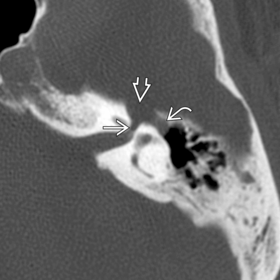
*Axial bone CT in a patient with CNVII paresis shows tubular enlargement of the distal labyrinthine segment , geniculate fossa , and anterior tympanic segment of the CNVII canal. Involvement of multiple segments of the facial nerve, as in this case, is highly suggestive of FNS.*
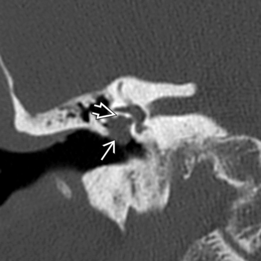
*Coronal bone CT in the same patient reveals the FNS involving the midtympanic segment of the facial nerve. Notice that the facial nerve bony canal "opens" into the middle ear mass .*
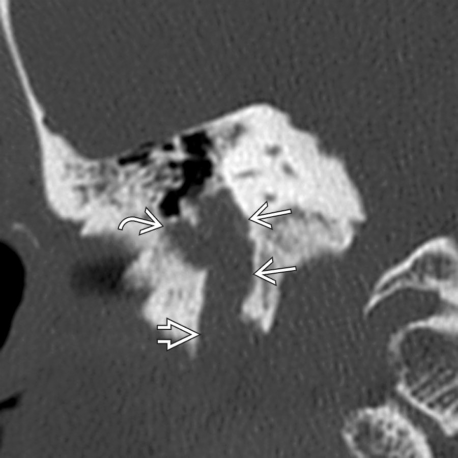
*Coronal bone CT in the same patient demonstrates that the FNS also involves the mastoid CNVII , exiting the enlarged stylomastoid foramen inferiorly. The tumor has broken into adjacent air cells on its lateral margin.*

*Axial temporal bone CT in a patient with a history of hearing loss and tinnitus shows a lobulated FNS extending along the course of the tympanic segment CNVII . The patient developed subjective right facial weakness not reproducible on clinical exam.*

*Axial T1 C+ FS MR in the same patient shows corresponding soft tissue enhancement along the tympanic segment of CNVII. Adjacent intrinsic mastoid hyperintensity was due to T1-hyperintense secretions, not enhancing tumor.*
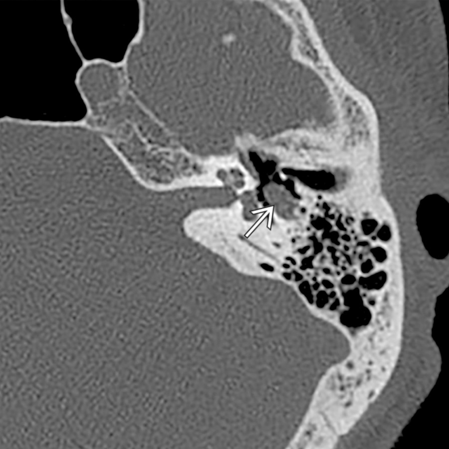
*Axial temporal bone CT in a patient with progressive partial left facial nerve paralysis associated with episodes of ipsilateral otalgia shows localized expansile enlargement of the tympanic segment of the facial nerve . The patient was initially treated with steroids for presumed Bell palsy, but subsequently underwent imaging when he did not respond.*
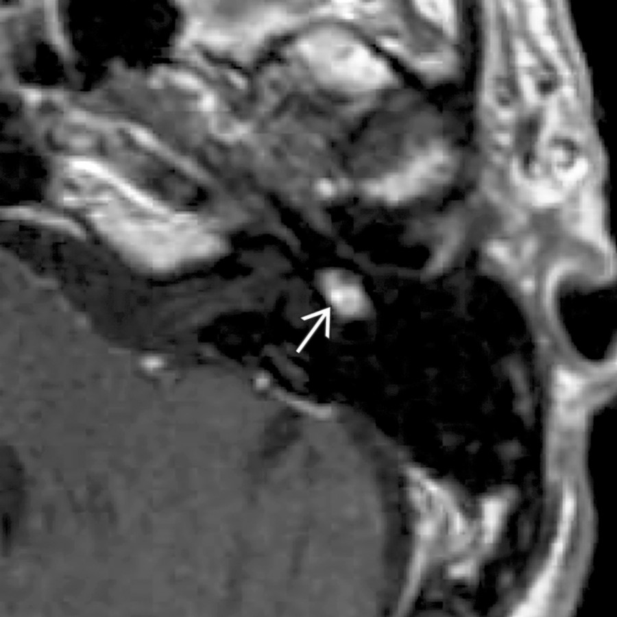
*Axial T1 C+ MR in the same patient shows enhancing schwannoma corresponding to the area of smooth osseous expansion on CT.*
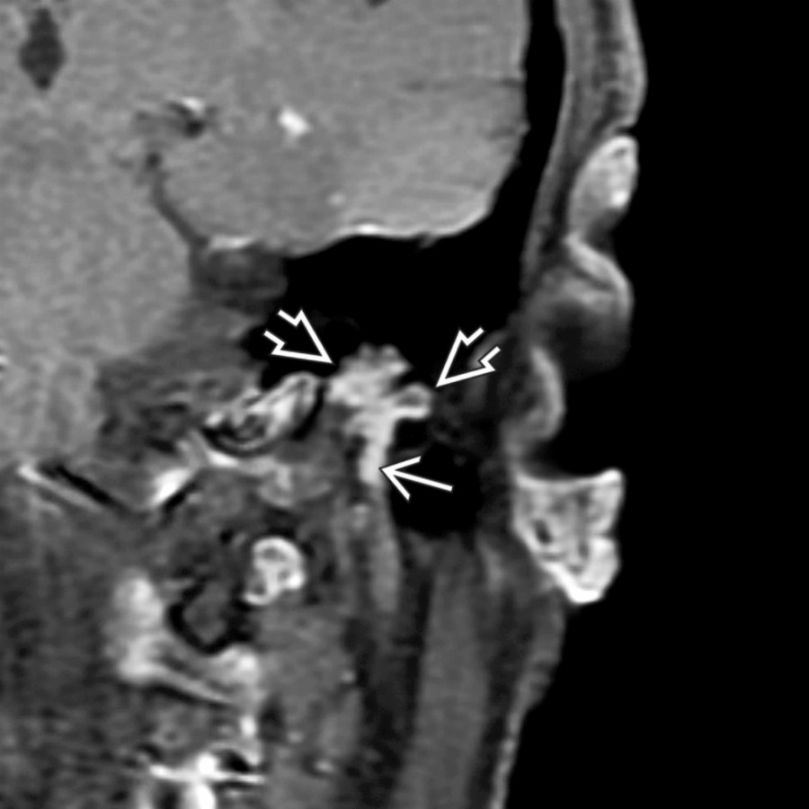
*Coronal T1 C+ FS MR in a patient with conductive hearing loss and facial twitching shows a multilobular, enhancing FNS that has broken into mastoid air cells and projects inferiorly along the mastoid CN segment .*
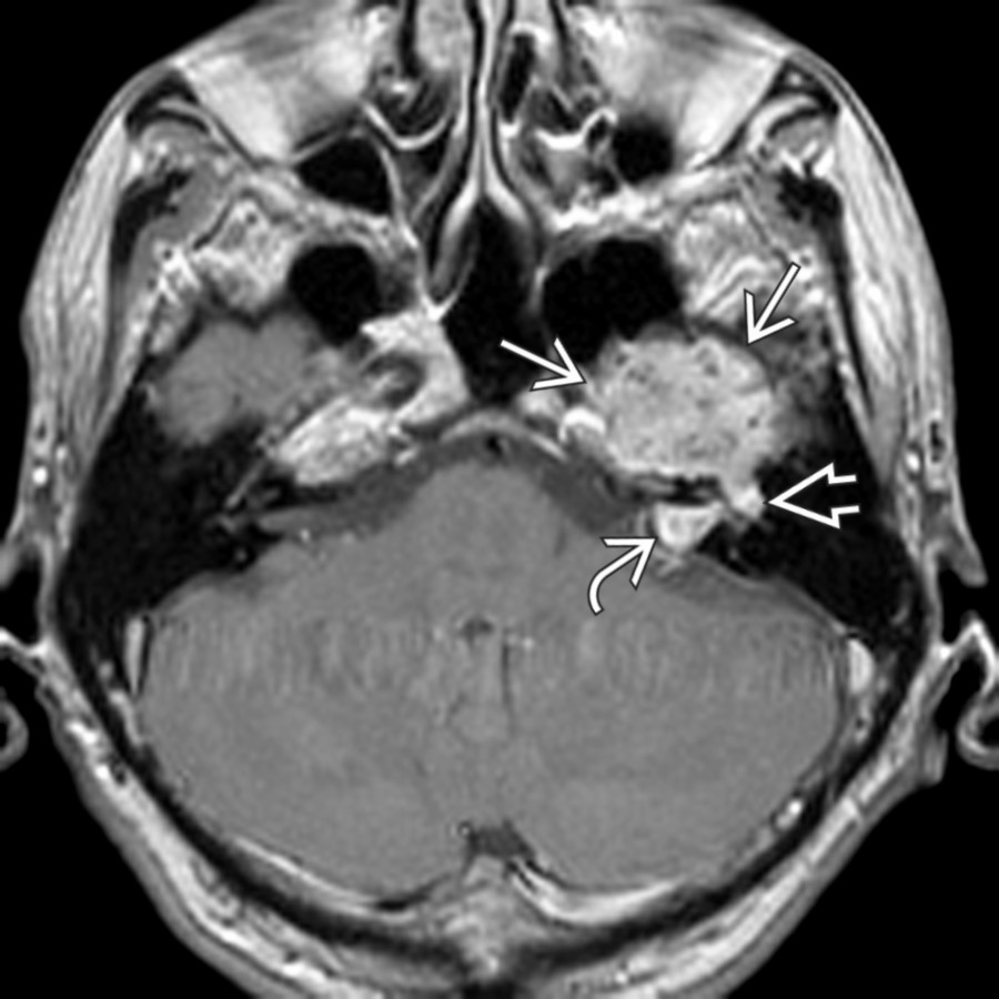
*Axial T1 C+ MR shows an enhancing mass projecting into the medial middle cranial fossa from the greater superficial petrosal nerve. FNS diagnosis is suggested if the projections along the tympanic CNVII and along the labyrinthine CNVII into the internal auditory canal are seen.*
### Additional Images

*Axial graphic shows a tubular facial nerve schwannoma (FNS) involving the labyrinthine segment, geniculate ganglion , and anterior tympanic segment of the intratemporal facial nerve.*

*Axial graphic shows a tubular facial nerve schwannoma (FNS) involving the labyrinthine segment, geniculate ganglion , and anterior tympanic segment of the intratemporal facial nerve.*

*Axial bone CT in a patient with CNVII paresis shows tubular enlargement of the distal labyrinthine segment , geniculate fossa , and anterior tympanic segment of the CNVII canal. Involvement of multiple segments of the facial nerve, as in this case, is highly suggestive of FNS.*

*Coronal bone CT in the same patient reveals the FNS involving the midtympanic segment of the facial nerve. Notice that the facial nerve bony canal "opens" into the middle ear mass .*

*Coronal bone CT in the same patient demonstrates that the FNS also involves the mastoid CNVII , exiting the enlarged stylomastoid foramen inferiorly. The tumor has broken into adjacent air cells on its lateral margin.*

*Axial temporal bone CT in a patient with a history of hearing loss and tinnitus shows a lobulated FNS extending along the course of the tympanic segment CNVII . The patient developed subjective right facial weakness not reproducible on clinical exam.*

*Axial T1 C+ FS MR in the same patient shows corresponding soft tissue enhancement along the tympanic segment of CNVII. Adjacent intrinsic mastoid hyperintensity was due to T1-hyperintense secretions, not enhancing tumor.*

*Axial temporal bone CT in a patient with progressive partial left facial nerve paralysis associated with episodes of ipsilateral otalgia shows localized expansile enlargement of the tympanic segment of the facial nerve . The patient was initially treated with steroids for presumed Bell palsy, but subsequently underwent imaging when he did not respond.*

*Axial T1 C+ MR in the same patient shows enhancing schwannoma corresponding to the area of smooth osseous expansion on CT.*

*Coronal T1 C+ FS MR in a patient with conductive hearing loss and facial twitching shows a multilobular, enhancing FNS that has broken into mastoid air cells and projects inferiorly along the mastoid CN segment .*

*Axial T1 C+ MR shows an enhancing mass projecting into the medial middle cranial fossa from the greater superficial petrosal nerve. FNS diagnosis is suggested if the projections along the tympanic CNVII and along the labyrinthine CNVII into the internal auditory canal are seen.*
### Additional Images
 .](67285cf5-0296-4375-ba37-782cb7da131e)
*Axial left ear temporal bone CT demonstrates FNS enlarging the labyrinthine and anterior tympanic segments of the CNVII canal, the geniculate fossa , and greater superficial petrosal nerve canal .*

*Coronal temporal bone CT of the right ear shows benign-appearing, smooth enlargement of the geniculate fossa by an ovoid-shaped FNS. Note that the roof of fossa is dehiscent .*

*Coronal T1WI C+ MR in the same patient reveals an avidly enhancing, ovoid FNS enlarging the geniculate ganglion. Notice cochlear signal just below the tumor .*

*Axial T1WI C+ MR reveals an enhancing FNS involving the geniculate ganglion and labyrinthine CNVII segment . The tumor has also invaded the subjacent cochlea .*

*Axial left ear temporal bone CT demonstrates FNS enlarging the labyrinthine and anterior tympanic segments of the CNVII canal, the geniculate fossa , and greater superficial petrosal nerve canal .*

*Coronal temporal bone CT of the right ear shows benign-appearing, smooth enlargement of the geniculate fossa by an ovoid-shaped FNS. Note that the roof of fossa is dehiscent .*

*Coronal T1WI C+ MR in the same patient reveals an avidly enhancing, ovoid FNS enlarging the geniculate ganglion. Notice cochlear signal just below the tumor .*

*Axial T1WI C+ MR reveals an enhancing FNS involving the geniculate ganglion and labyrinthine CNVII segment . The tumor has also invaded the subjacent cochlea .*
 . The nuclei have palisading.](53bff575-41b0-4097-9846-6ea9b9bfe264)
*Paraffin section, hematoxylin & eosin stain shows there is a juxtaposition and blending of cellular Antoni A areas with hypocellular Antoni B areas . The nuclei have palisading.*

*Paraffin section, S100 stain shows the neoplastic cells of a schwannoma are strongly and diffusely positive in both the nucleus and cytoplasm with S100 protein.*

*Axial bone CT in a patient with an external auditory canal (EAC) polyp shows an irregular mass centered in the area of the mastoid segment of CN that appears contiguous.*

*Axial T1WI C+ FS MR in the same patient reveals the enhancing FNS in the CNVII mastoid segment , projecting through a bony dehiscence into the EAC .*
. The nuclei have palisading.](53bff575-41b0-4097-9846-6ea9b9bfe264)
*Paraffin section, hematoxylin & eosin stain shows there is a juxtaposition and blending of cellular Antoni A areas with hypocellular Antoni B areas . The nuclei have palisading.*

*Paraffin section, S100 stain shows the neoplastic cells of a schwannoma are strongly and diffusely positive in both the nucleus and cytoplasm with S100 protein.*

*Axial bone CT in a patient with an external auditory canal (EAC) polyp shows an irregular mass centered in the area of the mastoid segment of CN that appears contiguous.*

*Axial T1WI C+ FS MR in the same patient reveals the enhancing FNS in the CNVII mastoid segment , projecting through a bony dehiscence into the EAC .*