---
title: "Ventriculomegaly"
docid: "f40bd6eb-e7e5-498a-8bde-ad6bcd546f21"
authors:
- key: "47381de4-c9fd-4999-8dd0-1808cd72db6b"
value: "Luke L. Linscott, MD"
breadcrumbs:
-
name: "Brain"
slug: "brain"
treeNodeId: "6d8829f1-14d7-45af-8675-255189aa526a"
-
name: "Differential Diagnosis"
slug: "differential-diagnosis"
treeNodeId: "a7fdd139-664e-4bb8-8d18-400e4733ff60"
-
name: "Ventricles, Periventricular Regions"
slug: "ventricles-periventricular-regions"
treeNodeId: "353c434a-a6fc-4ef1-8786-d30a1988a4dc"
-
name: "Generic Imaging Patterns"
slug: "generic-imaging-patterns"
treeNodeId: "969c31a2-ef56-4fc3-9125-05857cf9aac3"
-
name: "Ventriculomegaly"
slug: "ventriculomegaly"
treeNodeId: null
category: "Brain"
documentVersionId: "69dd2446-06ad-4f37-bba3-f59d4c5d40b8"
imageCount: 26
lastUpdated: "01/23/23"
pageDescription: "Ventriculomegaly"
pageKeywords: "Brain, Differential Diagnosis, Ventricles, Periventricular Regions, Generic Imaging Patterns, Ventriculomegaly"
pageTitle: "Ventriculomegaly | STATdx"
enhancedTitle: "Ventriculomegaly"
type: "DDX"
references: true
breadcrumbs:
- "Brain"
- "Differential Diagnosis"
- "Ventricles, Periventricular Regions"
- "Generic Imaging Patterns"
- "Ventriculomegaly"
---
# ESSENTIAL INFORMATION
- ## Key Differential Diagnosis Issues
- 2 main causes of ventriculomegaly
- ↑ CSF pressures → hydrocephalus
- ↓ brain volume → passive ventricular enlargement
- Clinical features to consider
- Head circumference is critical to interpreting significance of enlarged ventricles
- ↑ head size: Hydrocephalus
- ↓ head size: Brain parenchymal volume loss
- Patients may come to clinical attention during prenatal screening or well-child check-ups
- Imaging features of hydrocephalus
- Etiologies to consider
- Obstruction usually occurs at cerebral aqueduct, foramen of Monro, or 4th ventricular outlets
- e.g., aqueductal stenosis, obstructing tumor
- Communicating hydrocephalus is due to CSF overproduction or poor CSF resorption
- e.g., choroid plexus papilloma, benign enlargement of subarachnoid spaces
- Some etiologies can cause either or mixed forms
- e.g., posthemorrhagic, postinfectious, vein of Galen malformation (VGAM)
- Imaging signs of hydrocephalus
- Macrocephaly: ↑ craniofacial ratio
- Rounded margins of lateral ventricles, depressed floor of 3rd ventricle
- Transependymal edema: Periventricular white matter (WM) ↑ T2/FLAIR signal, greatest at frontal & occipital horns; effacement of cerebral sulci & basilar cisterns
- Imaging features of volume loss
- Etiologies to consider
- Diffuse vs. focal cortical ischemia
- e.g., hypoxic-ischemic encephalopathy (HIE) vs. arterial ischemic stroke
- Symmetric vs. focal WM volume loss
- e.g., periventricular leukomalacia vs. porencephaly
- Imaging signs of brain parenchymal volume loss
- Microcephaly: ↓ craniofacial ratio
- Angular margins of lateral ventricles
- Prominent sulci & basilar cisterns
- Modality considerations
- Fetal US for detection of in utero ventriculomegaly
- Fetal MR is excellent for underlying/associated anomalies
- US is important tool for initial evaluation of neonates/infants with suspected ventriculomegaly
- Requires open anterior fontanelle
- CT is often 1st-line modality in acute presentations
- MR is most definitive test for determination of underlying cause of ventriculomegaly
- Best test for determining cause of hydrocephalus
- Best evaluation of brain parenchyma to detect possible volume loss
- ## Helpful Clues for Common Diagnoses
- **Fetal Ventriculomegaly**
- Likelihood of normal neurologic development based on lateral ventricular size in utero
- Mild (10-12 mm): > 90%
- Moderate (13-15 mm): 80-93%
- Severe (> 15 mm): ~ 62%
- Underlying causes of fetal ventriculomegaly
- Infectious (e.g., CMV), genetic (e.g., trisomy), malformations (e.g., Chiari 2)
- CMV testing & amniocentesis typically performed
- Obstructive causes considered separately
- **Benign Enlargement of Subarachnoid Spaces**
- a.k.a. benign hydrocephalus of infancy, benign macrocrania of infancy, external hydrocephalus
- Enlarged subarachnoid spaces (SAS) ± mild ventriculomegaly
- Findings develop in 1st few months of life & usually resolve by ~ 2 years of age
- Often family history of macrocephaly
- Normal development or mild developmental delay
- Imaging: Prominent SAS over frontal & parietal convexities with normal vessels traversing SAS
- No displacement of arachnoid membrane from dura (i.e., no subdural fluid)
- **Chiari 2 Malformation**
- Brain malformation due to open neural tube defect (most commonly lumbosacral myelomeningocele)
- Hydrocephalus requiring shunting occurs almost universally without in utero surgical repair
- Imaging: Small posterior fossa, inferior migration of cerebellum/brainstem, beaked tectum, dysplastic corpus callosum, scalloping of dorsal clivus
- **Aqueductal Stenosis**
- Congenital atresia/stenosis of cerebral aqueduct
- Imaging: Lateral & 3rd ventricle dilation with normal 4th ventricle
- Narrowed/absent cerebral aqueduct on 3D SSFP MR
- Lack of flow void within cerebral aqueduct on T2
- **Acquired Hydrocephalus**
- 2 main types: Communicating & noncommunicating
- Communicating: ↑ production or ↓ resorption of CSF
- Obstructive: Blockage of ventricular outlets
- Depending on cause, may present enlarging head or signs of ↑ intracranial pressure
- May have signs of ↑ intracranial pressure
- Treatment: Shunting or 3rd ventriculostomy
- **Posthemorrhagic hydrocephalus**
- Most common in premature infants with germinal matrix/intraventricular hemorrhage (IVH)
- Look for hemosiderin along ventricles/brainstem
- SWI/GRE is most sensitive for detection of prior IVH
- **Acute infectious hydrocephalus**
- During acute bacterial infection, ↑ head size may occur due to ↑ extraaxial spaces from meningitis & subdural/epidural empyema
- **Postinfectious hydrocephalus**
- Obstruction may occur due to synechiae
- Communicating hydrocephalus may occur due to arachnoid granulation dysfunction
- **Obstructing tumor**
- Smaller tumors in critical locations may obstruct
- e.g., subependymal giant cell astrocytoma (SEGA) at foramen of Monro, tectal plate glioma at cerebral aqueduct
- Larger tumors anywhere in brain may obstruct, particularly in posterior fossa
- **CSF overproduction (choroid plexus tumors)**
- Choroid plexus papilloma/carcinoma causes CSF overproduction
- Inability to resorb excess CSF → ↑ ventricular size
- **Brain Volume Loss**
- Results in ventricular enlargement
- Focal (e.g., porencephaly, stroke)
- Symmetric (e.g., periventricular leukomalacia, metabolic disease)
- **Hypoxic-ischemic injury**
- Often results in symmetric or asymmetric brain volume loss & associated ventricular enlargement
- Look for characteristic patterns of injury (e.g., perirolandic or watershed)
- **Arterial ischemic stroke**
- Most often occurs in perinatal period or related to embolic phenomenon
- Encephalomalacia develops in arterial territory with focal ventricular enlargement
- **Periventricular leukomalacia**
- Sequelae of WM injury of prematurity
- Usually symmetric WM volume loss with little or no associated gliosis (↑ FLAIR signal)
- **Porencephaly**
- Most often results from grade 4 IVH, but any insult early in development may cause porencephaly
- Porencephaly develops rather than encephalomalacia as no astrocytic response occurs in very young brains
- **Metabolic brain disease**
- Numerous diseases cause significant brain volume loss, resulting in ventriculomegaly
- Usually symmetric; may be
- Frontal predominant (e.g., Alexander disease)
- Posterior predominant (e.g., X-linked adrenoleukodystrophy)
- Diffuse (e.g., metachromatic leukodystrophy)
- ## Helpful Clues for Less Common Diagnoses
- **Hemimegalencephaly**
- Unilateral abnormal neuronal proliferation & migration
- Neonate/infant: ↑ T1 & ↓ T2 in WM with blurred gray matter/WM junctions
- Often shows enlarged ipsilateral ventricle
- **Vein of Galen Aneurysmal Malformation**
- Ectatic vascular structure is median prosencephalic vein, not vein of Galen, which never forms
- High-flow vascular malformation due to direct communication between arteries & median prosencephalic vein
- Ventricular enlargement may occur due to
- Mass effect of malformation on cerebral aqueduct
- ↑ venous pressure & poor CSF resorption
- Parenchymal insult with volume loss
- After birth, ↑ in blood flow through malformation
- Up to 80% of left ventricular output may supply brain
- Leads to ↑ cardiac output & heart failure
- **Dandy-Walker Malformation**
- Vermian hypoplasia with large posterior fossa cyst that communicates with 4th ventricle but not SAS
- Lambdoid-torcular inversion
- Often associated with hydrocephalus
- **Hydranencephaly**
- In utero destruction of brain in internal carotid artery territories
- May cause poor CSF regulation & hydrocephalus, requiring shunting
- **Holoprosencephaly**
- Holoprosencephaly (alobar form) often develops ↑ size of monoventricle over time
- Typically treated with CSF shunting
## References
# Selected References
1. [Adiyaman D et al: Contribution of fetal magnetic resonance imaging in the evaluation of neurosonographically detected cases of isolated mild and moderate cerebral ventriculomegaly. J Obstet Gynaecol Res. 48(9):2314-24, 2022](http://www.ncbi.nlm.nih.gov/pubmed/?term=35778980%5Bpmid%5D)
1. [D'Addario V: Diagnostic approach to fetal ventriculomegaly. J Perinat Med. ePub, 2022](http://www.ncbi.nlm.nih.gov/pubmed/?term=36005554%5Bpmid%5D)
1. [Bauer DF et al: Congress of Neurological Surgeons Systematic Review and Evidence-Based Guidelines on the Treatment of Pediatric Hydrocephalus: Update of the 2014 Guidelines. Neurosurgery. 87(6):1071-75, 2020](http://www.ncbi.nlm.nih.gov/pubmed/?term=34791462%5Bpmid%5D)
1. [Di Mascio D et al: Systematic review and meta-analysis on the role of prenatal magnetic resonance imaging in the era of fetal neurosonography: mild and moderate ventriculomegaly. Ultrasound Obstet Gynecol. 54(2):164-71, 2019](http://www.ncbi.nlm.nih.gov/pubmed/?term=30549340%5Bpmid%5D)
1. [Katz JA et al: Utility of prenatal MRI in the evaluation and management of fetal ventriculomegaly. J Perinatol. 38(11):1444-52, 2018](http://www.ncbi.nlm.nih.gov/pubmed/?term=30158676%5Bpmid%5D)
1. [Orrù E et al: The child with macrocephaly: differential diagnosis and neuroimaging findings. AJR Am J Roentgenol. 210(4):848-59, 2018](http://www.ncbi.nlm.nih.gov/pubmed/?term=29470161%5Bpmid%5D)
1. [Rashid QT et al: Time trends and age-related etiologies of pediatric hydrocephalus: results of a groupwise analysis in a clinical cohort. Childs Nerv Syst. 28(2):221-7, 2012](http://www.ncbi.nlm.nih.gov/pubmed/?term=21818584%5Bpmid%5D)
1. [Kurihara Y et al: MR imaging of ventriculomegaly--a qualitative and quantitative comparison of communicating hydrocephalus, central atrophy, and normal studies. J Magn Reson Imaging. 5(4):451-6, 1995](http://www.ncbi.nlm.nih.gov/pubmed/?term=7549210%5Bpmid%5D)
## Images
### Selected Images
 (11 mm). If the ventriculomegaly worsens later in pregnancy, fetal or postnatal MR imaging should be obtained.](images/app.statdx.com_image_thumbnail_70468ab8-1e6a-4667-8825-a704fdbf86ce_annotated_true_size_900_quality_90_cfed8e44_20251018T165228Z.jpg)
**Fetal Ventriculomegaly**
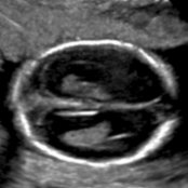
*Transverse oblique US in an 18-week fetus shows mild enlargement of the lateral ventricles (11 mm). If the ventriculomegaly worsens later in pregnancy, fetal or postnatal MR imaging should be obtained.*
**Fetal Ventriculomegaly**
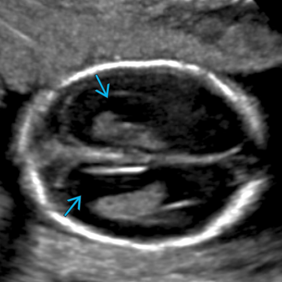
*Transverse oblique US in an 18-week fetus shows mild enlargement of the lateral ventricles (11 mm). If the ventriculomegaly worsens later in pregnancy, fetal or postnatal MR imaging should be obtained.*
**Fetal Ventriculomegaly**
*Transverse oblique US in an 18-week fetus shows mild enlargement of the lateral ventricles (11 mm). If the ventriculomegaly worsens later in pregnancy, fetal or postnatal MR imaging should be obtained.*

**Fetal Ventriculomegaly**
*Transverse oblique US in an 18-week fetus shows mild enlargement of the lateral ventricles (11 mm). If the ventriculomegaly worsens later in pregnancy, fetal or postnatal MR imaging should be obtained.*
 & thinning of the cerebrum
  . With severe fetal ventriculomegaly (particularly in the setting of aqueductal stenosis), the septum pellucidum (& even the cerebral mantle) may become disrupted.](images/app.statdx.com_image_thumbnail_82f99506-7e41-4801-843b-30dd71594c0b_annotated_true_size_900_quality_90_45cd65ce_20251018T165228Z.jpg)
**Fetal Ventriculomegaly**
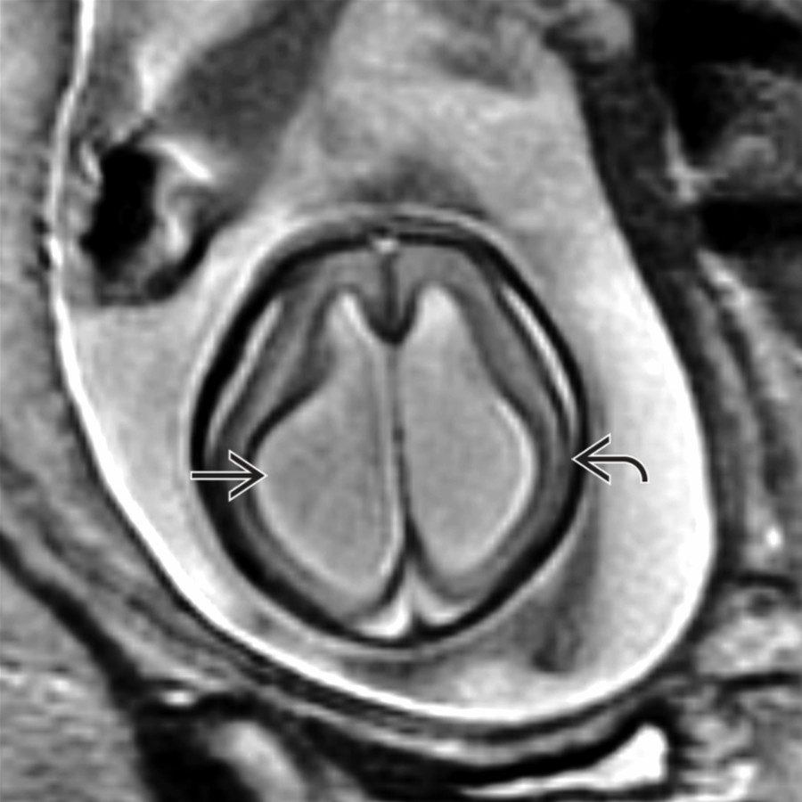
*Axial T2 SSFSE MR in a fetus shows marked enlargement (> 15 mm) of the lateral ventricles & thinning of the cerebrum . With severe fetal ventriculomegaly (particularly in the setting of aqueductal stenosis), the septum pellucidum (& even the cerebral mantle) may become disrupted.*
**Fetal Ventriculomegaly**
*Axial T2 SSFSE MR in a fetus shows marked enlargement (> 15 mm) of the lateral ventricles & thinning of the cerebrum . With severe fetal ventriculomegaly (particularly in the setting of aqueductal stenosis), the septum pellucidum (& even the cerebral mantle) may become disrupted.*
 ventricles with moderate enlargement of the bifrontal subarachnoid spaces (SAS), which are traversed by normal veins
  . Mild ventriculomegaly is often seen in benign enlargement of subarachnoid spaces (BESS).](images/app.statdx.com_image_thumbnail_0db6c91f-cab8-4b5c-ac67-4fb4207fefb1_annotated_true_size_900_quality_90_37c9b115_20251018T165228Z.jpg)
**Benign Enlargement of Subarachnoid Spaces**
*Coronal T2 MR in a 1-year-old with macrocephaly shows mild enlargement of the lateral & 3rd ventricles with moderate enlargement of the bifrontal subarachnoid spaces (SAS), which are traversed by normal veins . Mild ventriculomegaly is often seen in benign enlargement of subarachnoid spaces (BESS).*
**Benign Enlargement of Subarachnoid Spaces**
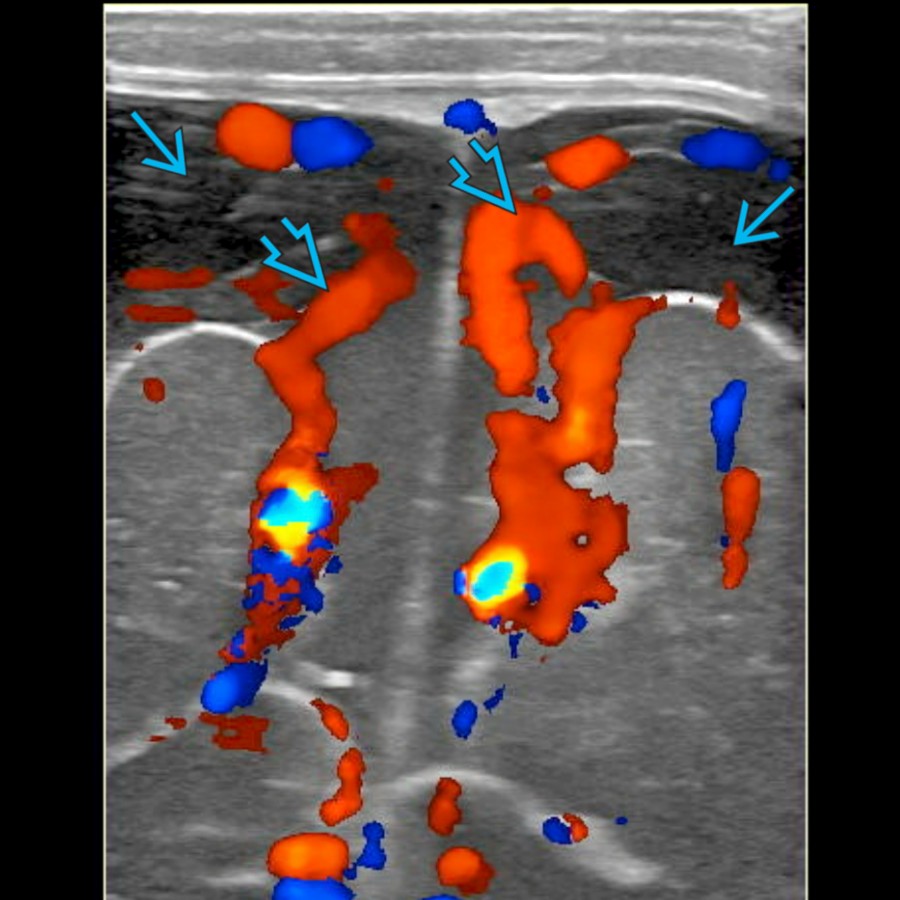
*Coronal color Doppler US in a 9-month-old with BESS shows normal vessels coursing through prominent fluid , an expected finding that helps differentiate the SAS from subdural collections.*
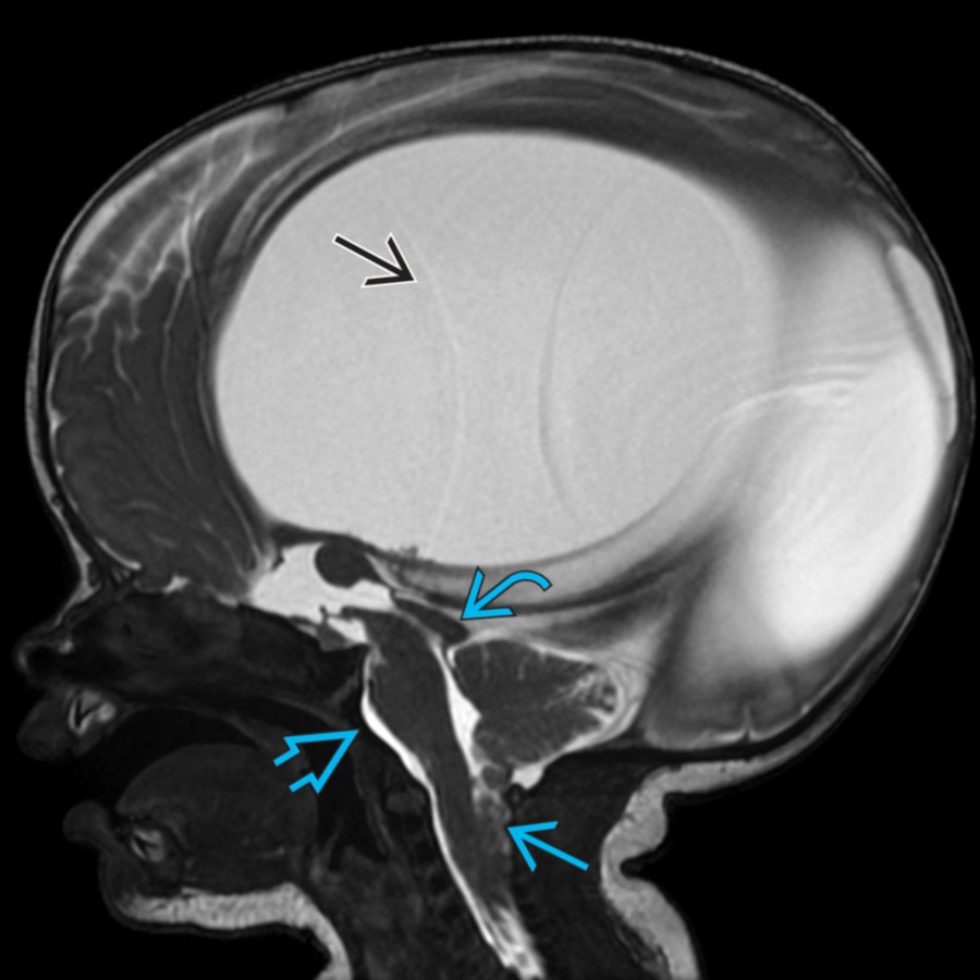
**Chiari 2 Malformation**
*Sagittal 3D SSFP MR in a neonate with a myelomeningocele shows marked enlargement of the lateral ventricles & characteristic features of Chiari 2 malformation: Small posterior fossa, brainstem & cerebellar descent , tectal beaking , & scalloped clivus .*
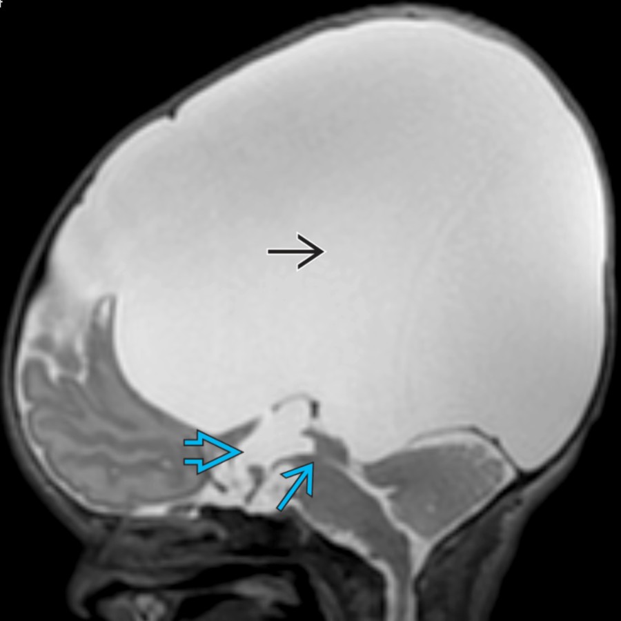
**Aqueductal Stenosis**
*Sagittal T2 MR in a neonate shows massive enlargement of the lateral ventricles due to obstruction at the level of the cerebral aqueduct . The 3rd ventricle is also enlarged , but the 4th ventricle is normal, typical of this disorder.*
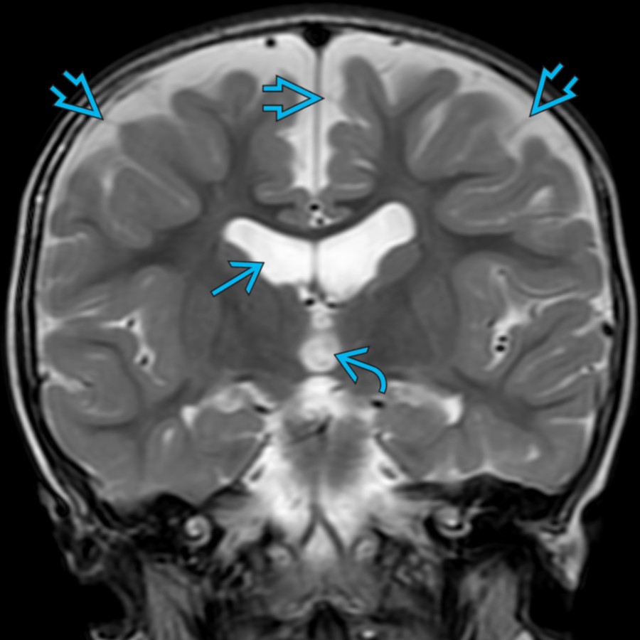
**Benign Enlargement of Subarachnoid Spaces**
*Coronal T2 MR in a 1-year-old with macrocephaly shows mild enlargement of the lateral & 3rd ventricles with moderate enlargement of the bifrontal subarachnoid spaces (SAS), which are traversed by normal veins . Mild ventriculomegaly is often seen in benign enlargement of subarachnoid spaces (BESS).*

**Benign Enlargement of Subarachnoid Spaces**
*Coronal color Doppler US in a 9-month-old with BESS shows normal vessels coursing through prominent fluid , an expected finding that helps differentiate the SAS from subdural collections.*

**Chiari 2 Malformation**
*Sagittal 3D SSFP MR in a neonate with a myelomeningocele shows marked enlargement of the lateral ventricles & characteristic features of Chiari 2 malformation: Small posterior fossa, brainstem & cerebellar descent , tectal beaking , & scalloped clivus .*

**Aqueductal Stenosis**
*Sagittal T2 MR in a neonate shows massive enlargement of the lateral ventricles due to obstruction at the level of the cerebral aqueduct . The 3rd ventricle is also enlarged , but the 4th ventricle is normal, typical of this disorder.*
 , & 4th ventricles. Note the dark hemosiderin lining the pial surface of the brainstem from prior intraventricular hemorrhage (IVH).](images/app.statdx.com_image_thumbnail_1507c0c1-9157-4c19-89cf-df575996cf50_annotated_true_size_900_quality_90_5c58dea8_20251018T165228Z.jpg)
**Posthemorrhagic Hydrocephalus**
*Sagittal T2 MR in a 1-month-old former premature infant with posthemorrhagic hydrocephalus shows marked enlargement of the lateral , 3rd , & 4th ventricles. Note the dark hemosiderin lining the pial surface of the brainstem from prior intraventricular hemorrhage (IVH).*
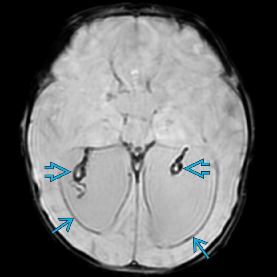
**Posthemorrhagic Hydrocephalus**
*Axial SWI MR in a 3-month-old former premature infant with posthemorrhagic hydrocephalus shows signal loss along the ependymal margins of the ventricles & choroid plexus , consistent with prior IVH.*
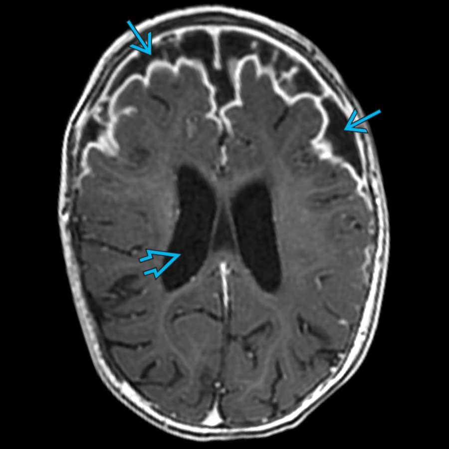
**Acute Infectious Hydrocephalus**
*Axial T1 C+ MR in a 7-year-old with Haemophilus influenzae meningitis shows marked expansion of the bifrontal SAS & mild enlargement of the lateral ventricles .*
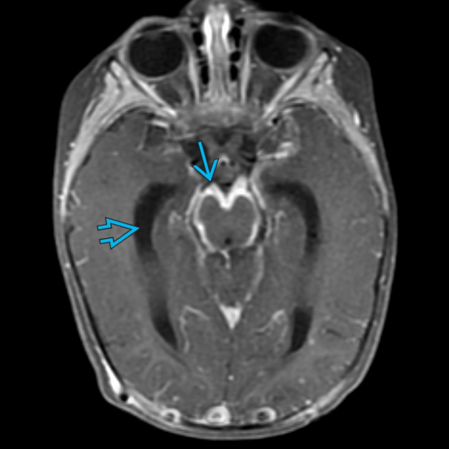
**Postinfectious Hydrocephalus**
*Axial T1 C+ FS MR in an 8-month-old with tuberculous meningitis shows extensive basilar leptomeningeal enhancement & enlargement of the lateral ventricles , resulting in macrocephaly. Granulomatous infections are more likely to result in hydrocephalus compared to other bacterial meningitis.*
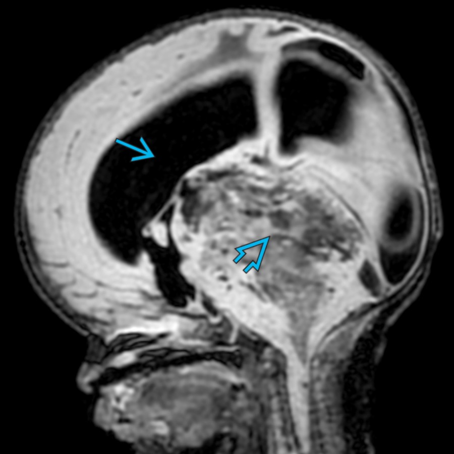
**Obstructing Tumor**
*Sagittal FLAIR MR in a neonate with a large, obstructing, hemorrhagic posterior fossa mass shows enlargement of the lateral ventricles & posterior fossa. Note the ↑ craniofacial ratio.*
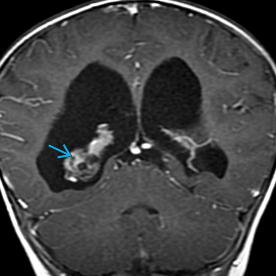
**CSF Overproduction (Choroid Plexus Tumor)**
*Coronal T1 C+ MR in a 1-year-old with a choroid plexus papilloma shows an enhancing mass in the right choroid plexus. The lateral ventricles are enlarged without evidence of obstruction. Hydrocephalus in this case is due to overproduction of CSF by the tumor.*
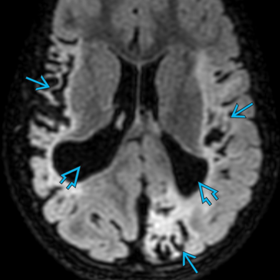
**Hypoxic-Ischemic Injury**
*Axial FLAIR MR in a 4-year-old with a history of perinatal hypoxic-ischemic injury (HII) shows extensive areas of cortical encephalomalacia . Note the localized areas of ventriculomegaly due to overlying brain volume loss.*
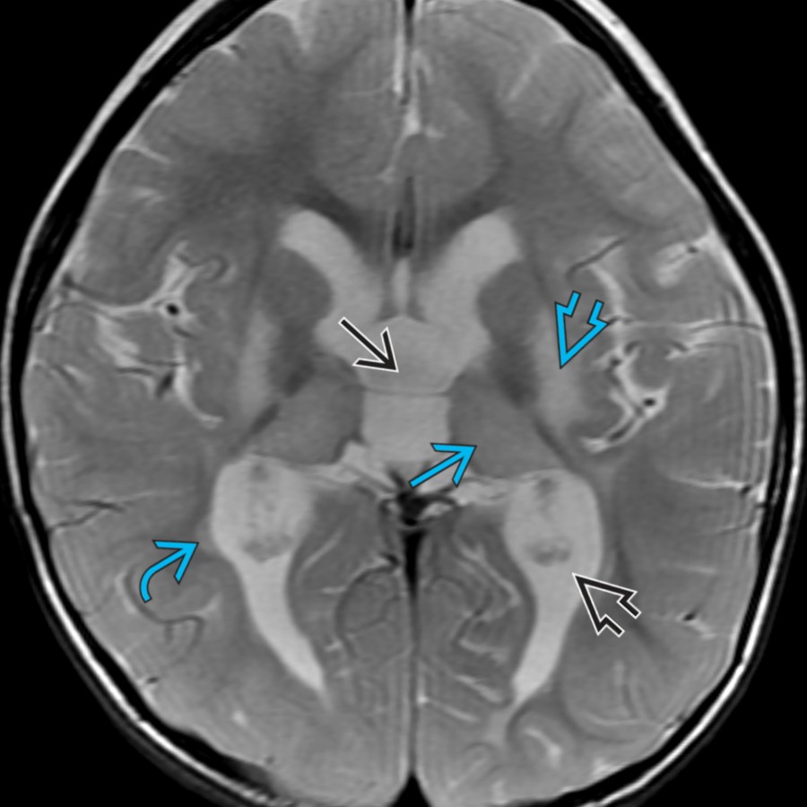
**Hypoxic-Ischemic Injury**
*Axial T2 MR in a 2-year-old with a history of perinatal HII shows symmetric areas of signal abnormality & volume loss involving the thalami , putamina , & periventricular white matter , resulting in enlargement of the lateral & 3rd ventricles.*

**Arterial Ischemic Stroke**
*Axial T2 MR in a 9-month-old with previous infarction shows extensive cystic encephalomalacia in right middle cerebral artery (MCA) territory with resultant asymmetric enlargement of the right lateral ventricle .*

**Periventricular Leukomalacia**
*Axial FLAIR MR in a 7-year-old with a history of extreme prematurity & periventricular leukomalacia (PVL) shows symmetric focal enlargement of the atria with adjacent white matter volume loss. The relative lack of abnormal FLAIR signal compared to the degree of volume loss is typical of PVL.*

**Porencephaly**
*Coronal US at 2 days of life in an extremely premature (23-week) infant shows a large right germinal matrix hemorrhage with associated hemorrhagic venous infarction in the right frontoparietal white matter.*

**Porencephaly**
*Coronal T2 MR in the same patient 3 months later shows the expected development of right parietal porencephaly & focal ventricular enlargement. Note the rim of T2 hypointensity , related to hemosiderin, which will eventually resolve.*

**Metabolic Brain Disease**
*Axial T2 MR in a teenager with metachromatic leukodystrophy shows ↑ signal & volume loss in the periventricular & deep white matter with sparing of the subcortical white matter, characteristic of this disease. Note the enlargement of the lateral ventricles & sulci due to the brain volume loss.*

**Metabolic Brain Disease**
*Axial T2 MR in a teenager with vanishing white matter disease shows extensive ↑ signal intensity & volume loss in the white matter with associated enlargement of the lateral & 3rd ventricles & sulci .*

**Hemimegalencephaly**
*Axial T1 MR in a neonate with seizures & hemimegalencephaly shows ↑ size of the left parietooccipital hemisphere with loss of normal sulcation & markedly abnormal neuronal organization . Also note enlargement of the ipsilateral occipital horn .*

**Hemimegalencephaly**
*Axial T1 MR in a 3-day-old with left hemimegalencephaly shows marked occipital horn enlargement . Note the abnormal white matter in the left frontal lobe. Enlargement of the ipsilateral occipital horn is common in this disease.*

**Vein of Galen Aneurysmal Malformation**
*Coronal T2 MR in a neonate with VGAM shows a markedly enlarged central vein with numerous enlarged choroidal & pericallosal feeding arteries. Note the enlarged ventricles , which are likely due to ↓ resorption of CSF due to ↑ venous pressures.*

**Dandy-Walker Malformation**
*Sagittal 3D SSFP MR in a 2-month-old with Dandy-Walker malformation shows a small cerebellar vermis & large posterior fossa cyst that is continuous with the 4th ventricle. There is elevation of the tentorium & torcular Herophili .*

**Hydranencephaly**
*Axial NECT in a 4-year-old with hydranencephaly shows porencephaly in the bilateral MCA & left anterior cerebral artery (ACA) territories in continuity with the lateral ventricles. Note the intact falx . Patients with hydranencephaly typically become macrocephalic due to poor CSF regulation.*

**Holoprosencephaly**
*Sagittal T2 MR in a neonate with holoprosencephaly shows an enlarged monoventricle . However, the patient is microcephalic overall due to the ↓ brain parenchymal volume.*
, & 4th ventricles. Note the dark hemosiderin lining the pial surface of the brainstem from prior intraventricular hemorrhage (IVH).](images/app.statdx.com_image_thumbnail_1507c0c1-9157-4c19-89cf-df575996cf50_annotated_true_size_900_quality_90_5c58dea8_20251018T165228Z.jpg)
**Posthemorrhagic Hydrocephalus**
*Sagittal T2 MR in a 1-month-old former premature infant with posthemorrhagic hydrocephalus shows marked enlargement of the lateral , 3rd , & 4th ventricles. Note the dark hemosiderin lining the pial surface of the brainstem from prior intraventricular hemorrhage (IVH).*

**Posthemorrhagic Hydrocephalus**
*Axial SWI MR in a 3-month-old former premature infant with posthemorrhagic hydrocephalus shows signal loss along the ependymal margins of the ventricles & choroid plexus , consistent with prior IVH.*

**Acute Infectious Hydrocephalus**
*Axial T1 C+ MR in a 7-year-old with Haemophilus influenzae meningitis shows marked expansion of the bifrontal SAS & mild enlargement of the lateral ventricles .*

**Postinfectious Hydrocephalus**
*Axial T1 C+ FS MR in an 8-month-old with tuberculous meningitis shows extensive basilar leptomeningeal enhancement & enlargement of the lateral ventricles , resulting in macrocephaly. Granulomatous infections are more likely to result in hydrocephalus compared to other bacterial meningitis.*

**Obstructing Tumor**
*Sagittal FLAIR MR in a neonate with a large, obstructing, hemorrhagic posterior fossa mass shows enlargement of the lateral ventricles & posterior fossa. Note the ↑ craniofacial ratio.*

**CSF Overproduction (Choroid Plexus Tumor)**
*Coronal T1 C+ MR in a 1-year-old with a choroid plexus papilloma shows an enhancing mass in the right choroid plexus. The lateral ventricles are enlarged without evidence of obstruction. Hydrocephalus in this case is due to overproduction of CSF by the tumor.*

**Hypoxic-Ischemic Injury**
*Axial FLAIR MR in a 4-year-old with a history of perinatal hypoxic-ischemic injury (HII) shows extensive areas of cortical encephalomalacia . Note the localized areas of ventriculomegaly due to overlying brain volume loss.*

**Hypoxic-Ischemic Injury**
*Axial T2 MR in a 2-year-old with a history of perinatal HII shows symmetric areas of signal abnormality & volume loss involving the thalami , putamina , & periventricular white matter , resulting in enlargement of the lateral & 3rd ventricles.*

**Arterial Ischemic Stroke**
*Axial T2 MR in a 9-month-old with previous infarction shows extensive cystic encephalomalacia in right middle cerebral artery (MCA) territory with resultant asymmetric enlargement of the right lateral ventricle .*

**Periventricular Leukomalacia**
*Axial FLAIR MR in a 7-year-old with a history of extreme prematurity & periventricular leukomalacia (PVL) shows symmetric focal enlargement of the atria with adjacent white matter volume loss. The relative lack of abnormal FLAIR signal compared to the degree of volume loss is typical of PVL.*

**Porencephaly**
*Coronal US at 2 days of life in an extremely premature (23-week) infant shows a large right germinal matrix hemorrhage with associated hemorrhagic venous infarction in the right frontoparietal white matter.*

**Porencephaly**
*Coronal T2 MR in the same patient 3 months later shows the expected development of right parietal porencephaly & focal ventricular enlargement. Note the rim of T2 hypointensity , related to hemosiderin, which will eventually resolve.*

**Metabolic Brain Disease**
*Axial T2 MR in a teenager with metachromatic leukodystrophy shows ↑ signal & volume loss in the periventricular & deep white matter with sparing of the subcortical white matter, characteristic of this disease. Note the enlargement of the lateral ventricles & sulci due to the brain volume loss.*

**Metabolic Brain Disease**
*Axial T2 MR in a teenager with vanishing white matter disease shows extensive ↑ signal intensity & volume loss in the white matter with associated enlargement of the lateral & 3rd ventricles & sulci .*

**Hemimegalencephaly**
*Axial T1 MR in a neonate with seizures & hemimegalencephaly shows ↑ size of the left parietooccipital hemisphere with loss of normal sulcation & markedly abnormal neuronal organization . Also note enlargement of the ipsilateral occipital horn .*

**Hemimegalencephaly**
*Axial T1 MR in a 3-day-old with left hemimegalencephaly shows marked occipital horn enlargement . Note the abnormal white matter in the left frontal lobe. Enlargement of the ipsilateral occipital horn is common in this disease.*

**Vein of Galen Aneurysmal Malformation**
*Coronal T2 MR in a neonate with VGAM shows a markedly enlarged central vein with numerous enlarged choroidal & pericallosal feeding arteries. Note the enlarged ventricles , which are likely due to ↓ resorption of CSF due to ↑ venous pressures.*

**Dandy-Walker Malformation**
*Sagittal 3D SSFP MR in a 2-month-old with Dandy-Walker malformation shows a small cerebellar vermis & large posterior fossa cyst that is continuous with the 4th ventricle. There is elevation of the tentorium & torcular Herophili .*

**Hydranencephaly**
*Axial NECT in a 4-year-old with hydranencephaly shows porencephaly in the bilateral MCA & left anterior cerebral artery (ACA) territories in continuity with the lateral ventricles. Note the intact falx . Patients with hydranencephaly typically become macrocephalic due to poor CSF regulation.*

**Holoprosencephaly**
*Sagittal T2 MR in a neonate with holoprosencephaly shows an enlarged monoventricle . However, the patient is microcephalic overall due to the ↓ brain parenchymal volume.*